For decades, the standard scientific narrative of the brain has been dominated by the study of genetics, proteins, and the electrical firing of neurons. While neuroscientists have mapped the brain’s connectome—the complex web of wiring that facilitates communication—and the transcriptome—the blueprint of gene expression—a critical, albeit elusive, component has remained largely in the shadows: the lipidome. Lipids, the diverse class of fatty molecules that constitute the structural bedrock of cellular membranes and the insulating myelin sheaths of nerve fibers, have historically been viewed as mere passive building blocks. However, a landmark study published in the journal Nature has fundamentally challenged this perspective, providing the first high-resolution, 3D atlas of lipid distribution in the mammalian brain.
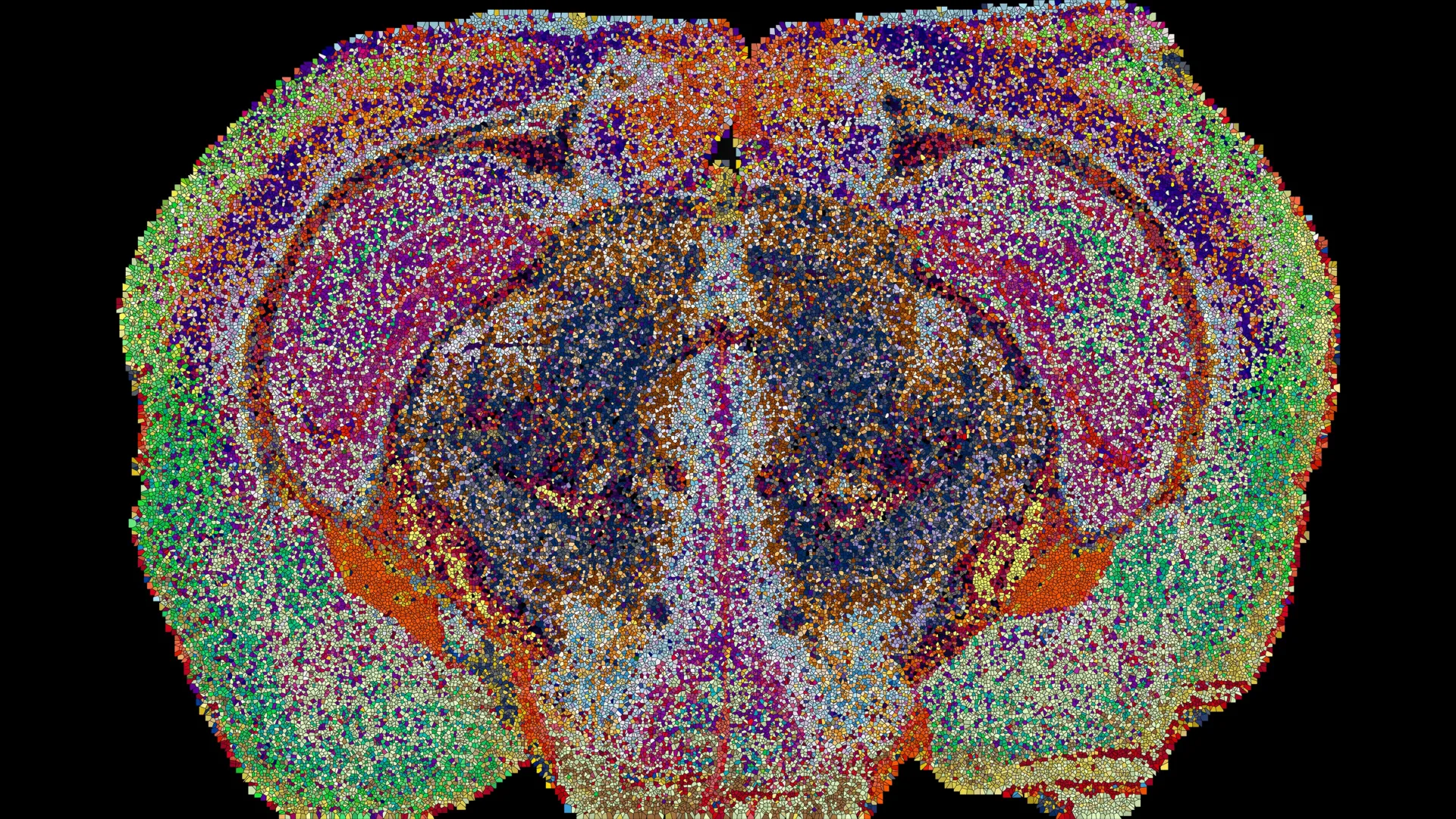
Led by researchers at the École Polytechnique Fédérale de Lausanne (EPFL), including Giovanni D’Angelo, Luca Fusar Bassini, and Gioele La Manno, the project represents a significant technological leap. By integrating mass spectrometry imaging with advanced machine learning, the team has identified 539 chemically distinct territories within the mouse brain, a discovery that suggests the brain is organized by a sophisticated, previously invisible chemical code.
The Technological Breakthrough: Beyond Traditional Imaging
The difficulty in mapping brain lipids has long been a technical hurdle. Conventional histology and imaging techniques, such as fluorescent tagging or standard MRI, are excellent for visualizing cellular architecture or blood flow, but they are notoriously poor at distinguishing between the vast, structurally similar families of lipids. Because lipids lack the unique genetic sequence of DNA or the complex folding patterns of proteins, they were often dismissed as secondary players in neurological function.
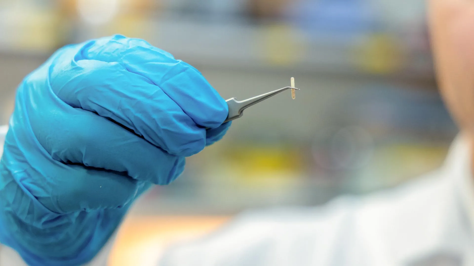
To overcome this, the EPFL team employed a cutting-edge approach: mass spectrometry imaging (MSI). In this process, a laser systematically scans ultra-thin sections of brain tissue, vaporizing molecules at specific coordinates to measure their precise mass-to-charge ratios. This allowed the researchers to create a spatial inventory of 172 different lipid species across 109 brain slices.
The scale of the data collection was unprecedented. The team processed approximately 7 million individual measurements, feeding the raw data into a machine learning pipeline designed to reconstruct the chemical topography of the brain in three dimensions. The result is the "Lipid Brain Atlas," an interactive digital resource that reveals the brain not as a uniform mass of fat, but as a highly structured, chemically partitioned landscape.
Defining the "Lipizones"
The atlas introduced a new term to the neuroscientific lexicon: "lipizones." These 539 identified territories represent regions of the brain characterized by a consistent and unique "lipid signature." This finding carries profound implications for how we understand brain organization.
Previously, anatomical maps were derived from cellular morphology or gene expression patterns. The Lipid Brain Atlas, however, reveals that these lipids act as a form of biological shorthand. As Professor Giovanni D’Angelo explains, lipids function effectively like "postal codes," providing a chemical address that coordinates disparate regions of the brain. When these chemical signatures were analyzed, the researchers found that they could identify the specific origin of a tissue sample simply by its lipid profile, confirming that these chemical boundaries are consistent and highly regulated across individuals.
Challenging Conventional Anatomical Boundaries
One of the most startling revelations of the study concerns the white matter of the brain. Traditionally, white matter—the pathways of myelinated axons that facilitate communication between gray matter regions—was viewed as a relatively homogeneous conductive material. The Lipid Brain Atlas shattered this assumption, revealing that white matter is partitioned into numerous, chemically distinct zones.
This suggests that the oligodendrocytes—the specialized cells responsible for producing myelin—are far more chemically diverse than their gene expression profiles would indicate. "This shows that the cells which make myelin are far more varied in their chemistry than their genes alone would suggest," noted Luca Fusar Bassini. This chemical heterogeneity implies that different segments of the brain’s "cabling" may have distinct metabolic requirements or functional properties, a nuance that has been entirely absent from current neurological models.
Furthermore, the team identified unexpected chemical boundaries within the choroid plexus and the lining of the brain’s ventricles. These areas, which are vital for the production and circulation of cerebrospinal fluid, displayed precise chemical gradients that do not align with traditional anatomical delineations. These findings indicate that the brain’s chemical organization is a multilayered system, operating in parallel with the genetic and structural maps that researchers have relied upon for decades.
Pregnancy and the Plasticity of the Lipidome
Beyond mapping the static brain, the researchers utilized the atlas to observe dynamic shifts in chemical composition under specific physiological conditions. By comparing the brains of male and female mice, and specifically examining the effects of pregnancy, the team uncovered a level of neuroplasticity that was previously unappreciated.
The data revealed that pregnancy triggers widespread, systematic changes in the lipid composition of the brain, particularly within the cortex. These shifts were found to be more significant than the baseline lipid differences observed between sexes. Of particular interest was the dramatic increase in galactosyl ceramide, a critical lipid component of myelin, throughout the white matter.
Gioele La Manno, co-author of the study, posits that these changes represent the brain’s active effort to "retune its own wiring." By adjusting the lipid environment, the brain may be optimizing neural pathways to adapt to the physiological and behavioral demands of pregnancy. This discovery opens a new frontier in neuroendocrinology, suggesting that the brain is not a static organ, but a dynamic, lipid-responsive system that adjusts its internal chemistry to meet the body’s changing requirements.
Implications for Human Health and Future Research
The release of the Lipid Brain Atlas as a freely available online tool marks a pivotal moment for the global research community. By providing a detailed reference for the healthy mammalian brain, the atlas serves as a control against which diseased tissue can be measured.
The medical implications are substantial. Changes in lipid metabolism are increasingly linked to a wide array of neurodegenerative and psychiatric disorders. In Alzheimer’s disease, for instance, the breakdown of myelin and the accumulation of lipid-related plaques are hallmarks of pathology. In depression and other mood disorders, the integrity of neuronal membranes—which rely on specific fatty acid compositions—is often compromised.
While the current map is based on mouse models, the methodology developed by the EPFL team provides a roadmap for human studies. Future research will likely focus on creating a human lipid atlas, which could provide biomarkers for early detection of neurological diseases. If researchers can identify the "lipid signature" of a healthy brain, they may eventually be able to identify the specific chemical deviations that precede the onset of clinical symptoms in conditions like Alzheimer’s, Parkinson’s, or multiple sclerosis.
Conclusion: A New Era of Lipidomics
The work of the EPFL team serves as a corrective to the "gene-centric" bias that has defined neuroscience for the past half-century. While DNA and proteins are the architects of the brain, lipids are the environment in which that architecture lives and functions. By revealing that the brain is organized into hundreds of chemically distinct "lipizones," the researchers have added a new dimension to our understanding of neurobiology.
As the scientific community begins to integrate the Lipid Brain Atlas into existing research frameworks, the potential for discovery is vast. From understanding the nuances of brain aging to unraveling the chemical underpinnings of cognitive adaptation, the study of lipids is no longer a niche interest—it is a central pillar of modern neuroscience. As Luca Fusar Bassini aptly summarized, the significance of the project lies not just in the map itself, but in the realization that the brain is a highly organized chemical entity. With this new, clearer picture of the brain’s lipid architecture, the scientific community is now better equipped than ever to navigate the complex, fatty landscape that dictates our thoughts, memories, and behaviors.