
In a landmark study published in the journal Science, a multidisciplinary team led by the Massachusetts Institute of Technology (MIT) has achieved the first-ever three-dimensional mapping of the atomic structure of relaxor ferroelectrics. These complex materials, which have been fundamental to the operation of ultrasound imaging, sonar, and high-precision sensors for decades, have long eluded precise structural characterization due to their inherent atomic disorder. By utilizing cutting-edge electron microscopy techniques, the researchers have provided a definitive look at the internal arrangements of these materials, effectively bridging a gap between theoretical models and physical reality that has persisted for over half a century.
The research, spearheaded by James LeBeau, the Kyocera Professor of Materials Science and Engineering at MIT, alongside collaborators from the University of Alabama at Birmingham, the Korea Advanced Institute of Science and Technology (KAIST), Rice University, and the University of Pennsylvania, represents a significant leap forward in the field of condensed matter physics. The findings offer a robust foundation for the next generation of computing systems, energy storage devices, and advanced actuators by allowing scientists to predict material behavior with unprecedented accuracy.
The Decades-Long Mystery of Relaxor Ferroelectrics
To understand the significance of this breakthrough, one must look at the history of ferroelectric materials. Discovered in the mid-20th century, ferroelectrics are characterized by a spontaneous electric polarization that can be reversed by the application of an external electric field. Relaxor ferroelectrics are a specific, more complex subclass. Unlike standard ferroelectrics, which undergo a sharp phase transition at a specific temperature, relaxors exhibit a broad, frequency-dependent peak in their dielectric permittivity.
This "relaxing" behavior makes them incredibly efficient at converting electrical energy into mechanical strain (and vice versa), a property known as piezoelectricity. This efficiency is why they are the "gold standard" for medical ultrasound transducers and naval sonar arrays. However, the source of this efficiency—a phenomenon known as "polar nanoregions"—has remained largely theoretical. For years, scientists have relied on bulk measurements and indirect observations to guess how the atoms within these nanoregions were organized.
The primary obstacle has been the material’s "chemical disorder." In alloys like lead magnesium niobate-lead titanate (PMN-PT), different types of atoms occupy the same sites in the crystal lattice in a seemingly random fashion. This randomness creates local electric fields that fluctuate throughout the material, frustrating the long-range order found in simpler crystals. Until now, imaging these fluctuations in three dimensions at the atomic scale was considered an impossible task for traditional microscopy.
Multi-Slice Electron Ptychography: A New Window into the Nanoscale
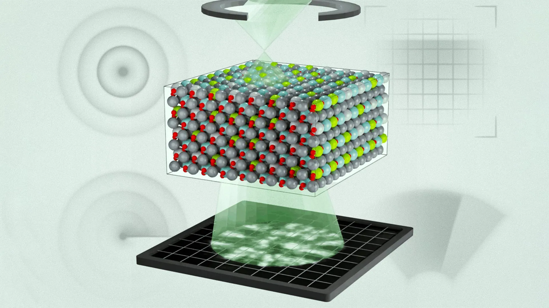
The breakthrough was made possible through the application of an advanced imaging technique known as multi-slice electron ptychography (MEP). Standard transmission electron microscopy (TEM) produces a two-dimensional projection of a three-dimensional object, much like a shadow on a wall. While useful, this "flattened" view loses critical information about the depth and specific orientation of atomic charges.
MEP functions differently. The researchers scanned a nanoscale beam of high-energy electrons across a sample of PMN-PT. As the electrons passed through the material, they were deflected by the electric fields of the atoms, creating complex diffraction patterns. By recording these patterns at every position and using an iterative algorithm to process the overlapping data, the team was able to reconstruct the three-dimensional electron wave function.
"We do this in a sequential way, and at each position, we acquire a diffraction pattern," explained Menglin Zhu, a postdoc at MIT and co-first author of the study. "That creates regions of overlap, and that overlap has enough information to use an algorithm to iteratively reconstruct three-dimensional information about the object."
This process allowed the team to see not just where the atoms were, but how their positive and negative charges were displaced relative to one another—the very definition of polarization.
Key Findings: Challenging Established Scientific Models
The results of the 3D mapping challenged several long-held assumptions in the materials science community. The researchers discovered a "layered hierarchy" of chemical and polar structures. This hierarchy extends from the scale of individual atoms up to mesoscopic features that span hundreds of nanometers.
One of the most surprising findings was the size of the polarized regions. Previous computer simulations and indirect measurements suggested that these regions were relatively large and uniform. However, the MEP data revealed that the regions of local polarization were significantly smaller than predicted. Furthermore, the team found that the chemical disorder—the "randomness" of how magnesium, niobium, and titanium atoms are distributed—plays a much more active role in modulating polarization than previously thought.
"We realized the chemical disorder we observed in our experiments was not fully considered previously," said Michael Xu, co-first author and MIT postdoc. "Working with our collaborators, we were able to merge the experimental observations with simulations to refine the models and better predict what we see in experiments."
By integrating these real-world observations into molecular dynamics (MD) simulations, the team was able to create a model that finally matched the physical behavior of the material. This "ground truth" data is essential for the future of material design, as it provides a benchmark against which all future simulations can be measured.
Impact on Defense, Medicine, and Energy
The implications of this research extend far beyond the laboratory. Relaxor ferroelectrics are critical components in several high-stakes industries:
- Medical Imaging: Improved understanding of PMN-PT structure can lead to ultrasound transducers with higher resolution and sensitivity, allowing for earlier detection of diseases and more precise diagnostic imaging.
- Naval and Defense Systems: Sonar technology relies on the piezoelectric properties of relaxors to detect underwater objects. More efficient materials mean quieter, more sensitive, and more durable sonar arrays for defense applications.
- Energy Storage: Relaxors are used in high-energy-density capacitors. Understanding the 3D charge distribution allows engineers to design capacitors that can store more energy and discharge it more reliably, which is vital for electric vehicle power electronics and renewable energy grids.
- Actuators and Robotics: These materials power the micro-actuators in high-end camera lenses and precision robotic tools. Better models will allow for the development of "smarter" materials that respond more predictably to electrical stimuli.
James LeBeau emphasized that as materials science moves toward AI-driven discovery, having accurate physical data is paramount. "Materials science is incorporating more complexity into the material design process—whether that’s for metal alloys or semiconductors—as AI has improved and our computational tools have become more advanced," LeBeau stated. "But if our models aren’t accurate enough and we have no way to validate them, it’s garbage in, garbage out. This technique helps us understand why the material behaves the way it does and validate our models."
A Global Collaborative Effort
The success of the project was the result of an extensive collaboration between several leading research institutions. While MIT provided the imaging expertise and facilities (utilizing the state-of-the-art MIT.nano center), other institutions contributed vital theoretical and material support.
The University of Pennsylvania and the University of Alabama at Birmingham provided the computational molecular dynamics frameworks necessary to interpret the massive amounts of data generated by the ptychography process. Rice University and KAIST contributed expertise in thin-film growth and material synthesis, ensuring the samples used were of the highest possible quality.
The study also acknowledges the support of the U.S. Army Research Laboratory, the U.S. Office of Naval Research, and the U.S. Department of Energy. The involvement of defense-related funding agencies underscores the strategic importance of mastering relaxor ferroelectrics for national security and advanced manufacturing.
Future Directions: Toward Tailored Electronic Properties
Looking ahead, the research team believes that multi-slice electron ptychography will become a standard tool for exploring other disordered materials. From high-entropy alloys to complex glass structures, the ability to map atomic arrangements in 3D opens up a new frontier in condensed matter physics.
The team’s next goal is to use this technique to observe how these 3D structures change in real-time when an electric field is applied. This "in-situ" imaging would allow scientists to watch the polarization regions shift and grow, providing a "movie" of the material’s function at the atomic level.
"This study is the first time in the electron microscope that we’ve been able to directly connect the three-dimensional polar structure of relaxor ferroelectrics with molecular dynamics calculations," Xu concluded. "It further proves you can get three-dimensional information out of the sample using this technique."
As the scientific community moves toward creating lead-free alternatives to PMN-PT to meet environmental regulations, the insights gained from this 3D mapping will serve as a roadmap. By understanding exactly how lead-based relaxors achieve their high performance, researchers can more effectively engineer sustainable materials that mimic those properties, ensuring that the next generation of technology is both high-performing and environmentally responsible.


Leave a Reply