
Cells organize many of their most important activities using structures known as biomolecular condensates. Unlike traditional compartments in the cell, these droplet-like clusters are not enclosed by membranes. They help control how genetic instructions in DNA are converted into proteins, assist in clearing away cellular waste that could otherwise become toxic, and can even play a role in suppressing tumor growth. Because condensates behave like liquids that can fuse, flow, and quickly exchange components, scientists long believed they were simple, unstructured droplets. However, groundbreaking research published on February 2, 2026, in Nature Structural and Molecular Biology challenges this long-standing view, revealing that some of these essential cellular structures possess a complex internal architecture crucial for their function.
Unveiling the Hidden Architecture of Cellular Condensates
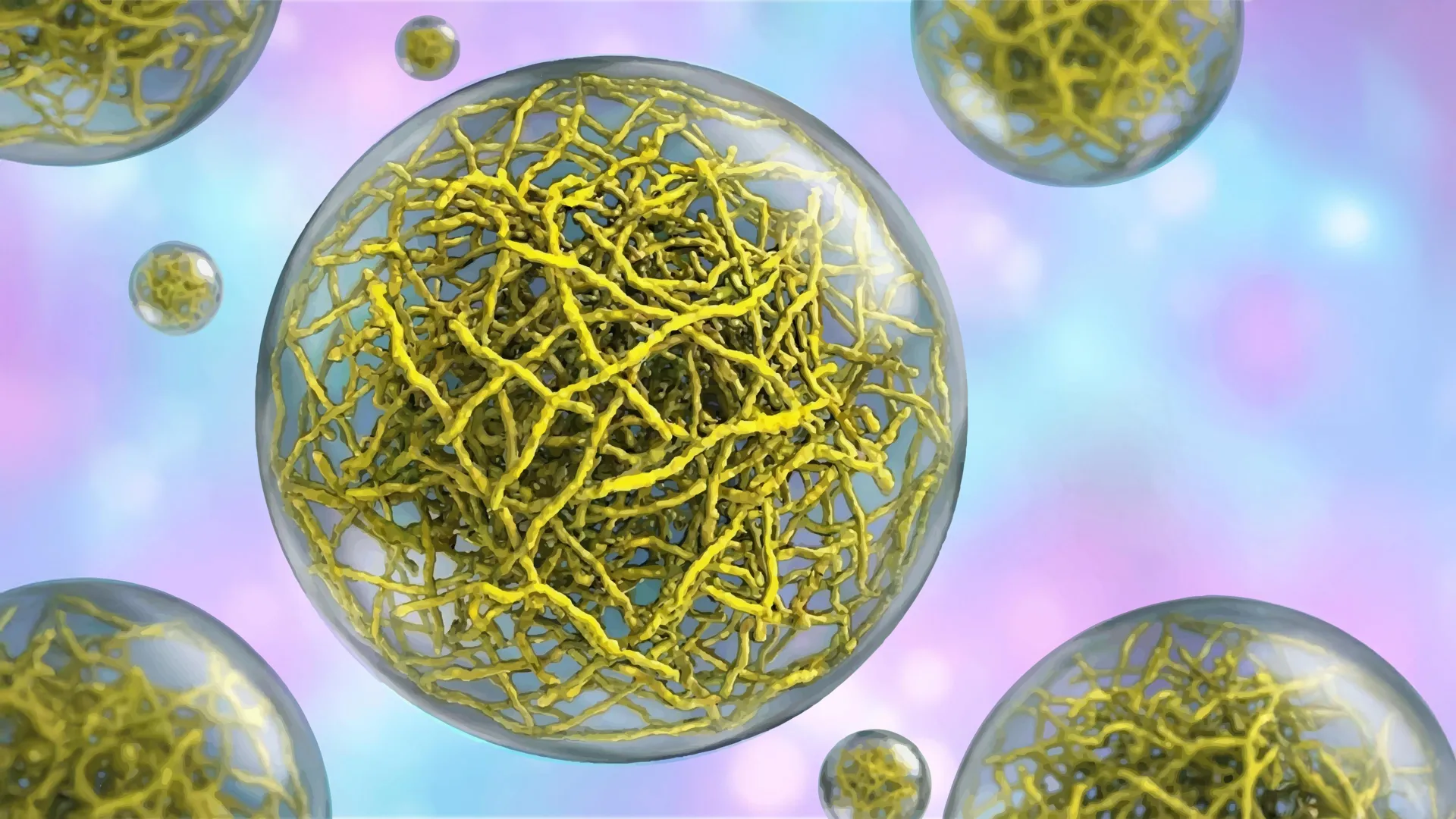
A team at Scripps Research has made a significant discovery, demonstrating that certain biomolecular condensates are not mere amorphous blobs but are meticulously constructed from intricate networks of thin, thread-like protein filaments. This internal scaffolding provides these droplet-like assemblies with a defined architecture that is paramount to their operational capabilities. This paradigm shift in understanding has profound implications for developing novel therapeutic strategies for a range of debilitating diseases, including cancer and neurodegenerative disorders.
"Ever since we realized that disruptions in condensate formation are at the heart of many diseases, it has been challenging to target them therapeutically because they appeared to lack structure — there were no specific features for a drug to latch onto," stated Keren Lasker, associate professor at Scripps Research and senior author of the study. "This work changes that. We can now see that some condensates have an internal architecture, and that, importantly, this structure is required for function, opening the door to targeting these membrane-less assemblies much like we target individual proteins."
The research builds upon decades of inquiry into the nature of cellular organization. For years, the cell’s internal landscape was primarily understood through the lens of membrane-bound organelles—distinct compartments like the nucleus or mitochondria that compartmentalize cellular processes. The discovery of biomolecular condensates offered a new perspective, highlighting a mechanism of organization that transcends the need for physical membranes. These non-membranous organelles, often formed through liquid-liquid phase separation (LLPS), were initially characterized by their dynamic, fluid-like properties, leading to the prevailing notion of their inherent simplicity.
The PopZ Protein: A Window into Condensate Architecture
To unravel the mystery of how membrane-less condensates function as effective cellular compartments, Lasker’s laboratory turned their attention to a bacterial protein known as PopZ. In certain species of rod-shaped bacteria, PopZ plays a critical role by accumulating at the cell poles, the rounded extremities of the bacterial cell. Here, it forms condensates that are instrumental in organizing other proteins essential for the vital process of cell division.
In collaboration with Scripps Research professor Ashok Deniz and assistant professor Raphael Park, who co-led the study, the team employed cryo-electron tomography (cryo-ET). This sophisticated imaging technique, often described as a molecular-scale CT scan, allows researchers to visualize cellular structures with unprecedented detail. The cryo-ET analysis provided a revelatory glimpse into the internal composition of PopZ condensates.
The images produced by cryo-ET clearly indicated that PopZ proteins do not aggregate randomly. Instead, they assemble into highly organized filaments through a precise, step-by-step process. These filaments then collectively form a robust scaffold, an internal framework that dictates the physical characteristics and overall architecture of the condensate. This discovery marked a pivotal moment, suggesting that the fluidity and dynamic nature of condensates did not preclude them from possessing an underlying, ordered structure.
Dynamic Protein Conformations Dictate Function
Beyond mapping the structural assembly of the filaments, the researchers delved deeper to understand the behavior of individual PopZ molecules within and outside these condensates. To achieve this, they utilized single-molecule Förster resonance energy transfer (FRET). FRET is a powerful biophysical technique that measures minuscule changes in distance within proteins by detecting energy transfer between fluorescently tagged molecules.
Through these FRET experiments, the team made a crucial observation: the conformation, or three-dimensional shape, of individual PopZ proteins is not static. Instead, it is highly dependent on their location within the cell. PopZ proteins adopt one specific shape when they are situated outside of a condensate and a distinctly different conformation when they are incorporated into the filamentary network of the condensate.
"Realizing that protein conformation depends on location gives us multiple ways to engineer cellular function," remarked Daniel Scholl, the study’s first author and a former postdoctoral researcher in the Lasker and Deniz labs. This finding underscores the intricate interplay between protein structure, cellular location, and functional output, suggesting that changes in protein shape are not arbitrary but are integral to the condensate’s activity.
The Indispensable Role of Filamentous Structure
To definitively ascertain whether the observed filamentous structure was merely an incidental detail or a fundamental requirement for the condensate’s biological role, the researchers conducted a series of critical experiments. They engineered a mutant version of the PopZ protein that was incapable of forming filaments. When these altered PopZ proteins were introduced into bacteria, the resulting condensates exhibited significantly altered physical properties. They became markedly more fluid, and their surface tension was considerably reduced.
The functional consequences of these structural and physical changes were dramatic. When these modified condensates were present in living bacteria, the cells exhibited a profound inability to grow and failed to properly segregate their DNA during cell division. This experimental outcome provided irrefutable evidence that the structural integrity of the condensate, specifically its filamentous scaffold, is not just a passive architectural feature but is absolutely vital for normal cellular function. The physical properties conferred by the filament network, rather than solely the chemical composition of the condensate’s components, are essential for life.
This aspect of the research is particularly significant because it moves beyond understanding condensates as simple aggregations of molecules to recognizing them as dynamic, structured entities whose physical attributes directly impact cellular processes. The fluidity and surface tension of a condensate are not just emergent properties of its liquid-like state but are actively regulated by its internal architecture, which in turn influences cellular mechanics and genetic regulation.
Broad Implications for Human Health: Cancer and Neurodegeneration
While the experiments meticulously focused on the bacterial PopZ protein, the implications of these findings extend far beyond prokaryotic cells, holding significant relevance for human health. In human cells, filament-based condensates are known to perform two critical functions: the meticulous clearing of damaged or misfolded proteins, a process essential for maintaining cellular homeostasis, and the precise control of cell growth.
When these protein-clearing condensates malfunction or break down, a buildup of toxic proteins can occur. This accumulation is a hallmark pathology observed in a spectrum of devastating neurodegenerative diseases, including amyotrophic lateral sclerosis (ALS), Alzheimer’s disease, and Parkinson’s disease. The intricate network of filaments within these condensates likely provides the necessary scaffolding for the efficient recruitment and degradation of protein aggregates.
Similarly, if condensates responsible for regulating cell growth fail in their structural integrity, the cellular mechanisms that prevent uncontrolled proliferation can collapse. This breakdown is a significant contributing factor to the development of various cancers, such as prostate, breast, and endometrial cancers. The ordered structure of these growth-regulating condensates is hypothesized to be critical for sequestering or organizing proteins involved in cell cycle checkpoints and signaling pathways.
"By demonstrating that condensate architecture is both definable and functionally critical, the work raises the possibility of designing therapies that act directly on condensate structure and correct the underlying disorganization that allows disease to take hold," Professor Lasker elaborated. This opens up a new frontier in drug discovery, shifting the focus from targeting individual proteins to modulating the structural properties of entire cellular assemblies.
Future Directions and Therapeutic Potential
The discovery that biomolecular condensates possess a crucial, functionally relevant internal architecture represents a significant leap forward in our understanding of cellular biology. It challenges the long-held perception of these structures as simple, unstructured liquid droplets. The ability to visualize and manipulate the filamentous scaffold within condensates provides a tangible target for therapeutic intervention.
Researchers are now exploring ways to develop drugs that can specifically interact with and stabilize or destabilize these protein filament networks. For instance, in neurodegenerative diseases, a therapeutic strategy might involve designing molecules that enhance the stability of protein-clearing condensates, thereby facilitating the removal of toxic protein aggregates. Conversely, in cancer, therapies could aim to disrupt the structure of growth-regulating condensates, thereby hindering tumor cell proliferation.
The study also highlights the importance of interdisciplinary research, combining advanced imaging techniques like cryo-ET with biophysical methods such as single-molecule FRET. This synergy of approaches has been instrumental in revealing the complex molecular dynamics at play within these fundamental cellular structures.
The research was supported by substantial funding from various national and international bodies, including the National Institutes of Health (NIH) and the National Science Foundation (NSF), underscoring the perceived importance and potential impact of this line of inquiry. The collaborative efforts of a broad team of scientists, including Tumara Boyd, Andrew P. Latham, Alexandra Salazar, Asma Khan, Steven Boeynaems, Alex S. Holehouse, Gabriel C. Lander, and Andrej Sali, were crucial to the success of this multifaceted investigation.
As scientists continue to probe the intricate world of biomolecular condensates, this latest finding serves as a powerful reminder that even seemingly simple cellular structures can harbor remarkable complexity, offering new avenues for understanding and combating human disease. The era of targeting condensate architecture for therapeutic benefit has officially begun.


Leave a Reply