

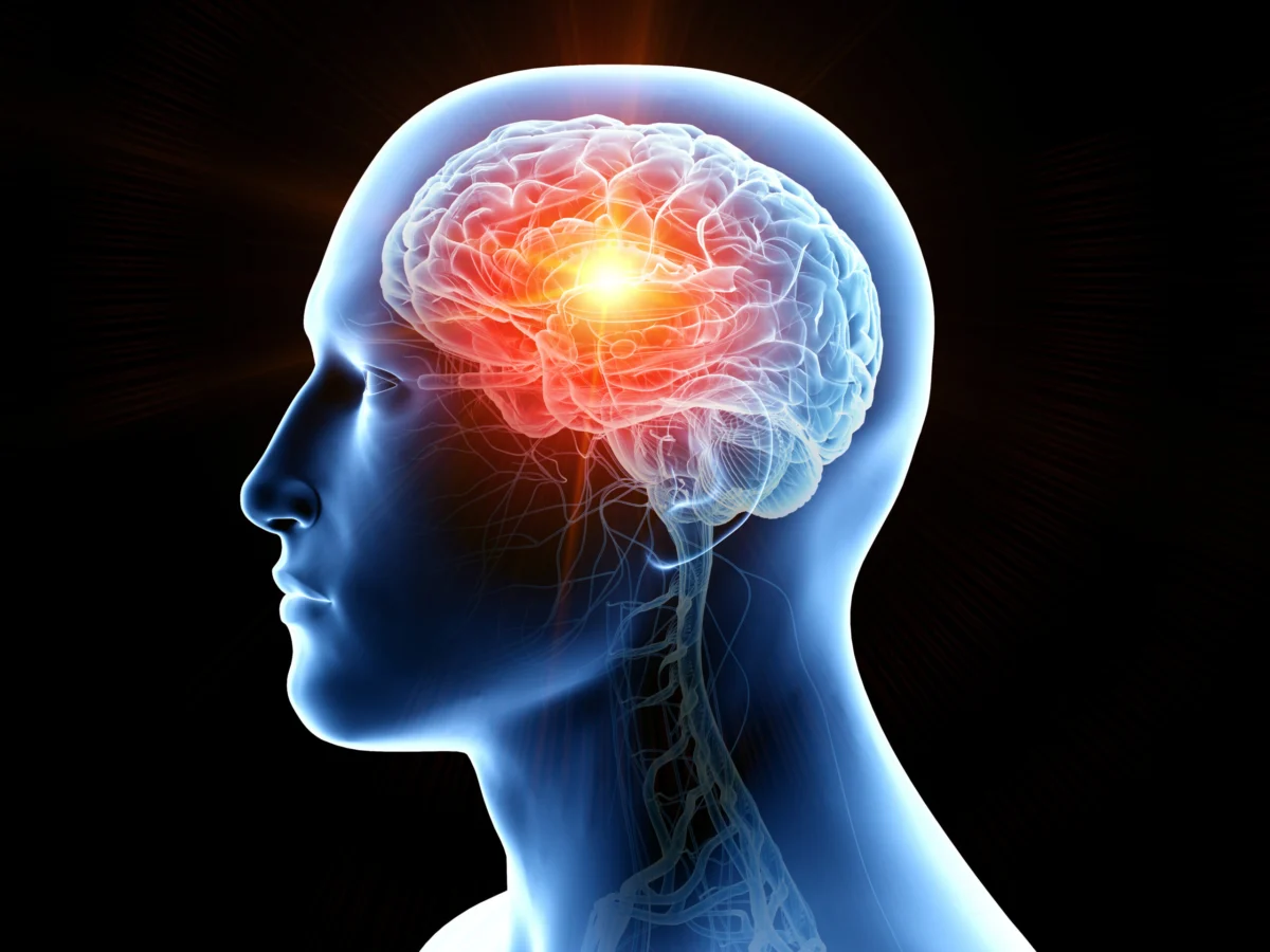
Traumatic brain injury (TBI) stands as a profound and pervasive global health crisis, impacting an estimated 69 million individuals each year across diverse demographics. This staggering incidence underscores TBI as a leading cause of disability and mortality worldwide. While often associated with high-impact events, the highest burden of TBI-related injuries is disproportionately observed in older adults, where falls represent the primary etiology. Beyond this demographic, contact sports, road traffic accidents, and interpersonal violence contribute significantly to TBI cases across all age groups and populations, highlighting the multifaceted nature of its origins. The immediate aftermath of a TBI is often chaotic, with medical efforts primarily focused on acute management, such as stabilizing intracranial pressure and ensuring adequate cerebral perfusion. However, these interventions, while critical for survival, largely address the immediate physical trauma. A critical unmet need persists in addressing the complex secondary pathophysiological processes that are initiated by the initial injury and can continue to unfold long after the acute phase. These processes, including neuroinflammation, progressive neurodegeneration, oxidative stress, and proteinopathy, are not merely transient complications but insidious drivers of long-term neurological impairment and significantly heighten the risk of developing chronic neurodegenerative conditions, notably Alzheimer’s disease (AD). The chronic persistence of neuroinflammation and progressive neurodegeneration, in particular, is increasingly recognized as a key factor contributing to prolonged symptoms, including cognitive decline, mood disturbances, and motor deficits, that can emerge years or even decades after the initial brain trauma.
The Silent Aftermath: Secondary Injury and the Path to Alzheimer’s
The journey from an acute TBI to the potential development of neurodegenerative diseases like Alzheimer’s is complex and characterized by a cascade of secondary injury mechanisms. Immediately following the mechanical insult, a rapid inflammatory response is triggered within the brain. While initially protective, prolonged or dysregulated neuroinflammation becomes detrimental, leading to widespread neuronal damage and synaptic dysfunction. Microglia, the brain’s resident immune cells, and astrocytes, crucial support cells, shift from a neuroprotective to a neurotoxic phenotype, releasing pro-inflammatory cytokines, chemokines, and reactive oxygen species. This creates a hostile microenvironment that perpetuates cellular stress and neurodegeneration. Oxidative stress, another critical component, involves an imbalance between the production of reactive oxygen species and the brain’s ability to detoxify them, leading to cellular damage and further exacerbating inflammation. Concurrently, TBI can initiate or accelerate proteinopathies, the abnormal aggregation of proteins within brain cells. These misfolded proteins, such as tau and amyloid-beta, are hallmarks of AD and other neurodegenerative conditions. The secondary injury processes are not isolated events but interact dynamically, forming a vicious cycle that contributes to the progressive nature of post-TBI pathology. Current therapies predominantly offer supportive care, managing symptoms rather than targeting these underlying disease-modifying pathways. This highlights a critical therapeutic gap, particularly for interventions that can interrupt or reverse these chronic processes, thereby preventing or mitigating long-term cognitive decline.
Shared Pathologies: Unveiling the TBI-AD Connection
The intricate link between TBI and AD is not merely epidemiological but deeply rooted in shared molecular and cellular pathologies. A growing body of evidence demonstrates that following TBI, the brain often exhibits pathological features remarkably similar to those observed in AD. These include the hyperphosphorylation of tau protein, leading to the formation of neurofibrillary tangles, and the accumulation of amyloid-beta (Aβ) peptides, which aggregate into characteristic amyloid plaques. Both tau hyperphosphorylation and Aβ accumulation are considered central to the pathogenesis of AD. The observation of these specific proteinopathies in post-TBI brains provides compelling mechanistic support for the link between brain trauma and subsequent neurodegenerative disease.
Furthermore, repetitive or severe TBI has been definitively associated with the development of Chronic Traumatic Encephalopathy (CTE), a distinct progressive neurodegenerative tauopathy initially recognized in athletes exposed to recurrent head impacts. CTE shares clinical and pathological overlaps with AD, particularly in the widespread aggregation of hyperphosphorylated tau. This connection underscores that mechanical brain trauma can directly initiate progressive neurodegenerative proteinopathy, reinforcing the idea that TBI acts as a significant risk factor, and potentially an accelerant, for neurodegenerative processes akin to those seen in AD. The persistence of neuroinflammation, a common denominator in both conditions, serves as a crucial bridge, fostering an environment conducive to protein aggregation and neuronal dysfunction.
Biomarkers: Diagnostic Windows into Brain Health
The shared pathological landscape between TBI and AD extends to key inflammatory and neuronal injury biomarkers, offering promising avenues for diagnosis, prognosis, and monitoring therapeutic efficacy. Several biomarkers, measurable in blood, have emerged as critical indicators of brain injury and disease progression in both conditions.
- Glial Fibrillary Acidic Protein (GFAP): GFAP is an intermediate filament protein primarily expressed in astrocytes, the star-shaped glial cells that provide structural and metabolic support to neurons. In response to brain injury or disease, astrocytes become reactive, leading to an upregulation and release of GFAP. Markedly elevated levels of GFAP are observed in the acute phase of TBI, reflecting astrocytic activation and injury. Importantly, GFAP levels are also chronically elevated in AD patients, correlating with disease severity and cognitive decline. Its presence indicates ongoing astrogliosis, a hallmark of neuroinflammation and neurodegeneration.
- Neurofilament Light Chain (NF-L): NF-L is a structural protein found within the axons of neurons. When neurons are damaged, NF-L is released into the cerebrospinal fluid and subsequently into the bloodstream. It serves as a highly sensitive and specific biomarker for axonal injury. Following TBI, NF-L levels increase significantly, reflecting the extent of neuronal damage. Similarly, NF-L is consistently elevated in AD, indicating widespread neuronal degeneration and correlating with cognitive impairment. Its utility lies in its ability to quantify ongoing neuronal loss in both acute injury and chronic neurodegeneration.
- Ubiquitin C-terminal hydrolase L1 (UCH-L1): UCH-L1 is a neuronal-specific enzyme involved in the ubiquitin-proteasome system, a crucial pathway for protein degradation and recycling within cells. Dysregulation of this system is implicated in many neurodegenerative diseases. In TBI, UCH-L1 is released from damaged neurons and serves as an early indicator of neuronal injury. More profoundly, UCH-L1 is intimately associated with the pathological mechanisms of AD. Elevated UCH-L1 levels are linked to increased phosphorylated tau, which forms neurofibrillary tangles, and the abnormal accumulation of amyloid-beta plaques. It does this, in part, by affecting β-secretase activity, an enzyme involved in amyloid-beta production. Furthermore, UCH-L1 has been shown to deplete Triggering Receptor Expressed on Myeloid cells 2 (TREM2), a receptor crucial for microglial function and the clearance of amyloid-beta, thereby exacerbating neuroinflammation. Consequently, UCH-L1 is considered a vital biomarker reflecting both neuronal damage and the specific proteinopathy and inflammatory pathways common to AD and TBI.
- S100 Calcium-Binding Protein B (S100B): S100B is another astrocytic protein, similar to GFAP, that is released upon astrocyte activation or damage. It acts as a pro-inflammatory mediator, especially at high concentrations. S100B is a well-established biomarker for TBI, showing acute upregulation proportional to injury severity. In the context of AD, S100B is chronically elevated and has been linked to increased neuroinflammation and accelerated cognitive decline.
(Figure 1, depicting the kinetics of blood biomarkers from acute to chronic phase following traumatic brain injury, illustrates how these biomarkers evolve over time post-injury, providing a dynamic view of the ongoing pathological processes.)
The significant overlap in these key inflammatory and neurodegenerative biomarkers between TBI and AD not only strengthens the mechanistic link between the conditions but also suggests that therapeutic interventions capable of modulating these pathophysiological processes or their associated biomarker expression could offer broad therapeutic potential across both devastating disorders.
A Novel Therapeutic Frontier: High-Dose NK Cell Therapy
Given the limitations of current supportive therapies, the scientific community is actively exploring novel disease-modifying strategies. One particularly intriguing and dramatically responsive approach has emerged from the field of immunology: the administration of massive doses of autologous natural killer (NK) cells. NK cells are a vital component of the innate immune system, serving as rapid responders against stressed or infected cells and playing a crucial role in immune surveillance. Beyond their well-known anti-cancer properties, NK cells are increasingly recognized for their involvement in clearing pathological material and regulating inflammatory responses within the central nervous system.
The serendipitous discovery of NK cells’ potential in neurodegeneration arose from a study conducted by researchers at NKGen Biotech, Inc. Initially, the company was investigating the use of high-dose autologous NK cell infusions to mitigate immune suppression in oncology patients undergoing chemotherapy. During this trial, a remarkable observation was made: patients who also suffered from co-existing Alzheimer’s disease demonstrated unexpected cognitive stabilization and, in some cases, even noticeable improvement. This unanticipated outcome sparked a dedicated investigation into NK cells as a therapy for AD.
A subsequent Phase 1 open-label study was launched to specifically investigate the effects of repeated intravenous administration of high doses (approximately 6 billion cells per infusion) of expanded autologous NK cells in patients diagnosed with mild-to-severe AD. The results were compelling. Across various cognitive measures, a striking 90% of the treated patients showed either no decline in their cognitive function or, more remarkably, demonstrated a marked improvement over observation periods ranging from 3 to 12 months. This cognitive stabilization and improvement were paralleled by significant reductions in key biomarkers associated with neuroinflammation and proteinopathy. Specifically, patients exhibited decreased levels of GFAP (indicating reduced astrogliosis), phosphorylated tau (suggesting a reduction in neurofibrillary tangle pathology), and α-synuclein (a protein implicated in other neurodegenerative conditions like Parkinson’s disease, whose reduction here hints at broader protein clearance capabilities). These findings provided robust preliminary evidence for the disease-modifying potential of high-dose NK cell therapy in AD.
Mechanism of Action: How NK Cells Tackle Neurodegeneration
While the precise and comprehensive mechanisms underlying the observed cognitive improvements and biomarker reductions in AD patients treated with NK cells are still under active investigation, several plausible pathways have been proposed. NK cells appear to exert their therapeutic effects through a multi-pronged approach, targeting both the protein aggregation and the neuroinflammatory aspects of neurodegeneration.
One key mechanism involves the direct clearance of neurotoxic protein aggregates. NK cells are believed to be capable of internalizing and subsequently degrading misfolded proteins, such as amyloid-beta and tau, through lysosomal pathways. This direct scavenging and breakdown of pathological proteins could significantly reduce the burden of plaques and tangles, thereby alleviating cellular stress and improving neuronal function.
Beyond direct clearance, NK cells are potent immunomodulators, capable of reshaping the brain’s inflammatory environment. Proposed immunomodulatory effects include:
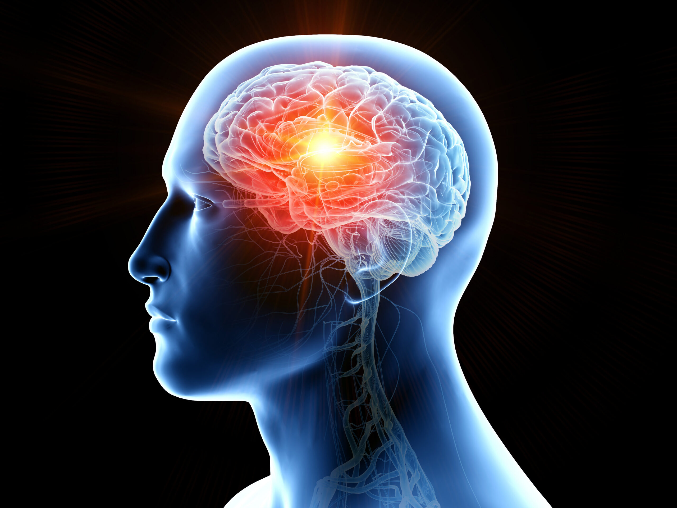
- Suppression of Pro-inflammatory Microglial Activity: In neurodegenerative diseases, microglia often adopt a chronically activated, pro-inflammatory state that contributes to neuronal damage. NK cells may interact with microglia, pushing them towards a more anti-inflammatory, neuroprotective phenotype.
- Production of Anti-inflammatory Cytokines: NK cells can produce a range of cytokines. While some are pro-inflammatory, under certain conditions or with specific priming, they can secrete anti-inflammatory cytokines that help to quell the chronic inflammatory response in the brain.
- Elimination of Autoreactive T-cells: In some neurodegenerative contexts, autoreactive T-cells may contribute to pathological inflammation. NK cells are known for their ability to eliminate target cells, and this function could extend to clearing harmful immune cells from the brain environment.
Such a multi-target mechanism, addressing both proteinopathy and chronic inflammation, is particularly advantageous in complex neurological conditions like TBI and AD, where numerous secondary injury pathways occur simultaneously and interact dynamically over extended periods. This holistic approach offers a distinct advantage over single-target therapies, which may fail to adequately address the multifaceted nature of these diseases.
Overcoming Challenges: The Rise of NK Cell-Derived Extracellular Vesicles (NK-EVs)
Despite the compelling promise of whole-cell NK therapies, their widespread clinical translation presents considerable logistical and practical challenges. Administering whole, living cells involves complex and costly manufacturing processes, difficulties in achieving scalability for large patient populations, the need for repeated intravenous dosing, and the inherent attrition of cells before they can effectively reach and cross the formidable blood-brain barrier (BBB). These obstacles necessitate innovative solutions to harness the therapeutic power of NK cells more efficiently.
This is where Extracellular Vesicles (EVs) emerge as a revolutionary "cell-free" therapeutic alternative. EVs are nanoscale (typically 30-150 nm) membrane-bound particles naturally released by virtually all cells into the extracellular space. They function as crucial mediators of intercellular communication, carrying a biologically active cargo of proteins, messenger RNAs (mRNAs), microRNAs (miRNAs), lipids, and signaling molecules. By delivering this cargo to recipient cells, EVs can profoundly influence their behavior and phenotype. Crucially, the composition and functional properties of EVs largely reflect those of their parent cells, meaning EVs derived from NK cells (NK-EVs) can retain many of the immunomodulatory and signaling characteristics of the original NK cells, but in a cell-free format.
Evinco Therapeutics: Pioneering Cell-Free Solutions
Australian company Evinco Therapeutics is at the forefront of developing NK-EVs for neurological disorders. Preliminary findings from their research indicate that EVs recovered from cultured NK cells exert a powerful anti-inflammatory effect on brain-resident immune cells, specifically microglia and astrocytes. This suggests that NK-EVs can directly modulate the detrimental neuroinflammatory responses observed in TBI and AD. Even more significantly, Evinco’s studies show that NK-EVs strongly induce microglia to internalize and degrade amyloid-beta. This finding is critical because it suggests that the direct clearance of neurotoxic protein aggregates, a key benefit of whole NK cells, can be achieved without the NK cells themselves being present in the brain. In essence, the immunomodulatory and protein-clearing components, critical for AD and TBI treatment, can be effectively leveraged by NK-EVs, offering a potent and targeted therapeutic delivery system.
The Promise of Intranasal Delivery and Scalability
Beyond their intrinsic biological advantages, NK-EVs offer substantial logistical and translational benefits over whole-cell therapies. Unlike living NK cells, NK-EVs are remarkably stable. They can be freeze-dried, enabling long-term storage and facile shipping at ambient temperatures, or maintained at room temperature for extended periods. This stability dramatically simplifies distribution and reduces cold chain requirements, which are often significant hurdles for cell therapies.
Furthermore, NK-EVs open the door to highly patient-friendly and effective delivery routes. They can be delivered via the nasal route as a spray, a non-invasive option that avoids intravenous injections and can potentially be self-administered at home. This intranasal administration also offers a more direct pathway to the brain than intravenous methods. EVs can move along the olfactory nerve bundle, bypassing the systemic circulation and, crucially, largely circumventing the blood-brain barrier (BBB). The BBB is a major physiological obstacle for many intravenously administered drugs and cell therapies, limiting their ability to achieve meaningful therapeutic concentrations in the central nervous system. The direct brain delivery offered by intranasal NK-EVs represents a significant advantage in treating neurological conditions.
Another transformative aspect of NK-EVs is their scalability and immunological profile. EVs are not recognized as foreign by a recipient’s immune system, even if derived from allogeneic (donor) sources. This allows for large-scale manufacturing from donor NK cell lines at a potentially very reasonable cost, eliminating the need for personalized autologous cell expansion for each patient. The freeze-dried product can be stored and simply reconstituted by adding a solution, making it a highly accessible and adaptable therapeutic option. Such scalability is particularly vital for conditions like TBI, given its enormous global incidence. The practical limitations associated with personalized cell therapies would make it challenging to meet the demand for millions of TBI patients annually; NK-EVs offer a path to widespread accessibility.
Charting the Path to Clinical Trials: Evinco’s Strategic Focus
Evinco Therapeutics is actively collaborating with a major pharmaceutical company to establish robust proof-of-concept for their NK-EV therapy in Alzheimer’s disease. Building on the profound overlap in pathologies and biomarkers between AD and TBI, Evinco is strategically interested in applying the same NK-EV product to TBI. TBI presents a particularly attractive target for early clinical development due to its defined onset, allowing for clearer intervention windows; quantifiable biomarker and behavioral readouts that can serve as objective efficacy endpoints; and potentially shorter trial times compared to the prolonged progression often seen in AD.
The present research roadmap for Evinco includes comprehensive preclinical studies focusing on safety, efficacy, and dose-response in both mouse and canine models. These rigorous studies are essential to fully characterize the therapeutic profile of NK-EVs and to gather the necessary data for regulatory submissions. Evinco anticipates having their product ready for "first-in-human" clinical studies within the next 15 months, contingent upon the successful completion of ongoing development and regulatory activities. The innovative nature of this cell-free, immunomodulatory approach, coupled with its potential for scalable and non-invasive delivery, has generated considerable interest across the neuroscientific and pharmaceutical communities, setting the stage for highly anticipated upcoming clinical studies.
Broader Implications and the Future of Neurodegenerative Treatment
Despite decades of extensive research and significant investment, there remains a critical and urgent need for disease-modifying therapies capable of truly repairing damaged neural tissue or fundamentally altering the underlying pathological processes that drive ongoing decline in conditions like TBI and AD. Current treatments largely offer symptomatic relief, failing to address core issues such as chronic neuroinflammation, protein aggregation, and synaptic loss. The persistent lack of curative or truly disease-modifying options underscores the imperative for innovative approaches that can restore brain health, halt progression, and significantly improve long-term outcomes for the millions affected by these devastating disorders.
The development of NK-EVs represents a paradigm shift in therapeutic strategy. By leveraging the natural immunomodulatory and protein-clearing capabilities of NK cells in a stable, scalable, and non-invasive cell-free format, Evinco Therapeutics and its collaborators are poised to potentially transform the treatment landscape for TBI, Alzheimer’s disease, and potentially other neurodegenerative conditions characterized by neuroinflammation and proteinopathy. The promise of an accessible, effective, and patient-friendly therapy that can target multiple pathological pathways offers a beacon of hope for patients and their families worldwide, heralding a new era in brain health intervention.
Dr. Karl Trounson is Scientific Advisor and Prof. Alan Trounson is Founder, CEO and Executive Chair of Evinco Therapeutics, a biotechnology company developing immune-based therapies for neurological disorders.
Author affiliations
Karl M. Trounson, PhD and Alan O. Trounson, AO, PhD















Leave a Reply