In a breakthrough that bridges the gap between advanced nanotechnology and molecular detection, a team of chemical and materials engineers at the University of Toronto has engineered a novel class of dye-sensitized nanoparticles capable of identifying trace chemical concentrations with unprecedented precision. Published recently in the Journal of the American Chemical Society, the discovery addresses one of analytical chemistry’s most persistent challenges: the ability to detect minute quantities of target molecules while reliably differentiating between structural isomers—molecules that share identical chemical formulas but possess vastly different spatial geometries.
Led by senior author Professor Kai Huang, alongside PhD student and lead paper author Jiaze Wu, the interdisciplinary research team successfully harnessed the physics of upconversion to develop nanoprobes up to 150 times brighter than conventional un-sensitized alternatives. This quantum leap in optical brightness, paired with a virtually zero-background signal environment, positions the technology for transformative applications across pharmaceutical manufacturing, environmental toxicology, and clinical diagnostics.
The Physics of Upconversion: Turning Low-Energy Photons into High-Energy Signals
For decades, analytical chemists have relied on organic molecules known as fluorophores to absorb light and re-emit it at longer wavelengths. This standard photoluminescent process, however, is fundamentally constrained by Stokes shift: the excitation frequency must invariably exceed the emission frequency, meaning high-energy photons are converted into lower-energy ones.
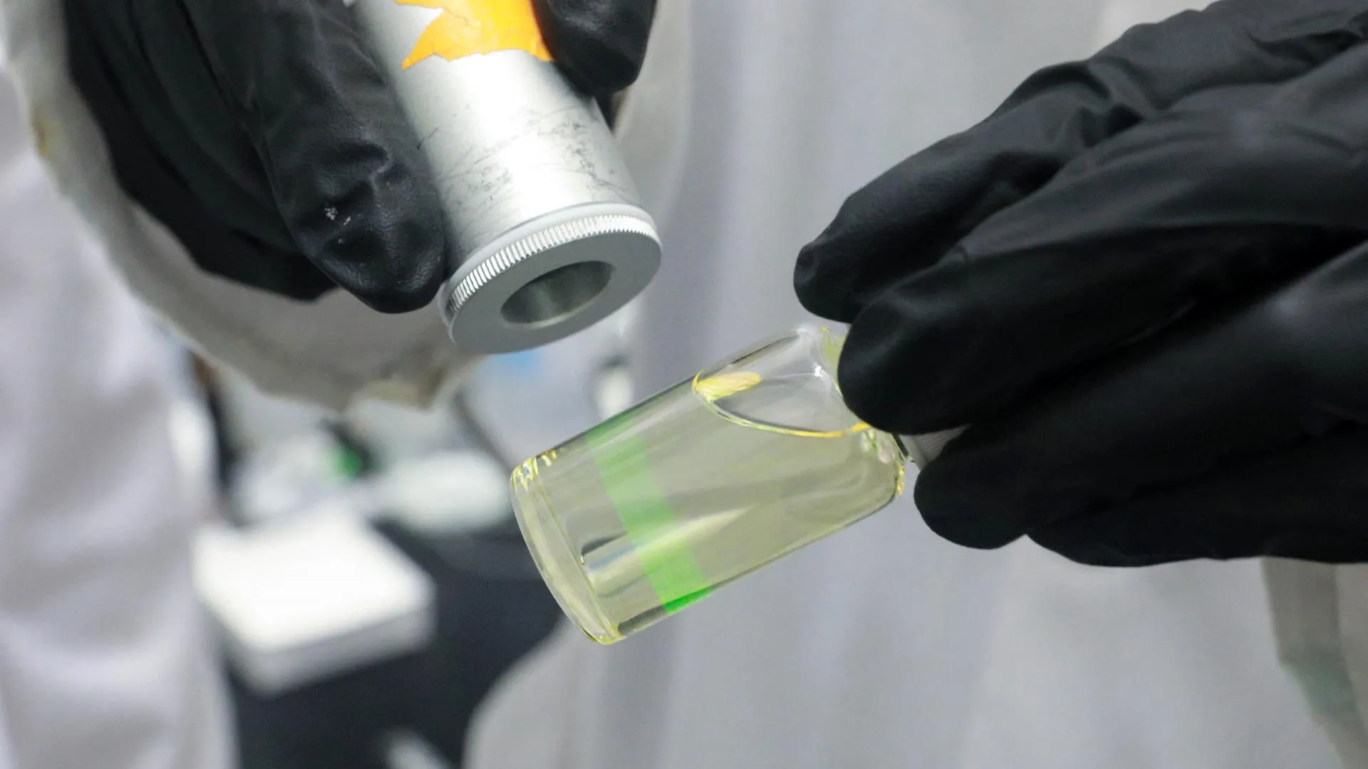
The University of Toronto team circumvented this limitation by designing nanoparticles capable of upconversion. Rather than absorbing ultraviolet or visible light, these engineered nanoprobes absorb low-energy photons in the near-infrared spectrum—a frequency easily and safely generated using economical, low-cost lasers—and emit them as high-energy, bright green light.
The advantages of this mechanism are twofold. First, near-infrared light penetrates biological tissues and complex chemical mixtures far more effectively than ultraviolet or visible light, minimizing photodamage and scattering. Second, because the excitation frequency differs drastically from the emission frequency, researchers can entirely eliminate autofluorescence, a common form of optical noise generated when sample matrices themselves absorb and scatter light.
Professor Huang illustrates the phenomenon through an astronomical analogy. "It’s like the difference between stargazing at night versus the daytime," he explains. "The stars shine the same brightness all the time, but during the day the sun is so powerful that it overwhelms them. Shifting the excitation frequency lower produces zero-autofluorescence background in the samples you are analyzing, while the luminescent nanoprobes keep shining; it is like turning off the sun, so you can see the stars better."
Chronology of Innovation: Overcoming the Back-Energy Trap
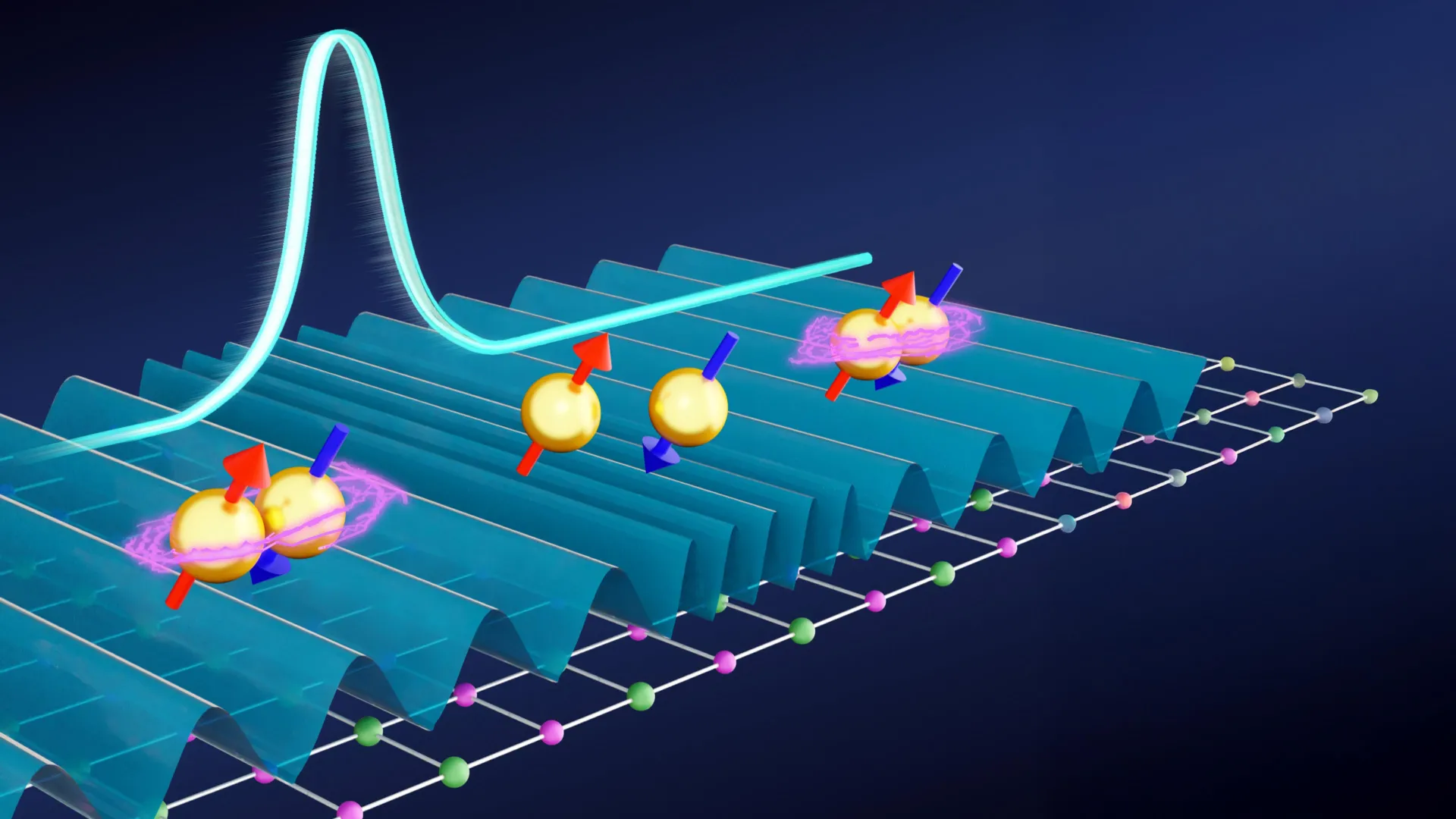
The foundation of the upconversion process rests on lanthanide ions, specifically ytterbium and erbium, embedded within a crystalline host matrix. In earlier iterations of such sensing technologies, these materials were manufactured as flat, hexagonal structures. Ytterbium ions served as sensitizers to capture incoming energy, transferring it sequentially to erbium ions, which ultimately performed the upconversion step to release green light.
Yet, scaling the brightness of these particles historically introduced a detrimental thermodynamic trade-off known as back-energy transfer. As researchers packed ytterbium atoms more densely into the host matrix to increase energy absorption, the atoms began to capture not only the incoming infrared energy, but also the outgoing green luminescence. Instead of reaching the surface to register as a detectable signal, the energy bounced back into the ytterbium relay and dissipated as heat.
To break this physical bottleneck, the Toronto research team initiated a rigorous multi-year development cycle. The project relied heavily on computational modeling before physical synthesis ever began in the laboratory. Undergraduate researcher Weixiang Ben spearheaded the computational efforts, utilizing advanced Monte Carlo simulations and density functional theory (DFT) to model atomic and subatomic energy transfers across dozens of hypothetical chemical formulations and physical geometries.
Through these simulations, the team abandoned traditional flat hexagonal architectures in favor of three-dimensional, diamond-shaped, core-shell-shell structures. Furthermore, they replaced the conventional sodium-yttrium-fluorine host matrix with a novel chemical composition built from lithium, lutetium, and fluorine.
This layered design established a chemical gradient in which the concentration of ytterbium ions increased progressively toward the core. The resulting architecture functioned as an atomic-scale one-directional energy tunnel, ensuring that incoming light energy flowed inexorably inward toward the erbium ions while preventing destructive back-energy transfer.
Quantitative Performance and Analytical Sensitivity
The resulting nanoparticles demonstrated extraordinary performance metrics during laboratory testing. Under identical excitation parameters, the newly engineered core-shell-shell nanoparticles achieved luminescence up to 150 times brighter than standard upconversion nanoparticles lacking dye-sensitization, and roughly 50 times brighter than the most highly optimized conventional structures documented in scientific literature to date.
This monumental increase in quantum yield translates directly to extreme analytical sensitivity. Because each individual nanoparticle emits a robust, highly concentrated optical signal, detectors can identify the presence of target molecules even when those molecules are present in exceedingly low concentrations.
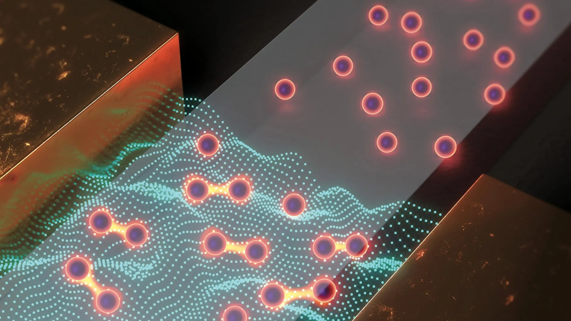
Beyond sheer sensitivity, the nanoprobes exhibit remarkable molecular selectivity. In chemical and pharmaceutical manufacturing, structural isomers present a formidable quality control hurdle. These compounds contain the exact same atoms bonded in different spatial configurations, meaning standard spectroscopic assays often fail to differentiate them without extensive, costly chromatographic separation. The Toronto nanoparticles, however, can be chemically functionalized to selectively bind with specific isomers, yielding distinct optical signatures that confirm molecular identity.
Industrial Implications: Transforming Pharmaceutical Quality Control
The commercial and industrial implications of this high-sensitivity, high-selectivity technology are far-reaching, particularly for the global pharmaceutical sector. Drug efficacy, pharmacokinetics, and toxicity profiles are frequently dictated by precise stereochemistry. If a synthetic batch of pharmaceuticals contains even a minor fraction of an unintended structural isomer—such as 10% impurity—the therapeutic outcome can be severely compromised, potentially triggering adverse clinical side effects.
Historically, detecting such trace isomeric impurities requires complex, time-consuming, and expensive analytical procedures, such as high-performance liquid chromatography coupled with mass spectrometry (HPLC-MS). Professor Huang’s team contends that their dye-sensitized nanoparticles could streamline this workflow dramatically.
"Let’s say you’re making a drug molecule, and your manufacturing process works fine, except that 10% of the batch is the wrong structural isomer," Wu notes. "That’s a huge problem: it can make the drug less effective, or worse, lead to side effects that you definitely don’t want. The current process for detecting this relies on very expensive analytical tests, but with these nanoparticles, you could do it using low-cost lasers and a very small sample."
Beyond the pharmaceutical cleanroom, environmental scientists face similar hurdles when monitoring trace chemical pollutants in natural aquatic ecosystems. Industrial solvents, endocrine disruptors, and agricultural runoff often contaminate groundwater reserves at parts-per-billion or parts-per-trillion concentrations. Traditional detection methods struggle with these trace thresholds due to complex organic backgrounds in natural water samples. The zero-autofluorescence capability of near-infrared upconversion nanoprobes offers a robust, field-deployable solution for real-time environmental monitoring.
Broader Economic and Scientific Analysis
Independent industry analysts observing the materials science sector have noted that the commercial viability of nanoparticle-based diagnostics hinges heavily on scalability and cost-efficiency. While the University of Toronto’s breakthrough currently stands as a proof-of-concept achievement within an academic setting, its heavy reliance on computational design significantly accelerates the R&D pipeline by eliminating trial-and-error synthesis cycles.
The substitution of lutetium and lithium into the host matrix also points toward future supply chain considerations, as rare-earth element sourcing remains a critical geopolitical and economic factor in advanced manufacturing. Nevertheless, the ability to utilize low-cost near-infrared laser diodes—widely manufactured for telecommunications and consumer electronics—rather than expensive ultraviolet laser systems significantly lowers the capital expenditure required for eventual point-of-use diagnostic instrumentation.
Future Outlook and Commercialization Roadmap
With the foundational physics validated and the peer-reviewed findings published in the Journal of the American Chemical Society, the research team is already pivoting toward the next phase of development: translational scalability.
Transitioning from gram-scale laboratory synthesis to industrial-grade manufacturing presents distinct engineering challenges, including batch-to-batch uniformity, surface functionalization stability, and regulatory compliance for medical and environmental deployment. Professor Huang remains pragmatic yet optimistic about the path forward.
"We’re working on this already, in fact," Huang affirms, discussing the roadmap toward commercial adoption. "We think it’s feasible, but it requires a very long roadmap. In the meantime, this model serves as proof-of-concept; with this technique, we can produce a very high-performance upconversion nanoparticle that could be customized to any molecule you might want to detect. That’s something entirely new."
As research institutions and commercial partners begin to explore licensing agreements and pilot-scale production, this University of Toronto innovation marks a foundational shift in optical biosensing, promising a future where detecting a single errant molecule in a complex chemical sea is no longer an analytical impossibility.