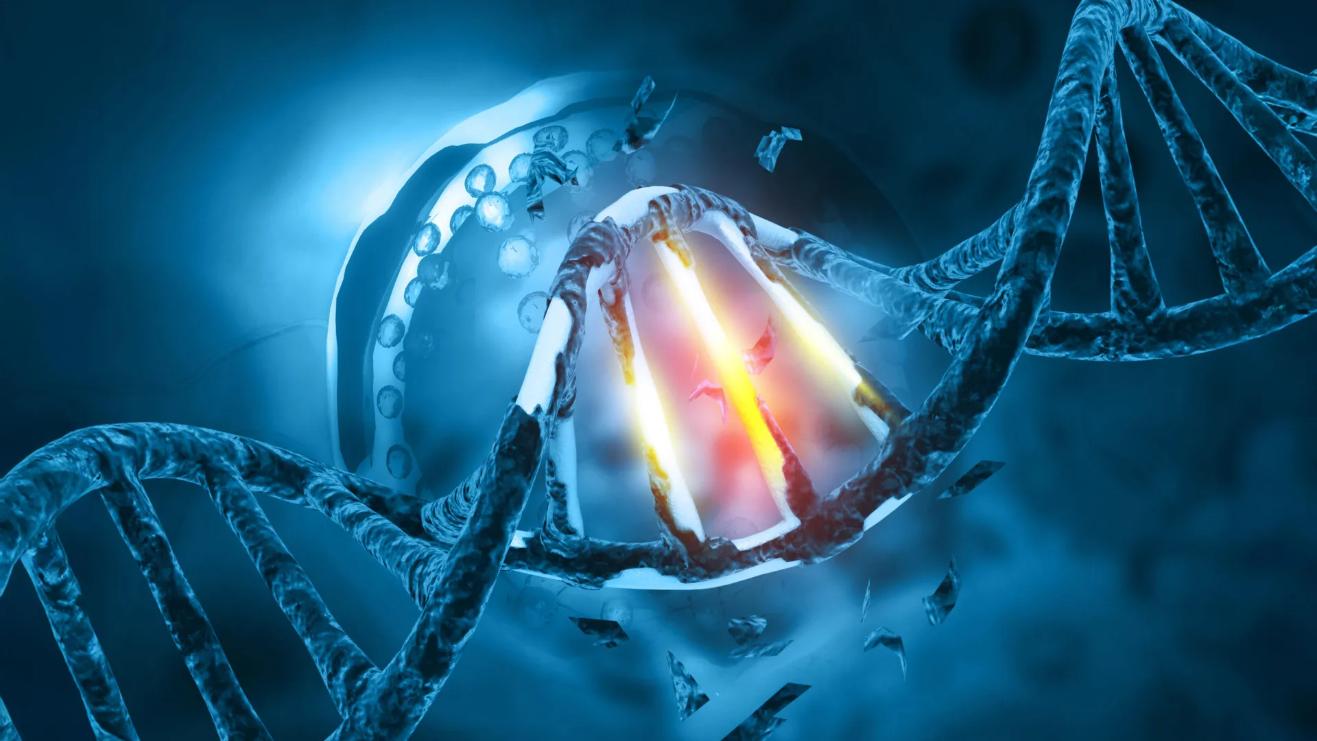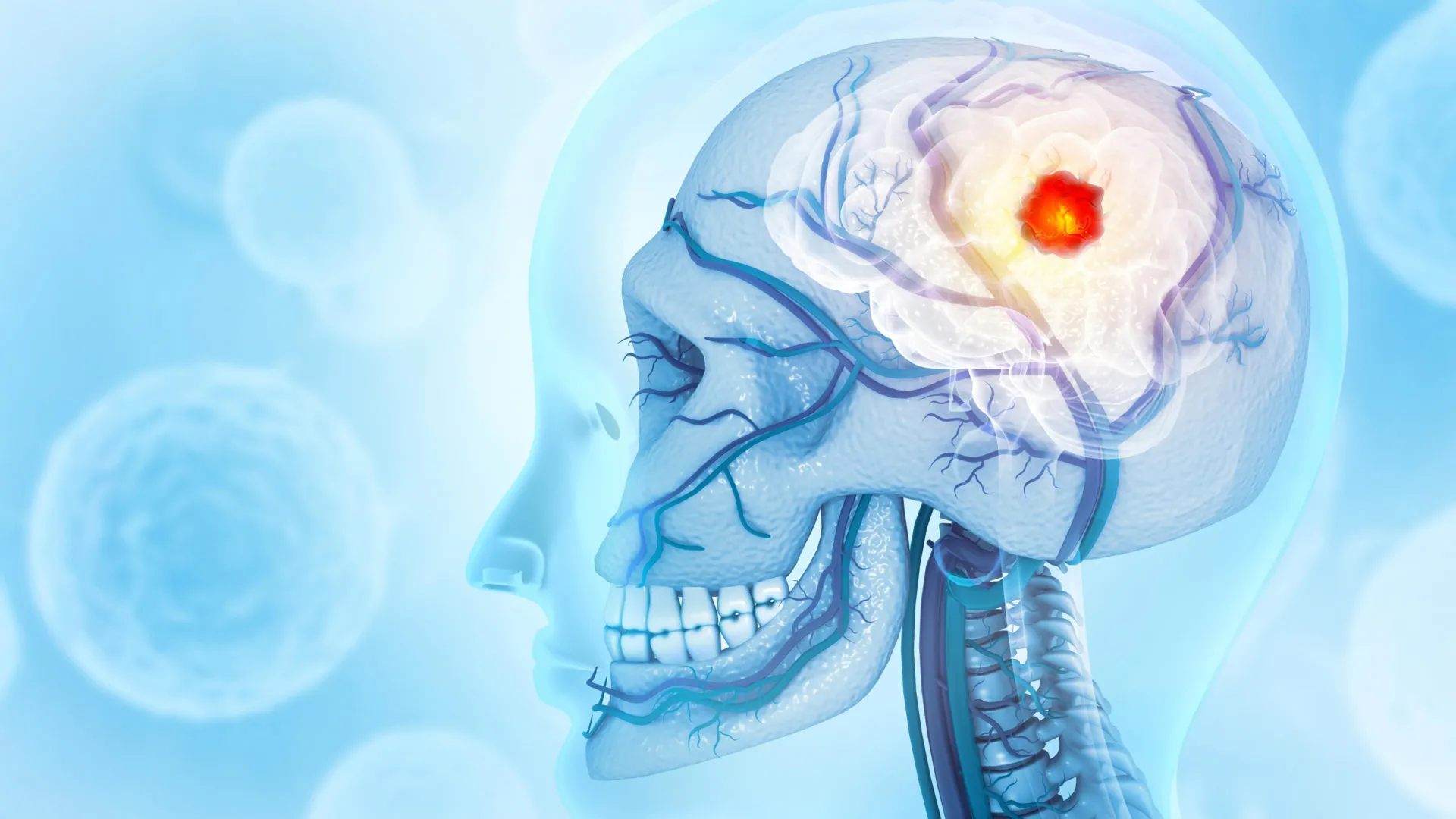
The medical community has long viewed glioblastoma multiforme (GBM) as a localized, albeit highly aggressive, neurological crisis. However, groundbreaking research from the Montefiore Einstein Comprehensive Cancer Center (MECCC) and the Albert Einstein College of Medicine suggests that this perspective may be dangerously narrow. In a study published on October 3 in the prestigious journal Nature Neuroscience, researchers have demonstrated that glioblastoma is not merely a tumor of the brain tissue; it is a systemic architect of destruction that actively reshapes the skull’s anatomy and hijacks the body’s immune reservoir.
The findings reveal that glioblastoma triggers significant erosion of the skull bone and fundamentally alters the composition of bone marrow within the cranium. Perhaps most alarmingly, the study found that certain common medications used to treat bone loss can inadvertently accelerate the progression of the cancer or neutralize the efficacy of immunotherapy. This discovery offers a potential explanation for why decades of localized treatments—including surgery, radiation, and chemotherapy—have largely failed to improve the long-term prognosis for patients diagnosed with this lethal disease.
The Lethal Landscape of Glioblastoma
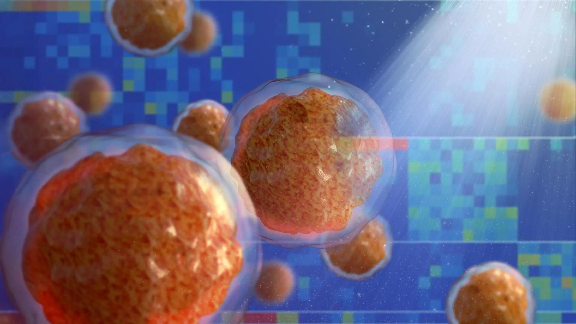
Glioblastoma is the most common and most aggressive primary brain tumor in adults. According to data from the National Cancer Institute (NCI), approximately 15,000 individuals in the United States are diagnosed with the condition annually. Despite advancements in neurosurgery and precision medicine, the median survival time remains a sobering 15 months for those receiving the standard of care.
The current therapeutic paradigm focuses almost exclusively on the brain. Surgeons attempt to resect as much of the tumor as possible, followed by Temozolomide (chemotherapy) and targeted radiation. However, the recurrence rate is nearly 100%. "Our discovery that this notoriously hard-to-treat brain cancer interacts with the body’s immune system may help explain why current therapies—all of them dealing with glioblastoma as a local disease—have failed," stated Jinan Behnan, Ph.D., the paper’s corresponding author and assistant professor at the Leo M. Davidoff Department of Neurological Surgery and the department of microbiology & immunology at Einstein.
Chronology of the Discovery: From Channels to Bone Loss
The research team, led by Dr. Behnan, began their investigation by examining the physical relationship between the brain and the skull. For decades, the skull was viewed as a static, protective shell. However, recent neuro-immunological discoveries have identified extremely thin, microscopic channels that connect the brain directly to the skull’s bone marrow. These channels serve as a "hotline" for the exchange of molecules and immune cells.
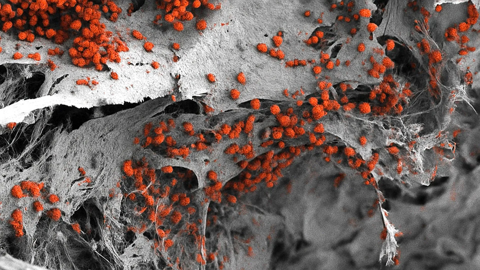
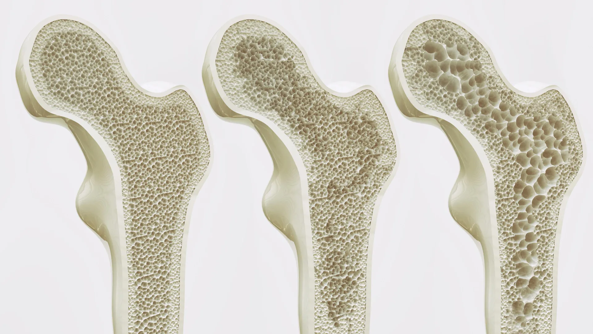
Using advanced high-resolution imaging tools, the researchers observed mice that had been implanted with two distinct types of glioblastoma. The results were startling: the presence of the tumor led to visible erosion of the skull bone. This degradation was most pronounced along the sutures—the fibrous joints where the different plates of the skull fuse together.
To determine if this bone loss was a general response to brain trauma or specific to glioblastoma, the team conducted a series of control experiments. They examined mice that had suffered strokes, various forms of traumatic brain injury (TBI), and mice with cancers originating in other parts of the body. In these cases, the skull remained intact. The erosion was a unique signature of aggressive brain tumors.
The researchers then transitioned from animal models to human clinical data. CT scans of human patients with glioblastoma confirmed the findings. Patients exhibited significant reductions in skull thickness in the same regions identified in the mice, suggesting that the tumor’s influence on the bone is a cross-species phenomenon.
The Mechanism: Enlarged Channels and Molecular Signaling
The physical erosion of the skull does more than weaken the bone; it fundamentally changes the "plumbing" between the brain and the immune system. The study found that as the bone eroded, the microscopic channels connecting the brain to the skull marrow increased in both number and diameter.
The scientists proposed that these enlarged conduits allow the tumor to export molecular signals directly into the skull marrow. In essence, the glioblastoma "reprograms" the marrow from a distance. By opening these floodgates, the tumor facilitates a two-way traffic system where it can send instructions to the immune system and receive a steady supply of cells that may actually help the tumor grow rather than fight it.
The Immune Shift: A Tilt Toward Inflammation
The most significant impact of these enlarged channels is the alteration of the immune landscape. Using single-cell RNA sequencing—a technology that allows researchers to see which genes are turned on or off in individual cells—the team mapped the immune cell population within the skull marrow.
They found that glioblastoma causes a dramatic shift toward a pro-inflammatory environment. The levels of inflammatory myeloid cells, particularly neutrophils, nearly doubled. Simultaneously, the marrow saw a near-total depletion of antibody-producing B cells and other lymphoid cells that typically help the body fight off threats.
"The skull-to-brain channels allow an influx of these numerous pro-inflammatory cells from the skull marrow to the tumor, rendering the glioblastoma increasingly aggressive and, all too often, untreatable," explained study co-author E. Richard Stanley, Ph.D., a professor of developmental and molecular biology at Einstein.
This influx of neutrophils creates a "protumorigenic" environment, where the inflammation actually shields the cancer from the body’s natural defenses and promotes the growth of new blood vessels to feed the tumor.
Systemic Disruption: Skull vs. Femur
One of the most surprising findings of the study was the divergent reaction of bone marrow in different parts of the body. While the marrow in the skull became a factory for pro-inflammatory cells, the marrow in the femur (the thigh bone) reacted in the opposite manner.
In the femur, glioblastoma appeared to suppress the genes necessary for producing various immune cells. This disparity underscores the fact that glioblastoma is a systemic disease that communicates differently with various "compartments" of the body. The skull marrow, due to its proximity and direct physical connection via the newly enlarged channels, is uniquely susceptible to the tumor’s manipulative signaling.
The Pharmaceutical Paradox: Osteoporosis Drugs and Cancer Risk
Given the observed bone erosion, the researchers investigated whether preventing this loss could slow the disease. They administered two FDA-approved anti-osteoporosis drugs to the mice: zoledronic acid (a bisphosphonate) and denosumab (a RANKL inhibitor).
While both drugs were successful in halting the erosion of the skull bone, the biological consequences were unexpected and concerning. In one type of glioblastoma, zoledronic acid actually fueled the progression of the tumor.
Furthermore, both drugs were found to interfere with the efficacy of immunotherapy. Specifically, they blocked the beneficial effects of anti-PD-L1, a class of drugs designed to "unmask" cancer cells so that T cells can attack them. By disrupting the immune landscape of the marrow, the bone-strengthening drugs inadvertently disabled the very treatments meant to save the patient.
"This indicates the need for treatments that restore the normal balance of immune cells in the skull marrow of people with glioblastoma," noted Dr. Stanley. The research suggests that simply stopping bone loss is not enough; the goal must be to restore the production of T and B cells while suppressing the production of inflammatory neutrophils.
Supporting Data and Collaborative Effort
The study was a massive collaborative effort involving experts from across the globe. In addition to the MECCC and Einstein teams, contributors included researchers from Osaka University in Japan, Karolinska Hospital in Sweden, Duke University Medical Center, the University of California San Francisco, and the German Rheumatism Research Center in Berlin.
Key data points from the study include:
- Sample Size and Scope: The study utilized multiple mouse models of GBM and analyzed high-resolution CT imaging from human patient cohorts.
- Genetic Mapping: Single-cell RNA sequencing provided a high-definition view of the myeloid-to-lymphoid shift, showing a 2x increase in neutrophils.
- Pharmacological Impact: The inhibition of anti-PD-L1 efficacy by bone-loss drugs was consistent across multiple trials, raising significant questions about the use of these drugs in oncology patients.
Implications for Future Treatment
The implications of this research are profound. It suggests that the failure of immunotherapy in glioblastoma—a major disappointment in the field over the last decade—may be due to the tumor’s ability to "source" immunosuppressive cells directly from the skull.
The findings point toward several new strategies for treatment:
- Targeting the Channels: If the physical channels between the skull and brain can be pharmacologically narrowed or blocked, it may prevent the influx of pro-inflammatory cells.
- Marrow-Focused Therapy: Future treatments might involve "re-balancing" the skull marrow, perhaps through local injections or targeted therapies that encourage B cell production while suppressing neutrophils.
- Re-evaluating Bone Health Protocols: Oncologists may need to exercise extreme caution when prescribing bisphosphonates or RANKL inhibitors to patients with primary brain tumors.
As the medical community digests these findings, the focus moves from the "local" to the "systemic." By understanding that the skull is a dynamic participant in the progression of glioblastoma, researchers have opened a new frontier in the fight against one of the world’s most devastating cancers. The hope is that by treating the whole environment—the brain, the bone, and the marrow—the 15-month survival barrier can finally be broken.