In a significant advancement for the field of molecular oncology, a multi-disciplinary team of investigators from the Johns Hopkins Kimmel Cancer Center and the Johns Hopkins Bloomberg School of Public Health has decoded a critical mechanism by which rearranged genes drive the progression of a rare and aggressive form of kidney cancer. The study, published on April 22 in the journal Cell Reports, provides the first comprehensive look at how fusion proteins in translocation renal cell carcinoma (tRCC) organize into microscopic liquid droplets to hijack cellular machinery. This discovery not only sheds light on a poorly understood malignancy but also suggests a novel therapeutic roadmap for various other cancers driven by similar genetic fusions, including certain leukemias and sarcomas.
Supported by the National Institutes of Health (NIH), the research team demonstrated that these "fusion genes" produce proteins that undergo a process known as liquid-liquid phase separation. This process results in the formation of condensates—concentrated, droplet-like structures within the cell nucleus. These droplets serve as specialized command centers where the cancer-driving proteins can efficiently turn on genes that promote tumor growth and suppress those that would otherwise halt cancer progression. By identifying the specific protein segments responsible for forming these droplets, the researchers have opened the door to a new class of treatments designed to "dissolve" the machinery of the cancer cell from the inside out.
The Clinical Challenge of Translocation Renal Cell Carcinoma
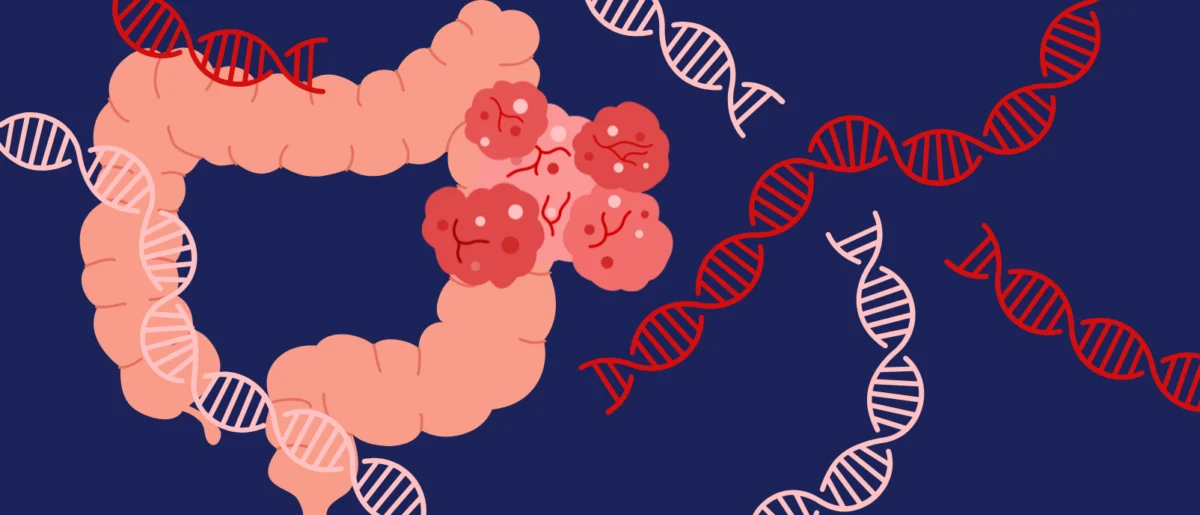
Translocation renal cell carcinoma represents a distinct and challenging subtype of kidney cancer. While it accounts for approximately 1% to 5% of all renal cell carcinomas in adults, it is significantly more prevalent in pediatric and young adult populations, making up as much as 50% of kidney cancer cases in children. Historically, tRCC has been difficult to treat because it often does not respond to the standard therapies used for more common types of kidney cancer, such as clear cell renal cell carcinoma.
The disease is characterized by a chromosomal translocation, a genetic event where a chromosome breaks and a portion of it reattaches to a different chromosome. In the case of tRCC, this rearrangement involves the TFE3 gene. The tail end of the TFE3 gene is fused with the beginning of one of several other "partner" genes, such as NONO, SFPQ, or PRCC. This genetic "chimera" leads to the production of TFE3 fusion proteins—hybrid molecules that do not exist in healthy cells.
"Scientists have known for some time that these TFE3 fusion proteins are the primary drivers of this rare kidney cancer," explained Danfeng "Dani" Cai, Ph.D., an assistant professor of biochemistry and molecular biology at the Johns Hopkins Bloomberg School of Public Health and the study’s senior author. "However, the exact ‘how’—the mechanical process by which these proteins actually transform a healthy cell into a malignant one—remained a mystery until now."
Unveiling the Mechanism: Liquid Condensates and Phase Separation
The breakthrough came when Dr. Cai’s laboratory, which specializes in the study of cellular condensates, applied the principles of phase separation to the TFE3 fusion proteins. Phase separation in a cell is analogous to oil droplets forming in water; certain proteins and molecules can spontaneously cluster together to form dense, liquid-like droplets that perform specific functions without the need for a physical membrane.
To observe this phenomenon, the research team employed advanced imaging techniques, attaching fluorescent "glowing tags" to the TFE3 fusion proteins in cells derived from kidney cancer patients. Under high-resolution microscopy, the fusion proteins did not spread evenly throughout the nucleus. Instead, they congregated into distinct, shimmering "dots" or condensates.
The team observed that these droplets were not just passive clusters of protein. They functioned as highly active hubs. Further investigation revealed that these condensates also contained marker proteins associated with gene activation and the recruitment of transcriptional machinery. This suggested that the TFE3 fusion proteins were using these liquid droplets to physically aggregate the tools necessary to over-express oncogenes—genes that drive cancer.
Strategic Collaboration and Chromatin Remodeling
To understand how these droplets interact with the cell’s genetic blueprint, Dr. Cai partnered with Eneda Toska, Ph.D., an assistant professor of oncology at the Johns Hopkins Kimmel Cancer Center. Dr. Toska’s expertise in epigenetics and chromatin biology allowed the team to map the physical changes the fusion proteins inflicted on the cell’s DNA.
In a healthy cell, DNA is packaged into a structure called chromatin, often described as "beads on a string." When the string is tightly wound around the beads (histones), the genes are "closed" and cannot be read. When the string is loose, the genes are "open" and active. The research revealed that TFE3 fusion proteins act as master architects of this landscape.
"We found that these fusion proteins open and close different sites on the chromatin by making specific chemical modifications," Dr. Toska stated. "They essentially bind to, regulate, and redesign the chromosome landscape. By doing so, they interact with target genes that promote cell proliferation and movement—the very functions a cancer cell needs to grow, invade surrounding tissue, and spread throughout the body."
The study specifically focused on two of the most common fusion partners, NONO and SFPQ, which together account for approximately 40% of all TFE3 fusion cases. The researchers found that the fusion proteins had a significantly stronger ability to control gene expression than the individual protein components would have on their own.
Identifying the Structural "Achilles’ Heel"
One of the most critical findings of the study involved identifying the specific structural component of the fusion protein that allows it to form these dangerous droplets. Through a series of molecular "editing" experiments, the team systematically removed different parts of the TFE3 fusion proteins to see which pieces were essential for condensate formation.
They discovered a small segment that creates a "coiled-coil" shape—a structure where multiple alpha-helices are wrapped around each other like the strands of a rope. This coiled-coil domain, located in the section of the protein that connects the TFE3 tail to its fusion partner, proved to be the linchpin. When this segment was removed, the TFE3 fusion proteins lost their ability to form liquid droplets. Consequently, they were no longer able to bind to the chromatin or activate the cancer-promoting genes.
This discovery is of paramount importance because it identifies a physical target for future drug development. If a small molecule or drug could be designed to disrupt the coiled-coil interaction or prevent the phase separation process, it could theoretically "de-program" the cancer cells, halting their growth without the need for traditional, highly toxic chemotherapy.
Chronology of the Research and Supporting Data
The study’s timeline reflects a rigorous multi-year effort involving several institutions. The research began with the characterization of patient-derived cell lines and moved through several phases:
- Initial Visualization: Identification of the "dots" in the nucleus via fluorescent tagging.
- Functional Analysis: Correlation of these dots with known gene-activation markers.
- Genomic Mapping: Collaboration with the Toska lab to perform ChIP-seq and other genomic assays to see exactly where on the DNA the fusion proteins were binding.
- Structural Dissection: The "editing" phase where the coiled-coil domain was identified as the structural driver of the condensates.
- Validation: Testing whether the disruption of droplets actually stopped the expression of oncogenes.
The data gathered during this process highlighted that the fusion proteins don’t just act as simple transcription factors; they act as "super-enhancers," creating an environment that is hyper-conducive to malignant gene expression. The study also benefited from the contributions of experts from the National Cancer Institute (NCI) and the National Human Genome Research Institute, underscoring the collaborative nature of modern oncology research.
Broader Implications for Oncology and Future Directions
While the current study focuses on translocation renal cell carcinoma, the implications reach far beyond this rare kidney cancer. Many other aggressive cancers are driven by similar gene fusions. For example, Ewing sarcoma, a bone cancer that primarily affects children and young adults, is driven by the EWS-FLI1 fusion. Similarly, various forms of leukemia are characterized by fusion genes like BCR-ABL.
"It is highly possible that these other fusion genes form similar droplets or condensates," said Dr. Cai. "If this mechanism is a universal feature of fusion-driven cancers, then the strategy of disrupting these liquid structures could lead to a whole new class of therapies across the oncology spectrum."
The Johns Hopkins team is already looking toward the next phase of their research. They hope to identify other proteins and molecules that are "recruited" into the TFE3 condensates. By understanding the full composition of these droplets, they can begin screening for existing drugs or developing new small molecules that can penetrate the droplets or prevent them from forming in the first place.
The research was a collaborative effort involving a large roster of scientists, including Choon Leng So, Ye Jin Lee, Wanlu Chen, and others from the Bloomberg School of Public Health, as well as Bujamin Vokshi from the Johns Hopkins University School of Medicine and W. Marston Linehan from the National Cancer Institute.
Funding for this groundbreaking work was provided by several branches of the NIH, including the National Institute of General Medical Sciences, the National Cancer Institute, and the National Human Genome Research Institute. Additional support came from the Department of Defense Kidney Cancer Idea Development Award, the Jayne Koskinas Ted Giovanis Foundation, and the Johns Hopkins Provost Catalyst Award.
As the medical community moves toward more personalized and molecularly targeted treatments, the findings from the Johns Hopkins team provide a vital piece of the puzzle in the fight against rare and recalcitrant cancers. By moving from a general understanding of genetic "errors" to a specific understanding of the physical "machinery" those errors create, researchers are paving the way for more effective, less toxic interventions for patients who currently have few options.