In a landmark achievement for regenerative medicine and hematology, a multidisciplinary team of scientists has successfully engineered a fully functional, three-dimensional model of the human bone marrow niche using exclusively human cells. This breakthrough, led by researchers at the University of Basel and University Hospital Basel, represents the first time the intricate architecture of the body’s "blood factory" has been recreated in vitro with such biological fidelity. By integrating bone cells, nerves, and blood vessels into a singular system, the researchers have opened a new frontier in the study of blood-related diseases, drug development, and the ethical pursuit of reducing animal experimentation in clinical research.
The study, published in the prestigious journal Cell Stem Cell, details how the research team, headed by Professor Ivan Martin and Dr. Andrés García-García from the Department of Biomedicine, overcame decades-long hurdles in bioengineering. For years, the scientific community has struggled to replicate the "endosteal niche"—a specific microenvironment located near the inner surface of the bone that is critical for the regulation of blood stem cells and is frequently implicated in the progression of hematological malignancies such as leukemia and multiple myeloma.
The Biological Significance of the Bone Marrow Niche
To appreciate the magnitude of this achievement, one must understand the role of bone marrow in human physiology. Bone marrow is not merely a filler for the skeletal system; it is a highly specialized organ responsible for hematopoiesis—the continuous production of red blood cells, white blood cells, and platelets. This process occurs within specialized "niches," or microenvironments, where various cell types interact to provide the chemical and physical signals necessary for stem cells to thrive or differentiate.
The endosteal niche is of particular interest to oncologists and biologists. It is here that hematopoietic stem cells (HSCs) reside in a state of quiescence until they are needed to replenish the blood supply. However, this niche also serves as a sanctuary for cancer cells. During chemotherapy, some malignant cells can "hide" within the endosteal niche, evading treatment and leading to later relapses. Until now, studying these interactions in humans was nearly impossible without invasive biopsies, and animal models often failed to accurately mirror human cellular responses due to fundamental species differences.
Methodology and the Chronology of Development
The development of this synthetic bone marrow followed a meticulous chronological process that combined materials science with advanced cellular reprogramming. The project began with the creation of a physical framework designed to mimic the porous structure of human bone. The team utilized hydroxyapatite, a naturally occurring mineral form of calcium apatite that constitutes the primary inorganic component of human bone and tooth enamel. This scaffold provided the necessary mechanical stability and biochemical cues to support cell growth.
Following the preparation of the scaffold, the researchers turned to molecular biology to source the necessary cellular components. Rather than harvesting multiple cell types from various donors—which would introduce significant biological variability—the team utilized human induced pluripotent stem cells (iPSCs). These cells, which can be reprogrammed from adult skin or blood cells, possess the unique ability to differentiate into any cell type in the human body.
By applying specific signaling molecules and growth factors at precise intervals, the team guided these stem cells through controlled developmental stages. Over several weeks, the pluripotent cells transitioned into the diverse array of specialized cells required for a functional niche, including osteoblasts (bone-forming cells), endothelial cells (which form blood vessels), and mesenchymal stromal cells (which support the marrow structure). The resulting 3D structure was not only biologically complex but also physically substantial, measuring eight millimeters in diameter and four millimeters in thickness—a significant scale-up from previous "organ-on-a-chip" models.
Supporting Data and Technical Validation
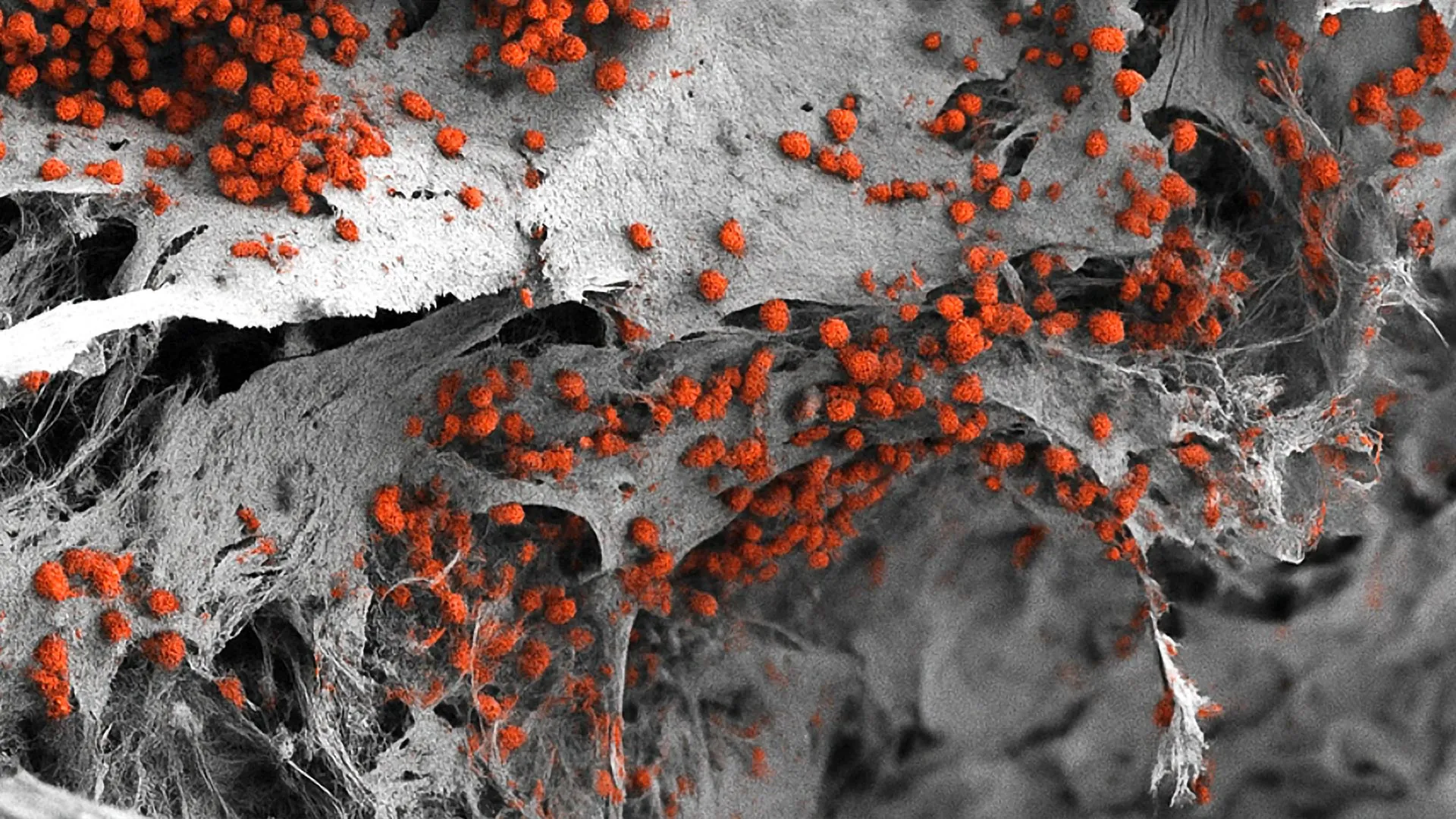
The technical validation of the model involved rigorous analysis to ensure the laboratory-grown tissue functioned like its natural counterpart. Using advanced imaging and transcriptomic analysis, the researchers confirmed that the spatial arrangement of cells within the hydroxyapatite scaffold closely matched the architecture found in human patients.
Key data points from the study include:
- Longevity: The model successfully maintained the formation and maturation of human blood cells for several weeks in a controlled laboratory environment, demonstrating sustained biological activity.
- Complexity: Unlike previous models that often featured only one or two cell types, this system integrated bone cells, vascular networks, and rudimentary nervous system components.
- Scale: At 8mm by 4mm, the model is large enough to allow for the observation of macro-scale tissue interactions while remaining small enough for laboratory manipulation.
- HSC Maintenance: The endosteal niche within the model successfully supported the "stemness" of hematopoietic stem cells, proving that the chemical signaling pathways were operational.
Institutional Perspectives and the 3R Principle
The implications of this research extend beyond the laboratory and into the realm of scientific ethics and regulatory policy. Professor Ivan Martin emphasized that while mouse models have provided the foundation for much of what is known about bone marrow, they are not perfect substitutes for human biology. Differences in cell surface markers, cytokine responses, and aging processes mean that many drugs that work in mice fail in human clinical trials.
"Our model brings us closer to the biology of the human organism," Professor Martin stated. He noted that the platform is designed to serve as a complement to animal experiments, aligning with the "3R Principle" supported by the University of Basel and international regulatory bodies: Replace, Reduce, and Refine. By providing a high-fidelity human alternative, the researchers hope to decrease the number of animals required for early-stage hematology research and toxicity testing.
The reaction from the broader scientific community has been one of cautious optimism. While the model is a breakthrough, experts note that integrating it into the standard drug development pipeline will require further refinement. Dr. Andrés García-García acknowledged that for high-throughput drug screening—where thousands of compounds are tested simultaneously—the current 8mm model might be too large and resource-intensive. Future iterations will likely focus on miniaturizing the system to allow for rapid, automated testing.
Broader Impact: From Drug Testing to Personalized Medicine
The potential applications for this technology are vast. In the immediate future, the pharmaceutical industry could utilize these human bone marrow models to test the toxicity of new chemotherapies. Many cancer drugs are limited by "myelosuppression," a side effect where the bone marrow’s ability to produce blood cells is dangerously impaired. Testing these drugs on a human-derived model before they reach clinical trials could help identify safety issues much earlier in the process.
Perhaps the most exciting long-term prospect is the realization of personalized medicine for blood cancer patients. The researchers envision a future where a patient’s own cells are used to create a "personalized niche" in the lab. Doctors could then introduce the patient’s specific cancer cells into this model and test a battery of different treatment combinations.
"In the future, this approach could help guide personalized treatment decisions," the research team noted. By identifying which therapy most effectively targets the cancer cells within the protective environment of the endosteal niche, clinicians could move away from a "one-size-fits-all" approach and toward precision oncology. This would be particularly transformative for patients with resistant forms of leukemia, where the standard of care often fails to prevent relapse.
Analysis of Implications for the Future of Hematology
The success of the Basel team marks a paradigm shift in how we approach tissue engineering. By proving that a complex, multi-lineage organ system can be grown from pluripotent stem cells within a synthetic mineral scaffold, the study provides a blueprint for recreating other complex tissues, such as the liver or the blood-brain barrier.
However, challenges remain. The current model, while impressive, lacks a fully integrated immune system and a systemic blood circulation, which are critical for understanding how the bone marrow interacts with the rest of the body. Furthermore, the cost of producing iPSC-derived models is currently high, which may limit its use to specialized research centers in the short term.
Despite these hurdles, the study represents a definitive step toward a new era of medical research—one that is less dependent on animal models and more focused on the specific nuances of human physiology. As the technology matures and the ability to scale and miniaturize these models improves, the "blood factory" in a lab may become a standard tool in the fight against some of the most challenging diseases known to medicine. The work of Professor Martin, Dr. García-García, and their colleagues ensures that the quiet work of the bone marrow will no longer go unnoticed, but will instead be observed, understood, and ultimately mastered for the benefit of patients worldwide.