The human brain is an extraordinary yet fragile organ, responsible for complex cognition, emotion, and bodily control, yet it possesses a notoriously poor capacity for self-repair. When neurons are destroyed by trauma, stroke, or neurodegenerative conditions such as Parkinson’s and Alzheimer’s disease, the damage is typically permanent, leaving patients with lifelong disabilities. However, nature offers extraordinary counterexamples of resilience. Certain organisms possess the remarkable ability to harness their own cellular machinery not merely to mend minor wounds, but to completely regenerate complex tissues, muscles, and even entirely functional brains.
Addressing this disparity in biological design, a team of researchers at the University of Georgia has successfully pinpointed specific genes that govern brain regeneration in freshwater flatworms known as planarians. Published in the journal Nature Communications, this breakthrough bridges a critical knowledge gap in regenerative biology. By identifying the exact genetic instructions that allow flatworms to replace lost neurons, scientists are laying the foundational science required to potentially awaken dormant regenerative pathways within human biology, offering a long-term beacon of hope for individuals suffering from currently incurable neurological disorders.
Understanding the Planarian Model: Masters of Biological Reconstitution
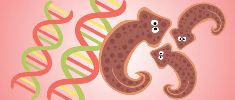
Planarians are unassuming creatures found globally in freshwater environments, marine habitats, and damp terrestrial soils. Despite lacking sophisticated organ networks such as circulatory or respiratory systems, these flatworms possess a master weapon in biological survival: an abundant population of adult stem cells known as neoblasts. These pluripotent stem cells hold the unique capacity to divide indefinitely and differentiate into any cell type required by the organism.
If a planarian is divided into minute fragments—sometimes as small as a single fraction of its original mass—its neoblasts migrate to the wound sites, proliferate, and meticulously reconstruct every missing anatomical structure, including a fully operational central nervous system complete with photoreceptors, a brain ganglion, and a coordinated neural network. This extraordinary biological feat has long fascinated developmental biologists, but the specific genetic recipes dictating how stem cells differentiate into specialized neurons and navigate to precise anatomical locations have remained elusive until now.
Cracking the Genetic Code of Dopaminergic Neurons
To understand how planarians orchestrate this cellular orchestration, the research team focused on the genesis of dopaminergic neurons. Dopamine is a critical neurotransmitter shared across the animal kingdom, functioning far beyond its colloquial reputation as the brain’s "pleasure and reward" chemical. In humans and other mammals, dopaminergic pathways are vital for regulating voluntary movement, mood, motivation, and cognitive focus. The degeneration of these specific neurons is the hallmark pathology of Parkinson’s disease, leading directly to debilitating tremors, muscle rigidity, and bradykinesia.
Using advanced genetic screening techniques, the University of Georgia researchers identified nearly a dozen distinct genes responsible for instructing planarian stem cells to mature into dopamine-producing neurons and directing those newly formed cells to migrate into their proper positions within the flatworm’s nervous system.
To validate the function of these newly discovered genes, the team performed loss-of-function experiments, systematically silencing or "knocking out" the target genes. The results were striking. Without the operational genetic blueprint, the planarians struggled significantly to generate new dopaminergic neurons. Consequently, the affected flatworms exhibited severe locomotor deficits, displaying slow and uncoordinated movement strikingly analogous to the motor dysfunctions observed in mammals experiencing dopamine deficiencies.
Contextualizing the Research: The Limits of Human Neuroregeneration
To fully appreciate the significance of the University of Georgia findings, one must examine the stark evolutionary and cellular differences dividing humans from simpler invertebrates. Both human and flatworm brains are fundamentally constructed from networks of specialized neurons that communicate via intricate electrical and chemical synapses. Both organisms harbor stem cell populations designed for maintenance and repair.
However, a profound divergence occurs in cellular programming. While human adult stem cells in the brain—primarily located in the subventricular zone and the subgranular zone of the dentate gyrus—retain some capacity for neurogenesis, their scope and efficiency are heavily restricted. Following an ischemic stroke, traumatic brain injury, or the progressive neuronal death characteristic of Alzheimer’s disease, the human central nervous system fails to mount a sufficient regenerative response. Instead, glial cells proliferate to form a glial scar, which, while sealing the wound and preventing further acute damage, effectively blocks axonal regrowth and new neuron integration.

As Rachel Roberts-Galbraith, corresponding author of the study and an associate professor in the University of Georgia’s Franklin College of Arts and Sciences, points out, humanity’s poor healing capacity is not an immutable law of biological evolution. "Big picture: We would like to come up with ideas for how to better empower the human brain to regenerate itself," Roberts-Galbraith stated. "The understanding of brain regeneration that we can develop using simple animals gives us a reason to be optimistic. It’s not an inherent property of brains that makes them bad at regeneration. It’s something specific to humans."
Implications for Clinical Therapeutics and Regenerative Medicine
The implications of mapping the planarian neuroregeneration cascade extend far beyond basic science, offering tangible pathways for translational medicine. Currently, therapeutic options for neurodegenerative diseases remain largely palliative. Treatments for Parkinson’s disease, such as levodopa administration or deep brain stimulation, manage symptoms but cannot halt the underlying loss of dopaminergic neurons or repair the damaged neural architecture.
Cell-based therapies have emerged as a promising frontier, involving the transplantation of lab-grown dopamine-producing neurons into the brains of patients. However, clinical progress has been stymied by the difficulty of generating pure, properly specified neuronal subtypes from human pluripotent stem cells and ensuring they integrate correctly into existing neural circuits.
By detailing the exact genetic recipe utilized by planarians, the University of Georgia study provides a biochemical roadmap that bioengineers and stem cell researchers can adapt. "We figured out the genetic recipe for making these cell types in planarians," Roberts-Galbraith noted. "We’re hoping this work helps others figure out how to create dopamine-producing neurons from stem cells that can be more effectively transplanted into patients."
Methodological Breakthroughs and Future Research Directions
The identification of these critical genes was made possible by recent advancements in high-throughput sequencing, RNA interference (RNAi), and single-cell transcriptomics. By analyzing gene expression changes in real-time as planarians regenerated their nervous systems following targeted amputations, the research team could isolate transient cellular states and map the exact transcriptional hierarchy required for neurogenesis.
Moving forward, the research team aims to expand their investigations beyond dopaminergic systems. The human brain relies on dozens of distinct neurotransmitter systems, including serotonergic, cholinergic, and GABAergic pathways, each playing vital roles in cognition, memory, and autonomic function. Determining whether similar genetic networks govern the regeneration of these other neuronal subtypes will be a crucial next step.
Furthermore, scientists face the complex challenge of identifying why human homologous genes fail to activate robust regenerative responses in the wake of injury. Comparative genomic analyses between planarians, zebrafish—which can regenerate portions of their spinal cords and hearts—and mammals will likely reveal evolutionary checkpoints or inhibitory signaling pathways that suppress neurogenesis in humans. Overcoming or bypassing these human-specific regulatory blocks represents the ultimate horizon in regenerative neurology.
A Paradigm Shift in Neurological Care
The publication of this study marks a conceptual shift in how neuroscientists view the boundaries of healing. For decades, the dogma of neuroscience dictated that the adult central nervous system was static and incapable of substantial structural repair. The remarkable abilities of planarians challenge this pessimism, proving that complex neural regeneration is biologically achievable within animal architectures.
While translating these invertebrate genetic insights into safe, effective clinical therapies for human patients will require years of rigorous preclinical testing, biocompatibility studies, and clinical trials, the foundation has been firmly established. By decoding the natural mechanisms of cellular regeneration in organisms like planarians, science moves one step closer to transforming therapies for traumatic brain injuries and neurodegenerative diseases from a distant aspiration into an achievable medical reality.