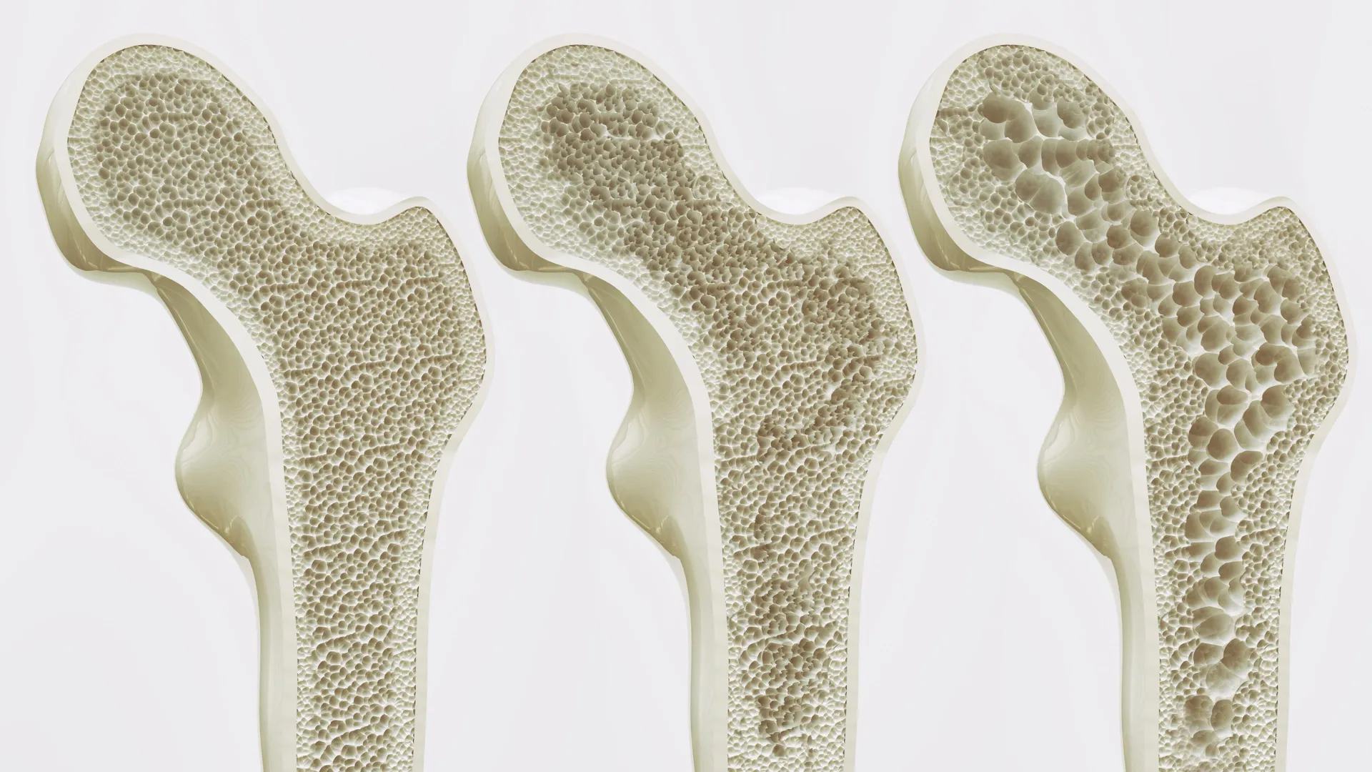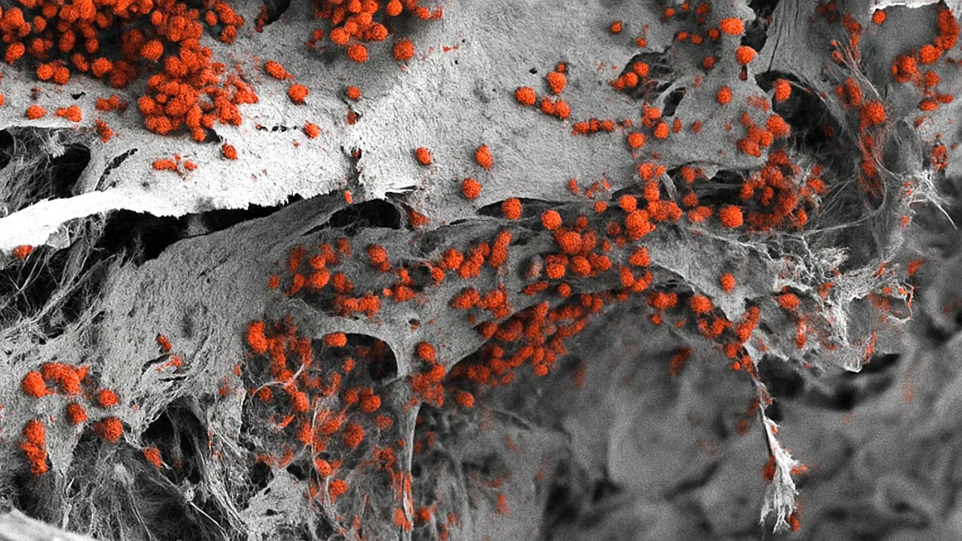
For decades, the intricate mechanics of the human body’s primary blood factory have remained largely hidden behind the limitations of traditional scientific models. Operating quietly within the skeletal framework, bone marrow is responsible for generating billions of red blood cells, white blood cells, and platelets every single day. Yet, studying this vital tissue—particularly when its delicate processes falter in conditions such as leukemia, lymphoma, and multiple myeloma—has posed a formidable challenge to medical researchers. Historically, scientists have had to rely heavily on murine models or overly simplified, two-dimensional cell cultures that fail to replicate the complex, three-dimensional ecosystem of human bone marrow.
That paradigm is now shifting. In a landmark achievement published in the prestigious journal Cell Stem Cell, a multidisciplinary team of researchers from the Department of Biomedicine at the University of Basel and University Hospital Basel has successfully recreated a fully functional, highly complex human bone marrow network in the laboratory. Constructed entirely from human cells, this pioneering three-dimensional platform marks a historic first in the field of hematology and tissue engineering. By bridging the gap between human physiology and laboratory experimentation, the new system offers an unprecedented window into normal blood formation, disease pathology, and the complex microenvironments that allow blood cancers to thrive. Furthermore, this breakthrough aligns with global scientific directives to minimize the reliance on animal testing, offering a viable, human-relevant alternative for preclinical pharmaceutical development and toxicological screening.
The Anatomy of the Breakthrough: Replicating the Endosteal Niche
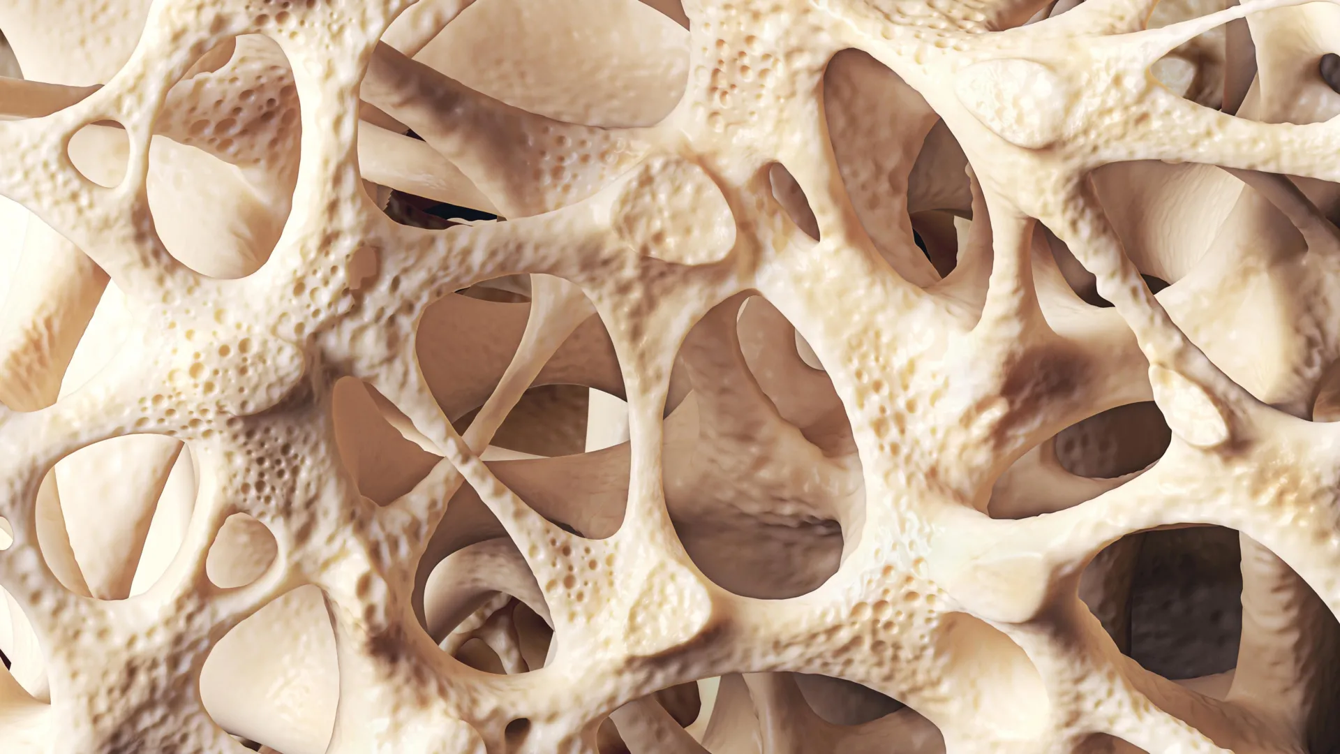
To understand the significance of the University of Basel’s achievement, one must examine the microarchitectural complexity of human bone marrow. The marrow does not exist as a uniform soup of cells; rather, it is organized into specialized microenvironments known as niches. Among the most critical of these is the endosteal niche, located near the inner surface of the bone. This specific region plays a central role in hematopoiesis—the production of blood cells—and has been increasingly implicated in the survival and treatment resistance of various blood cancers.
The endosteal niche is a cellular mosaic. It comprises a delicate interplay of specialized bone cells (such as osteoblasts and osteoclasts), intricate networks of blood vessels, peripheral nerve fibers, and various immune cell populations. Until now, no artificial system had successfully integrated all of these diverse biological components into a single, cohesive human model. Previous attempts were frequently hampered by the inability to sustain multiple distinct cell lineages simultaneously within a supportive structural matrix.
The Swiss research team, spearheaded by Professor Ivan Martin and Dr. Andrés García García, overcame this hurdle by adopting a sophisticated, biomimetic approach. Their methodology began with the creation of an artificial bone framework crafted from hydroxyapatite—a naturally occurring calcium phosphate mineral that serves as the primary inorganic matrix of human teeth and bones. This scaffold provided the necessary mechanical rigidity and chemical cues to mimic the natural skeletal environment.
Next, the researchers turned to human pluripotent stem cells. Utilizing advanced molecular biology techniques, somatic cells were reprogrammed into induced pluripotent stem cells (iPSCs), which possess the remarkable capacity to differentiate into virtually any cell type in the human body depending on the biochemical signals they receive. By carefully seeding these stem cells onto the hydroxyapatite scaffold and subjecting them to precisely controlled developmental cues, the team guided the cells through coordinated differentiation pathways.
The resulting structure was far more than a simple collection of cells; it was a living, breathing micro-organism analogue. Advanced analytical evaluations confirmed that the three-dimensional architecture closely mirrored the cellular composition, spatial organization, and biochemical signaling networks of the natural human endosteal niche. Crucially, the model achieved a notable physical scale—measuring eight millimeters in diameter and four millimeters in thickness—making it significantly larger and more robust than any previously engineered bone marrow surrogate. Within this expansive environment, the researchers successfully maintained human blood cell formation in vitro for several consecutive weeks, demonstrating sustained functional viability.
A Chronology of Innovation: The Path to Human-Centric Hematology Models
The journey toward a complete human bone marrow model has been decades in the making, reflecting broader technological leaps in stem cell biology, biomaterials science, and tissue engineering.
During the latter half of the 20th century, hematological research relied almost exclusively on animal models, predominantly mice. While murine models have yielded foundational insights into mammalian hematopoiesis, significant physiological differences between mice and humans have frequently bottlenecked the translation of laboratory discoveries into successful clinical therapies. Differences in immune system architecture, bone density, marrow composition, and drug metabolism mean that treatments showing immense promise in murine trials often fail or behave unpredictably in human clinical trials.
The twenty-first century ushered in the era of in vitro cell cultures, but these early iterations were largely restricted to two-dimensional Petri dishes. While useful for observing isolated cellular behaviors, 2D cultures strip cells of their natural physical cues, extracellular matrix interactions, and three-dimensional mechanical forces. Cells cultured on flat plastic surfaces rapidly lose their phenotype and functional capabilities, rendering them poor substitutes for living tissue.
The advent of induced pluripotent stem cell technology in 2006 marked a monumental turning point. For the first time, researchers could generate patient-specific or universal stem cell lines without the ethical controversies surrounding embryonic stem cells. Concurrently, advancements in biomaterials engineering allowed for the synthesis of porous, biocompatible scaffolds that could support complex cellular ingrowth.
Over the past ten years, bioengineers began combining iPSCs with advanced biomaterials to create "organ-on-a-chip" devices and simplified bone marrow mimics. However, these systems typically struggled to incorporate the full complement of vascular, neural, and endosteal components required to truly simulate the human marrow environment. The work conducted by Professor Martin’s team represents the culmination of these converging technological streams—successfully merging advanced biomaterials, iPSC differentiation protocols, and 3D co-culture techniques into a unified, highly functional platform.
Official Perspectives and Expert Analysis
The implications of this research extend far beyond academic curiosity, drawing commentary and analysis from leading figures in biomedical research, pharmaceutical development, and bioethics.
Reflecting on the limitations of historical methodologies, Professor Ivan Martin emphasized the profound shift this model represents for the scientific community. "We have learned a great deal about how bone marrow works from mouse studies," Martin noted. "However, our model brings us closer to the biology of the human organism. It could serve as a complement to many animal experiments in the study of blood formation in both healthy and diseased conditions."
This sentiment is strongly echoed by institutional mandates across Europe and North America, where regulatory bodies and academic institutions are increasingly enforcing the principle of the "Three Rs"—to Replace, Reduce, and Refine the use of animals in scientific research. By providing a human-based alternative that can accurately model complex hematopoietic processes, the University of Basel platform offers a tangible tool for laboratories seeking to curb animal usage without compromising scientific rigor.
At the same time, the research team remains pragmatic about the current limitations and necessary iterations of their technology. Dr. Andrés García García pointed out specific hurdles that must be cleared before the platform can be deployed in high-throughput industrial settings. "For drug development and screening purposes, the size of our bone marrow model might be too large," García García explained. In modern pharmaceutical research, high-throughput screening campaigns require miniaturized assay formats—often in 96-well or 384-well plates—capable of testing hundreds of distinct chemical compounds or dosage concentrations simultaneously. Scaling down the current eight-millimeter architecture while preserving its cellular complexity will be a primary objective for future engineering phases.
Broader Impact: Transforming Oncology, Pharmacology, and Personalized Medicine
The successful recreation of a human bone marrow niche opens up vast new horizons across multiple distinct sectors of healthcare and biotechnology.
In the realm of oncology, particularly concerning hematological malignancies such as acute myeloid leukemia (AML) and multiple myeloma, the model provides an unprecedented platform for dissecting disease mechanisms. Cancers of the blood often originate or find sanctuary within the bone marrow niches, where they co-opt resident cells to evade chemotherapeutic agents and immune surveillance. By utilizing the new human cell model, researchers can directly observe how malignant cells interact with vascular, neural, and endosteal components, potentially identifying novel therapeutic targets capable of disrupting treatment-resistant cancer stem cell niches.
In pharmacology, the platform promises to streamline the drug discovery pipeline. Preclinical toxicity testing is notoriously inefficient, with many candidate molecules failing in human trials despite successful animal testing due to unexpected human-specific toxicities. A physiologically accurate human bone marrow model could serve as a rigorous pre-screening filter, identifying bone marrow toxicity—such as drug-induced myelosuppression—much earlier in the development cycle, thereby saving time, capital, and reducing animal loss.
Looking toward the horizon, the most transformative application of this technology may lie in the domain of personalized medicine. The research team envisions a future where clinicians could harvest cells from an individual patient diagnosed with a blood disorder, reprogram them, and construct a personalized, patient-specific bone marrow model in the laboratory. Oncologists could then test a panel of diverse chemotherapeutic regimens directly on the patient’s custom marrow model to identify the most efficacious treatment course prior to administering therapy to the patient. While significant technical hurdles—including reducing production timeframes and standardizing scalability—must be overcome before personalized marrow platforms enter clinical workflows, the foundational proof-of-concept has now been established.
Conclusion and Future Outlook
The creation of the first fully human-cell-based bone marrow model by researchers at the University of Basel and University Hospital Basel marks a watershed moment in biomedical engineering and hematological research. By successfully integrating bone cells, neural components, vascular elements, and stem cell-derived hematopoietic networks onto a hydroxyapatite mineral scaffold, the Swiss team has bridged a decades-old chasm between animal models and human physiological reality.
As the scientific community continues to refine this technology—scaling it down for high-throughput pharmacological screening and adapting it for patient-specific diagnostic applications—the horizon of medical research appears increasingly human-centric. By offering a sophisticated, ethical, and highly accurate alternative to animal testing, this breakthrough not only honors the imperative to refine and reduce animal experimentation but also sets a new gold standard for how humanity studies, understands, and heals the very foundations of life within our bones.