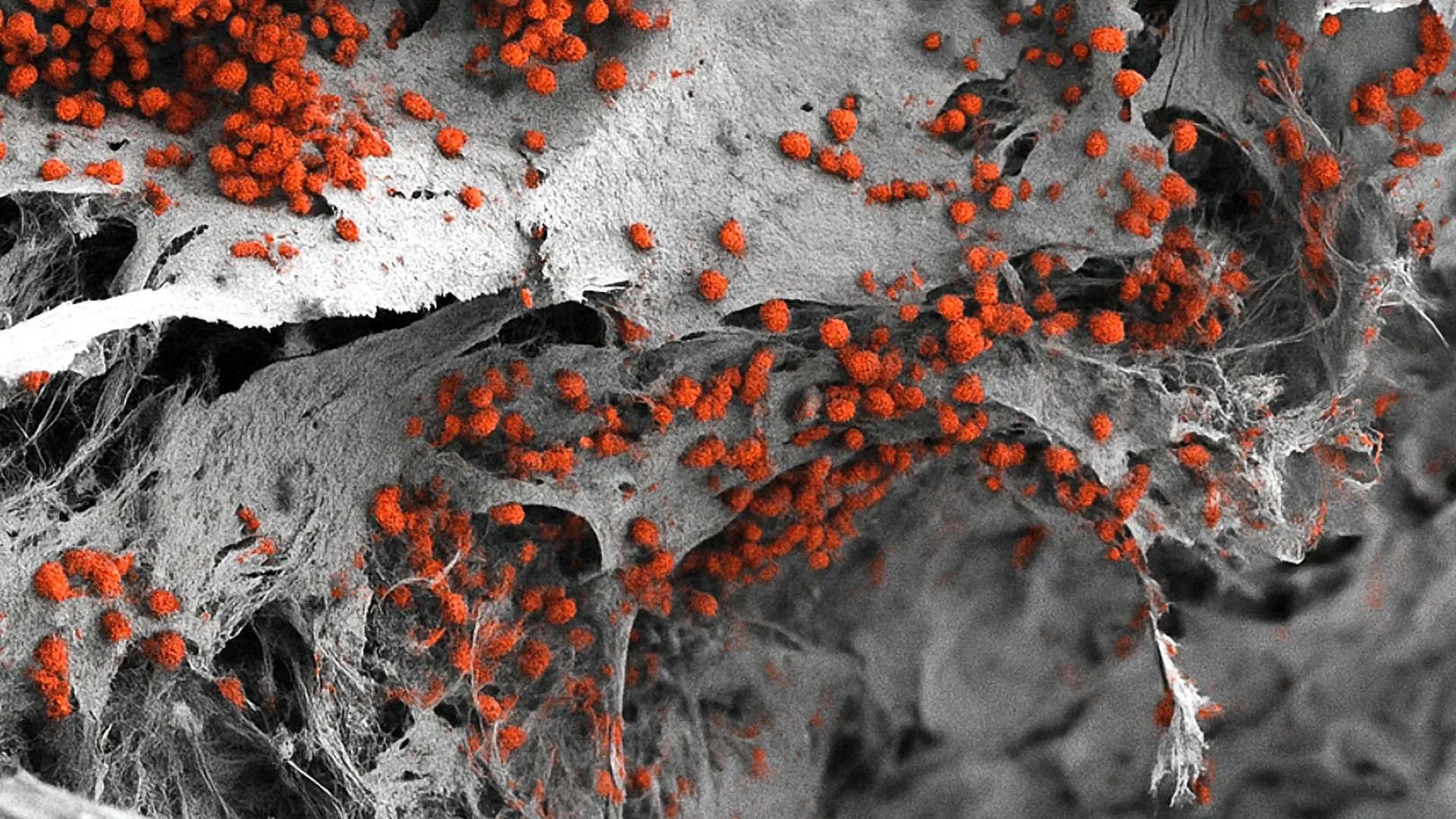
Scientists at the Indiana University School of Medicine have successfully engineered a transformative high-resolution imaging technique designed to examine mouse bone marrow within its native, undisturbed state. This unprecedented scientific achievement effectively surmounts historical barriers that have hindered biomedical research involving this notoriously difficult tissue. By utilizing advanced multiplex technology, the research team can now capture intricate cellular landscapes that were previously invisible to modern science. This methodological leap forward holds profound implications for the future development of targeted pharmaceuticals and novel therapeutic interventions directed at a wide spectrum of devastating pathologies, including hematological malignancies, severe autoimmune disorders, and complex musculoskeletal conditions.
The study, which details the breakthrough and its expansive potential, was recently published in the peer-reviewed scientific journal Leukemia. The research was spearheaded by a multidisciplinary team operating under the umbrella of the Indiana University Cooperative Center of Excellence in Hematology, bringing together experts from orthopedic surgery, pediatric research, and commercialization sectors to address one of the most persistent bottlenecks in modern biomedical investigation.
Unlocking the Intricacies of Bone Marrow
Bone marrow is arguably one of the most vital yet structurally challenging tissues in the mammalian body. Acting as the primary site of hematopoiesis—the continuous process through which all blood and immune cells are generated—bone marrow is also the exclusive sanctuary for hematopoietic stem cells. These specialized cells maintain the body’s entire blood and immune system throughout an organism’s lifespan.
However, examining this dynamic microenvironment has historically presented formidable obstacles for researchers. The tissue is simultaneously gelatinous and heavily encased within hard, mineralized cortical bone. Consequently, extracting or viewing the marrow without severely altering its structural integrity has remained a persistent challenge. Traditional laboratory techniques required researchers to either strip away the protective bone or physically homogenize the tissue, destroying the spatial relationships between cells in the process. Without knowing the exact neighborhood in which specific cells reside, understanding how diseases originate, progress, and respond to treatment has been severely limited.
The core of the new methodology relies on the adaptation of the Phenocycler 2.0 platform, a sophisticated multiplex imaging tool. While the device has previously been deployed with varying degrees of success to map softer, more accessible organs such as the spleen and kidneys, the IU research team is the first in the scientific community to successfully optimize and apply the technology to intact murine bone marrow.
Methodological Evolution: Overcoming Analytical Limitations
To fully appreciate the significance of the Indiana University innovation, it is necessary to examine the limitations of the methodologies that preceded it. For decades, the gold standards of tissue analysis in hematology and immunology laboratories have relied heavily on flow cytometry and standard fluorescence microscopy.
Flow cytometry is exceptionally adept at quantifying various cell populations within a sample. However, it requires the complete physical and chemical dissociation of the tissue. Researchers must grind down or enzymatically digest the bone marrow into a liquid suspension of single cells to pass them through a laser beam. While this provides quantitative data regarding cell types, it completely obliterates the spatial architecture of the marrow. The microscopic geography—such as how a stem cell interacts with its niche, blood vessels, or nerve endings—is permanently lost.
On the other hand, standard fluorescence microscopy preserves tissue architecture but suffers from severe technical constraints regarding spectral overlap. Traditional fluorescent tags typically limit researchers to visualizing no more than three or four cellular markers simultaneously within a single tissue section. Because the bone marrow microenvironment houses dozens of distinct cell types, signaling molecules, and structural components interacting concurrently, a three-marker snapshot offers an overwhelmingly incomplete picture.
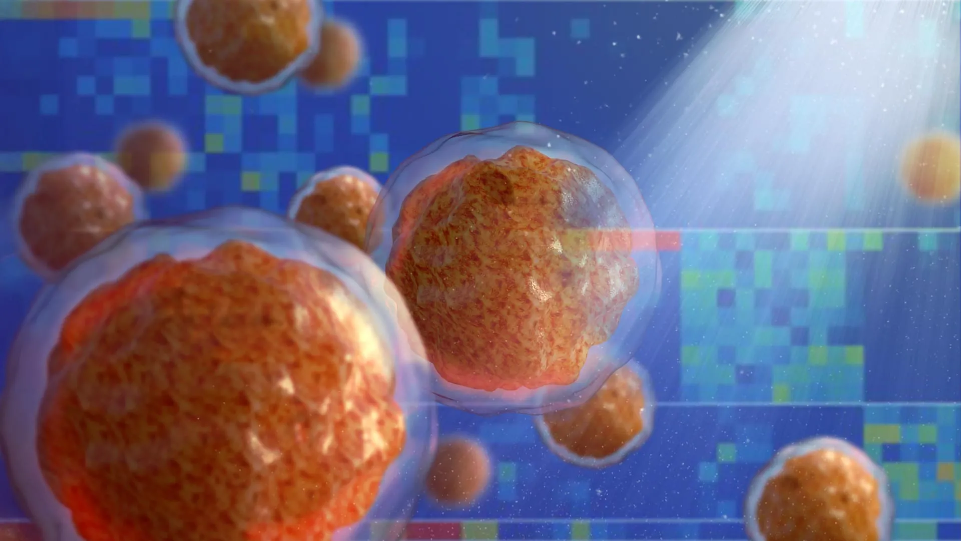
The newly refined Phenocycler 2.0 approach shatters these historical boundaries. By enabling the visualization of an unprecedented 25 distinct cellular markers simultaneously within a completely intact murine bone marrow tissue section, the IU team has provided the scientific community with a multi-dimensional spatial map. This capability allows researchers to observe cellular interactions in real time within their native biological context, establishing an entirely new baseline for tissue analysis.
Chronology of the Discovery and Institutional Collaboration
The journey toward this methodological breakthrough represents the culmination of extensive collaborative efforts within the Indiana University academic health center network. The project began as an initiative to solve specific analytical bottlenecks encountered by researchers studying bone marrow failure syndromes and leukemia models at the Herman B Wells Center for Pediatric Research.
Initial phases of the project, spanning many months, involved rigorous troubleshooting to adapt the Phenocycler platform to the unique chemical and physical properties of bone-encased soft tissue. Researchers had to design specialized protocols for tissue fixation, decalcification, and staining that would preserve fragile surface proteins while allowing antibodies to penetrate the dense matrix.
By mid-stage development, the team successfully pushed the marker threshold from traditional low-multiplex limits up to 15, and finally to 25 distinct markers. The validation of these markers against known biological controls confirmed that the spatial integrity of the tissue was maintained throughout the multi-cycle imaging process. Following successful peer review, the findings were formally accepted and published in Leukemia, marking a major milestone for the institution and the broader hematological research community.
Voices from the Research Front
The significance of the work has been underscored by lead figures within the Indiana University research apparatus, who emphasize both the technical achievement and its translational utility.
"Bone marrow is difficult to study because it is gelatinous and encased in hard bone," explained Sonali Karnik, PhD, assistant research professor of orthopedic surgery at the IU School of Medicine and co-lead author of the study. Highlighting the broader physiological relevance of the tissue, Karnik noted, "Since bone marrow plays an important role in blood and immune cell formation and houses valuable stem cells, our unique imaging approach offers a useful tool for a variety of research applications."
The utility of mouse models in preclinical testing makes this technological adaptation particularly potent for the global pharmaceutical and academic research sectors. Because murine physiology shares fundamental parallels with human disease progression, methodologies that succeed in mouse models often pave the way for human diagnostic and therapeutic applications.
"Because mouse models are widely used to study human diseases, this technique offers a promising new method for investigating a range of conditions like autoimmune diseases, leukemia and other disorders involving bone marrow," stated Reuben Kapur, PhD, co-senior author of the study. Kapur serves as the director of the IU School of Medicine’s Herman B Wells Center for Pediatric Research and co-director of the IU Cooperative Center of Excellence in Hematology. His perspective highlights the translational bridge this technology creates between basic bench science and clinical application.
Broad Implications for Drug Development and Disease Pathology
The implications of this high-plex spatial imaging technique extend far beyond academic curiosity, carrying direct economic and clinical relevance for the biotechnology and pharmaceutical sectors. Drug development for bone marrow-related disorders has historically suffered from high clinical trial attrition rates. A primary driver of these failures is an incomplete understanding of how candidate therapeutics affect the complex cellular neighborhoods within the bone marrow microenvironment.
When pharmaceutical companies test novel compounds designed to eradicate leukemia stem cells or stimulate tissue regeneration, they must understand whether the drug reaches its intended cellular target without inadvertently disrupting neighboring healthy cells. Traditional assay methods often provide contradictory or incomplete spatial data, leaving researchers blind to localized toxicities or off-target binding within the marrow cavity.
By deploying the new IU methodology, preclinical researchers can now map the exact distribution of therapeutic molecules and observe how resident cell populations—including immune cells, stromal cells, and vascular elements—respond to treatment over time. This granular level of spatial resolution can significantly streamline lead optimization during early-stage drug development, allowing scientists to eliminate ineffective or toxic compounds long before they reach expensive human clinical trials.
Furthermore, autoimmune diseases such as lupus and rheumatoid arthritis often involve aberrant immune cell generation within the bone marrow. The ability to track 25 or more markers simultaneously allows immunologists to dissect the precise signaling cascades and cellular recruitment patterns that drive these chronic conditions, potentially uncovering entirely new drug targets that were previously obscured by technological limitations.
Commercialization and Future Directions
Recognizing the immense commercial and scientific value of the innovation, the Indiana University Innovation and Commercialization Office has already taken decisive legal steps to protect the intellectual property generated by the research team. A provisional patent has been formally filed for the new imaging methodology, laying the groundwork for potential licensing agreements, industry partnerships, or the creation of specialized spin-out biotechnology enterprises.
Far from resting on their laurels, the research team is actively expanding the capabilities of the platform. Current laboratory efforts are focused on scaling up the marker panel significantly. Future iterations of the imaging protocol aim to incorporate architectural features that go beyond hematopoietic and immune cells, integrating markers for bone matrix components, peripheral nerves, smooth muscle tissues, and rare signaling cell subsets that orchestrate microenvironmental homeostasis.
In addition to expanding the marker repertoire, the IU Cooperative Center of Excellence in Hematology is establishing collaborative frameworks with external academic institutions and industry partners to test the methodology across an even wider array of pathological mouse models. These ongoing studies aim to standardize the protocol so that it can be readily adopted by standard molecular pathology laboratories worldwide.
Financial Support and Institutional Recognition
The rigorous scope and execution of this foundational research were made possible through substantial external backing, primarily driven by public research funding agencies. The project received direct financial support from the National Institutes of Health (NIH), reflecting the federal agency’s ongoing commitment to fostering innovative biomedical tools that accelerate the understanding and treatment of complex human diseases.
The collaborative success of the study is further evidenced by its extensive roster of co-authors from across the Indiana University academic ecosystem. In addition to Karnik and Kapur, the published research includes contributions from Connor Gulbronson, Paige C. Jordan, Rahul Kanumuri, Baskar Ramdas, Ramesh Kumar, Melissa L. Hartman, Izza Khurram, Drew M. Brown, Karen E. Pollok, Pratibha Singh, and Melissa A. Kacena. This diverse collective represents a blend of expertise spanning orthopedic surgery, pediatrics, molecular biology, pharmacology, and translational imaging.
As the scientific community digests the findings published in Leukemia, the Indiana University School of Medicine stands at the vanguard of a new era in hematological investigation. By illuminating the previously hidden architecture of bone marrow, this breakthrough technique not only resolves a decades-old analytical bottleneck but also provides a powerful lens through which the future of targeted medicine will be shaped.