
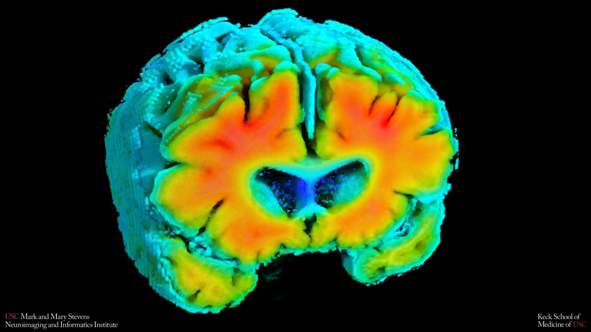
The intricate relationship between the brain’s vascular health and the onset of neurodegenerative conditions has reached a new milestone with recent findings from the Mark and Mary Stevens Neuroimaging and Informatics Institute (Stevens INI) at the Keck School of Medicine of USC. Researchers have identified that subtle fluctuations in how blood circulates through the brain and how oxygen is delivered to neural tissues serve as critical indicators for the risk of developing Alzheimer’s disease. Published in the prestigious journal Alzheimer’s and Dementia: The Journal of the Alzheimer’s Association, the study highlights a shift in the clinical understanding of the disease, moving beyond a singular focus on protein accumulation toward a more integrated view of the brain’s circulatory efficiency.
By examining a cohort of older adults across the cognitive spectrum, the USC team demonstrated that noninvasive measures of cerebrovascular function are directly linked to the biological hallmarks of Alzheimer’s, such as the accumulation of amyloid-beta plaques and the atrophy of the hippocampus. This research suggests that the physiological state of the brain’s blood vessels could be one of the earliest "canaries in the coal mine," providing a window into a patient’s future cognitive health long before memory loss or behavioral changes manifest.
The Evolution of the Vascular Hypothesis in Alzheimer’s Research
For decades, the "Amyloid Cascade Hypothesis" dominated the field of Alzheimer’s research, positing that the primary cause of the disease was the toxic buildup of amyloid-beta plaques, followed by tau tangles. However, the failure of several amyloid-clearing drugs in clinical trials led researchers to investigate secondary and tertiary factors. The "Two-Hit Vascular Hypothesis" of Alzheimer’s suggests that vascular impairment (Hit 1) acts in concert with amyloid-beta (Hit 2) to accelerate neurodegeneration.
The USC study provides empirical weight to this integrated theory. While amyloid and tau remain central to the diagnosis, the study’s lead author, Amaryllis A. Tsiknia, a PhD candidate at USC, emphasizes that blood flow and oxygen delivery are not merely secondary effects but are "critical players" in the disease’s progression. This perspective aligns with a growing body of evidence suggesting that the blood-brain barrier’s integrity and the brain’s ability to regulate its own blood supply (autoregulation) are foundational to maintaining cognitive resilience during aging.
Methodology: Noninvasive Innovation in Neuroimaging
The Stevens INI team utilized two primary noninvasive tools to capture the real-time dynamics of brain circulation: Transcranial Doppler (TCD) ultrasound and Near-infrared spectroscopy (NIRS). These tools represent a significant departure from the more invasive and expensive gold standards of neuroimaging, such as Positron Emission Tomography (PET) and Magnetic Resonance Imaging (MRI).
Transcranial Doppler ultrasound functions by emitting high-frequency sound waves that bounce off moving red blood cells, allowing clinicians to measure the velocity of blood flow through the brain’s major arteries, such as the middle cerebral artery. Near-infrared spectroscopy, on the other hand, utilizes light in the near-infrared spectrum to penetrate the skull and measure the oxygenation levels of the hemoglobin in the brain’s cortical surface.
The innovation of the USC study lies in the application of advanced mathematical modeling to these raw readings. By analyzing how blood flow and oxygen levels fluctuated in response to natural changes in blood pressure and carbon dioxide levels, the researchers created a composite "indicator of cerebrovascular function." This model essentially measures the brain’s "vascular fitness"—its ability to adapt to physiological stress and maintain a stable environment for neurons.
Supporting Data: Linking Vascular Health to Brain Structure
The study’s findings established a clear correlation between high-performing vascular indicators and positive brain structure. Participants whose vascular systems functioned similarly to those of "healthy agers" exhibited significantly lower levels of amyloid plaque. Furthermore, these individuals possessed larger hippocampal volumes. The hippocampus is the region of the brain responsible for forming new memories and is typically the first area to show significant shrinkage in Alzheimer’s patients.
Data from the study revealed a distinct gradient across the participant groups:
- Cognitively Normal Participants: This group showed the highest levels of cerebrovascular regulation, with efficient oxygen delivery and stable blood flow velocities.
- Mild Cognitive Impairment (MCI): Participants in this category demonstrated a measurable decline in vascular indicators, often preceding the most severe stages of brain atrophy.
- Dementia Patients: This group exhibited the most significant impairment in vascular function, suggesting that as the disease progresses, the brain’s ability to manage its own "life support system" of blood and oxygen breaks down entirely.
Meredith N. Braskie, PhD, the study’s senior author and assistant professor of neurology at the Keck School of Medicine, noted that these vascular measures align closely with what is typically seen on expensive MRI and PET scans. This alignment validates vascular monitoring as a legitimate proxy for measuring Alzheimer’s risk.
Chronology of the Research and Institutional Context
The Stevens Neuroimaging and Informatics Institute has long been at the forefront of mapping the human brain. The current study is part of a decade-long trajectory of research at USC aimed at identifying "biomarkers"—measurable biological indicators—that can predict Alzheimer’s years in advance.
- 2015-2018: Early pilot studies at USC began exploring the use of TCD to monitor blood flow in aging populations, noting that "pulsatility" in the arteries was linked to white matter lesions.
- 2019-2021: The integration of NIRS allowed researchers to look not just at flow, but at the actual consumption of oxygen by brain tissue.
- 2022-2023: Advanced mathematical models were developed to combine these disparate data points into a single, cohesive vascular health score.
- 2024: The publication of the current study in Alzheimer’s and Dementia marks the formal validation of these noninvasive methods against established Alzheimer’s markers like amyloid and hippocampal volume.
This research was supported by significant federal funding, including grants from the National Institute on Aging (NIA) and the Office of The Director at the National Institutes of Health (NIH). These investments reflect a broader federal mandate to find low-cost, scalable screening methods for an aging American population.
Official Responses and Expert Analysis
The implications of the study have drawn praise from leaders in the field of neurology. Arthur W. Toga, PhD, Director of the Stevens INI, highlighted that the findings "open new doors for early detection." By proving that vascular health is intertwined with classic neurodegeneration, Toga suggests that the medical community can now view Alzheimer’s through a more holistic lens.
Independent experts in geriatric medicine have noted that this study could lead to a "preventative neurology" model. If vascular health is a precursor to amyloid buildup, then interventions aimed at cardiovascular health—such as exercise, blood pressure management, and diet—could be framed specifically as Alzheimer’s prevention strategies.
"If we can track these signals over time, we may be able to identify people at higher risk earlier and test whether improving vascular health can slow or reduce Alzheimer’s-related brain changes," said Tsiknia. This proactive approach contrasts with the current reactive model, where patients are often only diagnosed after significant cognitive loss has occurred.
Broader Impact: Economic and Clinical Implications
One of the most significant impacts of the USC research is the potential for democratizing Alzheimer’s screening. Currently, a definitive diagnosis of Alzheimer’s often requires a PET scan, which can cost between $3,000 and $6,000 and involves the injection of radioactive tracers. Alternatively, a lumbar puncture (spinal tap) can be used to check for amyloid and tau, but it is invasive and often avoided by patients.
In contrast, TCD ultrasound and NIRS are:
- Cost-Effective: The equipment is significantly cheaper than PET or MRI machines and requires less specialized infrastructure.
- Noninvasive: There are no needles, no radiation, and no need for the patient to remain perfectly still in a claustrophobic tube for an hour.
- Scalable: These tests can be performed in a standard neurologist’s office or even a primary care clinic, making large-scale population screening feasible.
From a public health perspective, the ability to identify at-risk individuals in their 50s or 60s through a 20-minute vascular check-up could save the healthcare system billions of dollars in long-term care costs by delaying the onset of dementia.
Future Outlook and Longitudinal Goals
While the results of the study are groundbreaking, the authors maintain a degree of scientific caution. The current data represents a "snapshot in time," meaning it is a cross-sectional study that shows correlation rather than direct cause-and-effect. To address this, the Stevens INI team is currently engaged in longitudinal studies, tracking the same group of participants over several years.
These ongoing studies aim to answer critical questions: Does a decline in vascular function always precede amyloid buildup? Can an improvement in cardiovascular fitness through medication or lifestyle changes actually reverse a "risky" vascular signature?
As the global population ages, the urgency for accessible diagnostic tools grows. The USC study provides a roadmap for a future where Alzheimer’s is not a sudden, mysterious decline, but a manageable condition detected through the rhythmic pulse of blood and the steady flow of oxygen to the brain. Through the marriage of mathematical modeling and noninvasive technology, the Stevens INI has moved the needle closer to a world where the first signs of Alzheimer’s are caught not in a memory test, but in the very vessels that sustain human thought.


Leave a Reply