
Researchers at Johns Hopkins Medicine have unveiled a significant breakthrough in the quest to understand and eventually treat Alzheimer’s disease, identifying a specific protein in the brain that produces hydrogen sulfide as a critical regulator of cognitive health. The study, funded by the National Institutes of Health (NIH), centers on the enzyme Cystathionine γ-lyase, commonly referred to as CSE. While hydrogen sulfide is widely recognized as a toxic gas characterized by the pungent odor of rotten eggs, this research highlights its indispensable role in extremely small concentrations as a "gasotransmitter" that facilitates memory formation and protects the structural integrity of the brain.
The findings, published in the Proceedings of the National Academy of Sciences (PNAS), suggest that a deficiency in CSE and the subsequent loss of hydrogen sulfide production are directly linked to the hallmarks of neurodegeneration. Lead researcher Bindu Paul, M.S., Ph.D., an associate professor of pharmacology, psychiatry, and neuroscience at the Johns Hopkins University School of Medicine, and her colleagues utilized genetically engineered mouse models to demonstrate that without this protein, the brain undergoes a cascade of failure, including increased oxidative stress, DNA damage, and a breakdown of the blood-brain barrier.
The Biological Paradox of Hydrogen Sulfide
For decades, hydrogen sulfide (H2S) was viewed primarily as an environmental hazard and a metabolic byproduct. However, in recent years, it has joined nitric oxide and carbon monoxide in a class of molecules known as gasotransmitters—gaseous signaling molecules produced endogenously that regulate various physiological processes. In the brain, H2S is essential for long-term potentiation, the process by which synaptic connections strengthen, which is the cellular basis for learning and memory.
The challenge for modern medicine lies in the molecule’s inherent toxicity. In high concentrations, H2S is lethal; however, the brain requires precise, minute amounts to maintain neuronal health. The Johns Hopkins study focuses not on the external administration of the gas, which would be unsafe for human patients, but on the internal mechanism of its production. By understanding how the CSE enzyme functions, scientists hope to develop therapies that can stimulate the brain’s natural ability to produce H2S at safe, therapeutic levels.
A Chronology of Discovery: From Blood Pressure to Neurodegeneration
The recent findings are the culmination of nearly two decades of research conducted at the Johns Hopkins University School of Medicine, much of it initiated by Solomon Snyder, M.D., D.Sc., D.Phil., a pioneer in neuroscience and professor emeritus. The trajectory of this research illustrates the often-unexpected paths of scientific discovery:
- 2008: Cardiovascular Foundations. Researchers first developed the strain of mice lacking the CSE protein to study its role in the peripheral nervous system. This early work, published in Science, established that CSE was vital for regulating blood vessel function and maintaining healthy blood pressure levels.
- 2014: The Huntington’s Disease Connection. Snyder’s team expanded their scope to the central nervous system, reporting in Nature that CSE levels were significantly depleted in the brains of patients with Huntington’s disease. In animal models, restoring CSE activity helped mitigate some of the motor and cognitive symptoms associated with the condition.
- 2021: Initial Alzheimer’s Link. The group turned its attention to Alzheimer’s disease, finding that CSE activity was severely compromised in the brains of mice with Alzheimer’s-like pathology. They discovered that small, controlled injections of hydrogen sulfide-releasing compounds could improve cognitive function.
- Present Day: Isolating the Role of CSE. The latest research moves beyond examining CSE in the context of other genetic mutations. By focusing on mice where the only variable is the absence of the CSE protein, the team has proven that CSE deficiency alone is sufficient to trigger a progressive neurodegenerative state that mirrors Alzheimer’s disease.
"This most recent work indicates that CSE alone is a major player in cognitive function and could provide a new avenue for treatment pathways in Alzheimer’s disease," stated Dr. Snyder, who retired from the faculty in 2023 but continues to contribute to the field’s foundational knowledge.
Experimental Evidence: The Barnes Maze and Spatial Memory
To quantify the impact of CSE loss on behavior, the researchers employed the Barnes maze, a standardized tool for assessing spatial learning and memory in rodents. The maze consists of a circular platform with multiple holes around the perimeter, only one of which leads to a dark, recessed "escape box." Mice, which naturally prefer dark environments over bright, open spaces, must use visual cues to remember the location of the escape route.
The study compared "wild-type" (normal) mice with CSE-deficient mice at various stages of development. At the age of two months, both groups performed with high efficiency, finding the escape box in under three minutes. However, by the six-month mark—the mouse equivalent of middle age—the results diverged sharply. While the normal mice retained their ability to navigate the maze quickly, the CSE-deficient mice showed significant impairment, struggling to remember the location of the shelter and taking considerably longer to escape the light.
Suwarna Chakraborty, a researcher in Paul’s lab and the study’s first author, noted that this decline was not sudden but progressive. "The decline in spatial memory indicates a progressive onset of neurodegenerative disease that we can attribute to CSE loss," Chakraborty explained.
Cellular Collapse and the Blood-Brain Barrier
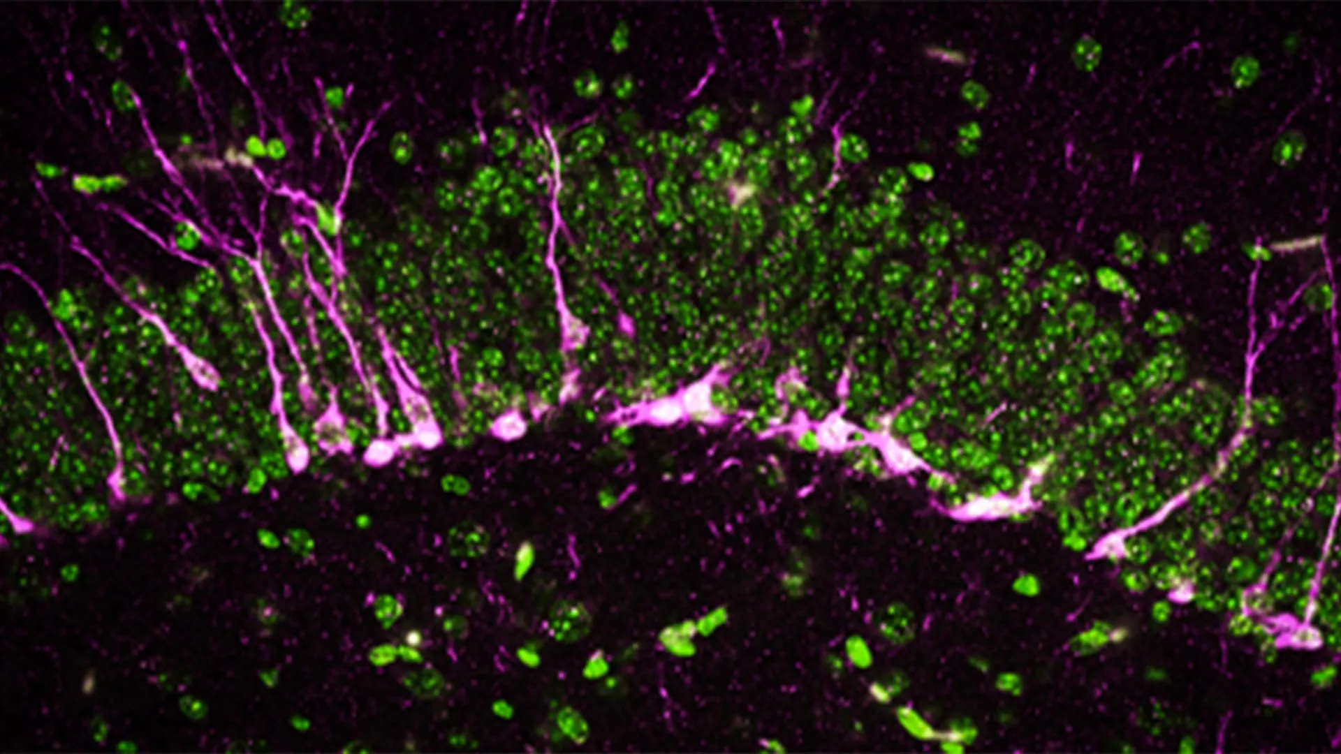
Beyond behavioral changes, the research team utilized high-powered electron microscopy and biochemical analysis to observe the physical state of the brain tissue. They focused on the hippocampus, the region primarily responsible for memory consolidation and the site where neurogenesis—the birth of new neurons—occurs.
The findings were stark. In mice lacking CSE, the proteins necessary for neurogenesis were either significantly reduced or entirely absent. Without the birth of new neurons, the hippocampus loses its plasticity, leading to the cognitive decline observed in the Barnes maze. Furthermore, the researchers discovered structural damage to the brain’s vasculature.
"The mice lacking CSE were compromised at multiple levels," said co-first author Sunil Jamuna Tripathi. The team observed large breaks in the blood vessels, indicating a failure of the blood-brain barrier (BBB). The BBB is a specialized system of cells that prevents toxins and pathogens in the blood from entering the brain while allowing essential nutrients through. When the BBB is compromised, as is common in Alzheimer’s patients, the brain becomes vulnerable to inflammation and oxidative stress, which further accelerates cell death.
Implications for Alzheimer’s Treatment and Future Research
The implications of this research are profound, particularly given the current state of Alzheimer’s therapeutics. According to the U.S. Centers for Disease Control and Prevention (CDC), more than 6 million Americans are currently living with Alzheimer’s, a number projected to nearly triple by 2060. Despite billions of dollars in research, most treatments currently on the market focus on clearing amyloid-beta plaques or tau tangles—hallmarks of the disease that many scientists now believe may be symptoms rather than the root cause.
The Johns Hopkins study suggests a shift in focus toward metabolic and gaseous signaling. If CSE deficiency is a primary driver of neurodegeneration, then pharmacological agents designed to boost CSE activity or safely mimic the effects of hydrogen sulfide could offer a disease-modifying therapy that stops or slows progression, rather than just managing symptoms.
Furthermore, the research highlights the importance of "oxidative stress" management. The lack of H2S leads to an accumulation of reactive oxygen species that damage DNA. By restoring the CSE pathway, clinicians might be able to bolster the brain’s internal defense mechanisms against this cellular "rusting."
Collaboration and Global Funding Support
The scale of this study reflects a massive collaborative effort involving multiple institutions and significant financial backing from both public and private sectors. The National Institutes of Health provided the bulk of the funding through various grants, reflecting the agency’s commitment to finding novel targets for Alzheimer’s research.
In addition to the Johns Hopkins team, the study involved contributors from Case Western Reserve University, the Leibniz Institute for Analytical Sciences in Germany, the Hollings Cancer Center, the Medical University of South Carolina, and the West Virginia University School of Medicine. Supporting organizations included the American Heart Association, the Department of Defense, and various private foundations such as the Valour Foundation and the Wick Foundation.
This broad support underscores the scientific community’s recognition that Alzheimer’s is a multifaceted disease requiring an interdisciplinary approach. By linking cardiovascular health (the 2008 study) to neurogenesis and cognitive function (the 2024 study), the researchers have provided a more holistic view of how the body’s systemic health influences the brain’s longevity.
As the research moves toward potential clinical applications, the next phase will likely involve identifying small-molecule drugs that can cross the blood-brain barrier to enhance CSE expression. While a cure for Alzheimer’s remains elusive, the discovery of the CSE-hydrogen sulfide pathway provides a critical new map for researchers to follow in the fight against this devastating condition.


Leave a Reply