
In a landmark study published in the journal Science, a multidisciplinary team of researchers led by the Massachusetts Institute of Technology (MIT) has achieved the first-ever three-dimensional mapping of the atomic structure of a relaxor ferroelectric. This breakthrough addresses a decades-old mystery in condensed matter physics and materials science, providing a definitive look at the internal arrangements that give these materials their extraordinary properties. By utilizing advanced electron microscopy and computational modeling, the team has successfully visualized the "chemical disorder" and polar structures that drive the performance of technologies ranging from medical ultrasound imaging to deep-sea sonar and advanced energy storage systems.
The research, headed by James LeBeau, the Kyocera Professor of Materials Science and Engineering at MIT, represents a significant shift in how scientists approach the design of complex materials. For more than half a century, the scientific community has relied on incomplete or theoretical models to explain why relaxor ferroelectrics exhibit such high levels of electromechanical coupling—the ability to convert electrical energy into mechanical strain and vice versa. With this new 3D roadmap, engineers can now move beyond trial-and-error experimentation toward a more predictive and precise era of materials engineering.
The Science of Relaxor Ferroelectrics: A Hidden Foundation of Modern Tech
To understand the significance of this mapping, one must first consider the unique nature of relaxor ferroelectrics. Unlike standard ferroelectrics, which possess a uniform internal polarization, relaxors are characterized by a high degree of structural and chemical "disorder." This disorder is not a defect but rather the source of their utility. When an electric field is applied to these materials, they undergo a massive change in shape or electrical state, making them ideal for transducers, actuators, and sensors.
The specific material studied by the MIT team was a lead magnesium niobate-lead titanate alloy, commonly referred to as PMN-PT. Since its discovery and refinement in the late 20th century, PMN-PT has become the gold standard for high-performance piezoelectric applications. In medical settings, it allows ultrasound probes to produce high-resolution images of internal organs and developing fetuses. In defense, it is the critical component in sonar arrays used for underwater navigation and detection. Despite its ubiquity, the exact mechanism by which its atoms interact to produce these effects remained obscured at the nanoscale.
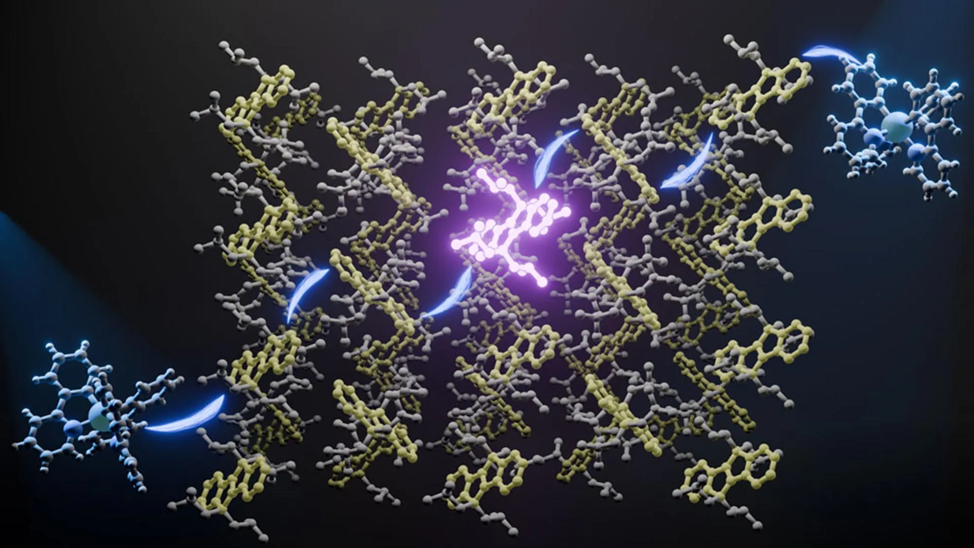
The challenge lay in the material’s complexity. In a typical crystal, atoms are arranged in a repeating, predictable lattice. In relaxors, different types of atoms—such as magnesium and niobium—are distributed in a seemingly random fashion. This randomness creates "polar nanoregions," tiny pockets where the electrical charge is aligned differently than in the surrounding material. Until now, these regions were too small and too buried within the 3D bulk of the material to be seen clearly with traditional imaging techniques.
Multi-Slice Electron Ptychography: A New Lens for the Atomic World
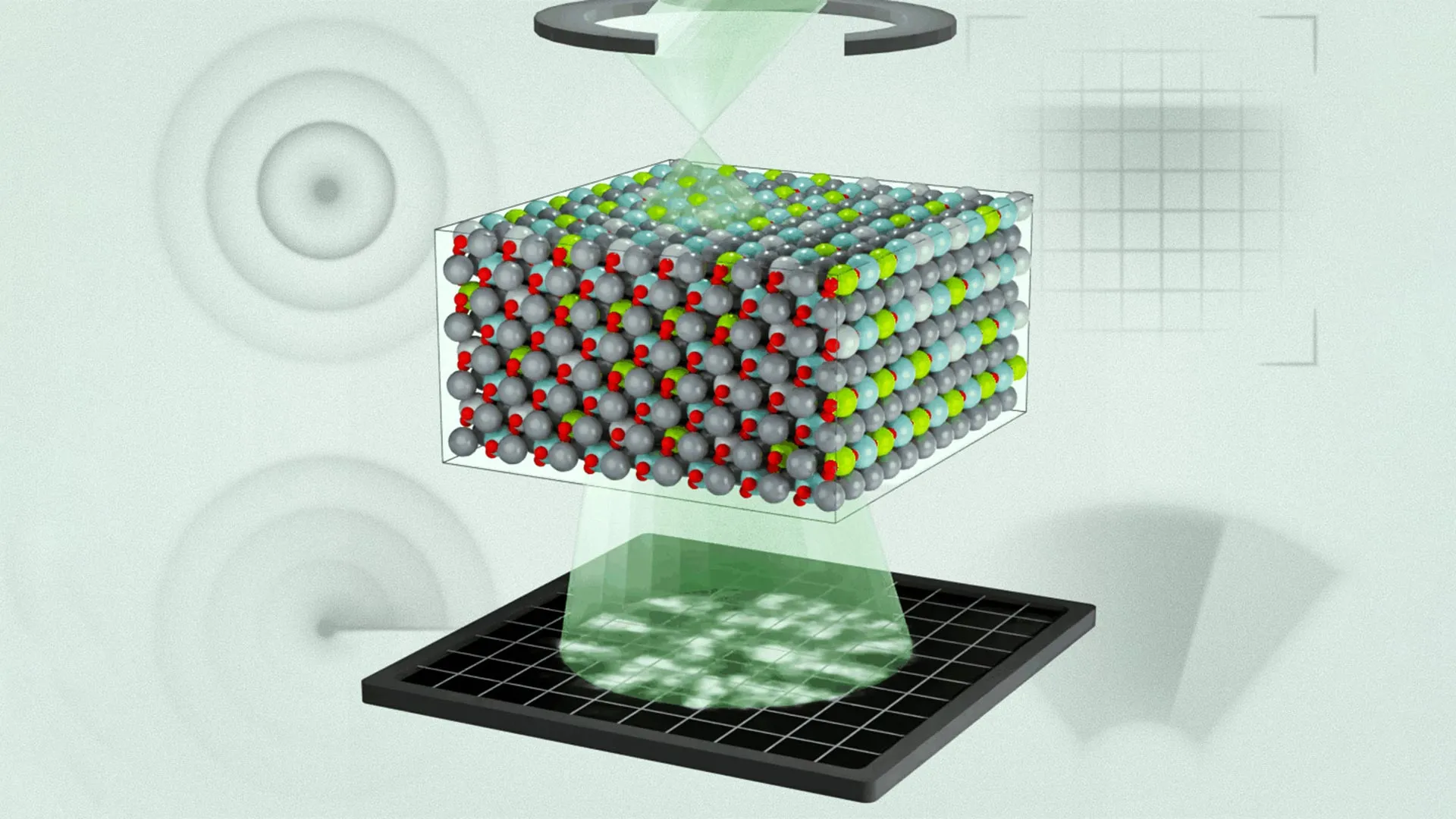
The breakthrough was made possible by an imaging technique known as multi-slice electron ptychography (MEP). While traditional electron microscopy provides a two-dimensional projection of a sample—essentially a shadow of the atoms—ptychography allows researchers to reconstruct the full three-dimensional volume.
The process involves scanning a concentrated beam of high-energy electrons across a sample in a sequential grid. As the beam passes through the material, the atoms deflect the electrons, creating a series of complex diffraction patterns. By overlapping these scans, the researchers collect a massive amount of data regarding how the electron wave function is altered by the material’s internal structure.
"We do this in a sequential way, and at each position, we acquire a diffraction pattern," explained Menglin Zhu, a postdoctoral researcher at MIT and co-first author of the study. "That creates regions of overlap, and that overlap has enough information to use an algorithm to iteratively reconstruct three-dimensional information about the object and the electron wave function."
This computational approach is what distinguishes ptychography from older methods. It requires immense processing power to solve the "phase problem"—recovering the lost information about the timing of the electron waves—to produce a clear 3D image. The result is a high-fidelity map that reveals not just where the atoms are, but how their positions correlate with the electric charges around them.
Challenging Established Models through Experimental Observation
One of the most significant outcomes of the study was the realization that previous computer simulations had fundamentally misjudged the scale and behavior of the material’s internal structures. For years, the "random site" model was the prevailing theory, suggesting that polarization regions were distributed in a relatively chaotic and independent manner.
However, the MIT team’s 3D mapping revealed a "layered hierarchy" of structures. They found that the regions of polarization were significantly smaller than what earlier simulations had predicted. Furthermore, they discovered that these regions are not independent; instead, they are highly correlated with the specific chemical species present in the lattice.
"We realized the chemical disorder we observed in our experiments was not fully considered previously," said Michael Xu, a postdoc at MIT and co-first author. "Previously, these models basically had random regions of polarization, but they didn’t tell you how those regions correlate with each other. Now we can tell you that information, and we can see how individual chemical species modulate polarization depending on the charge state of atoms."
By integrating these real-world observations back into their computational models, the researchers were able to achieve a level of predictive accuracy that was previously impossible. This feedback loop between experimental data and theoretical simulation is essential for the development of next-generation materials.
A Chronology of Discovery and Collaboration
The road to this discovery has been a multi-decade journey involving the evolution of electron microscopy and the steady refinement of ferroelectric theory.
- 1950s-1960s: Relaxor ferroelectrics were first discovered by Soviet scientists, who noted their unusual dielectric properties but lacked the tools to see their atomic structure.
- 1990s: The development of PMN-PT alloys revolutionized the field of piezoelectricity, leading to a surge in commercial and military applications.
- 2010s: The rise of ptychography in the scientific community began to offer hope for 3D atomic imaging, though it was initially limited to very thin or simple samples.
- 2020-2023: The MIT team, in collaboration with researchers from the University of Pennsylvania, Rice University, the University of Alabama at Birmingham, and the Korea Advanced Institute of Science and Technology (KAIST), began applying multi-slice ptychography to complex, disordered alloys.
- 2024: The publication of the team’s findings in Science marks the culmination of this effort, providing the first validated 3D model of a relaxor ferroelectric.
The collaboration was essential, as it required a mix of experimental expertise in microscopy and theoretical expertise in molecular dynamics. The research team included Colin Gilgenbach and Bridget R. Denzer from MIT; Yubo Qi (University of Alabama at Birmingham); Jieun Kim (KAIST); Jiahao Zhang and Andrew M. Rappe (University of Pennsylvania); and Lane W. Martin (Rice University). The work was supported by the U.S. Army Research Laboratory, the Office of Naval Research, and the National Science Foundation, highlighting the strategic importance of these materials to national security and industrial infrastructure.
Broader Implications: From Energy Storage to Artificial Intelligence
The ability to map and understand relaxor ferroelectrics in three dimensions has implications that extend far beyond the laboratory. As the world moves toward more energy-efficient technologies, the demand for materials that can store and convert energy with minimal loss is skyrocketing.
1. Energy and Power Systems:
Relaxor ferroelectrics are prized for their high energy density and efficiency in capacitors. By understanding the 3D atomic structure, scientists can engineer materials that can hold more charge and release it more reliably, which is critical for the power electronics used in electric vehicles and renewable energy grids.
2. Advanced Computing and AI:
As James LeBeau noted, the field of materials science is increasingly incorporating artificial intelligence to design new substances. However, AI models are only as good as the data they are trained on. "If our models aren’t accurate enough and we have no way to validate them, it’s garbage in, garbage out," LeBeau said. This new mapping technique provides the "ground truth" data needed to train AI to discover entirely new classes of electronic materials.
3. Next-Generation Sensors:
In the medical field, the findings could lead to ultrasound transducers that are more sensitive and capable of even higher resolution. In the realm of the "Internet of Things" (IoT), it could enable the creation of ultra-low-power sensors that harvest energy from their environment—such as vibrations or temperature changes—to power themselves indefinitely.
4. Defense and Infrastructure:
The Office of Naval Research’s interest in this study underscores the role of relaxors in sonar and maritime technology. More precise models will allow for the development of sonar systems that can operate at greater depths and with higher sensitivity, improving both navigation and security.
Conclusion: A New Standard for Materials Validation
The MIT-led study serves as a powerful proof of concept for the future of electron microscopy. It demonstrates that the internal, three-dimensional world of disordered materials is no longer a "black box." By providing a way to directly connect the 3D polar structure with molecular dynamics, the research team has set a new standard for how materials are validated and engineered.
As the scientific community continues to push the boundaries of what is possible at the atomic scale, the lessons learned from relaxor ferroelectrics will likely be applied to other complex systems, such as high-entropy alloys and quantum materials. The transition from observing behavior to understanding structure is the hallmark of a maturing science, and with this latest breakthrough, the future of high-performance materials looks clearer than ever.


Leave a Reply