Electron microscopy has long served as an indispensable pillar of modern scientific research, allowing physicists, biologists, and materials scientists to peer into the microscopic domain with unprecedented clarity. By substituting traditional photons of light with focused beams of high-energy electrons, contemporary instruments can resolve features measured in fractions of a nanometer. Yet, this high-resolution imaging capability exacts a heavy toll. Traditional electron microscopes rely almost exclusively on simple particle counting, tallying electrons as they collide with or pass through a specimen. In doing so, these conventional systems discard a wealth of subtle quantum-mechanical data inherently carried by each individual electron. Recognizing this vast, untapped reservoir of information, an interdisciplinary consortium of researchers in Austria has formulated an ambitious blueprint to fundamentally redefine the limits of electron microscopy by coupling it directly with a trapped-ion quantum computer.
This pioneering initiative brings together academic heavyweights from the Vienna University of Technology (TU Wien), the University of Vienna, Johannes Kepler University Linz (JKU Linz), and the University of Innsbruck. Operating under the auspices of the quantA Cluster of Excellence—an elite research network funded significantly by the Austrian Science Fund (FWF) alongside backing from the Gordon and Betty Moore Foundation—the collaboration seeks to harmonize two traditionally distinct technological paradigms. If successful, the resulting quantum-enhanced electron microscope could shatter longstanding statistical barriers in imaging physics, delivering sharper, higher-fidelity visual data while drastically minimizing the radiation dosage inflicted upon delicate, beam-sensitive samples.
The Core Dilemma: Radiation Damage Versus Atomic Resolution
To understand the profound implications of the TU Wien-led project, one must first examine the primary physical bottleneck constraining contemporary transmission electron microscopy (TEM). Modern instruments boast extraordinary resolving power, capable of distinguishing individual atomic columns and mapping crystal lattices with breathtaking precision. However, achieving this level of spatial fidelity necessitates bombarding the target specimen with an intense flux of electrons.
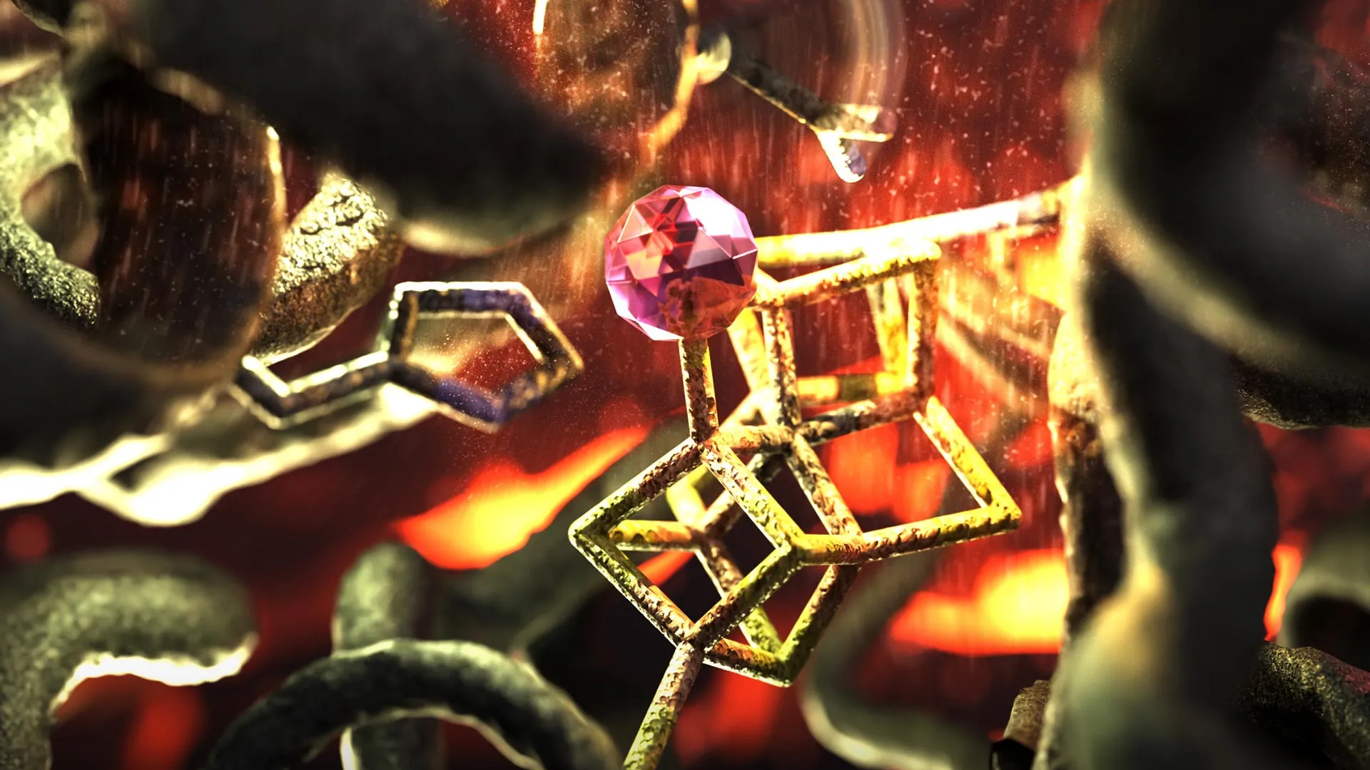
While hard metallic surfaces or inorganic crystals can easily withstand this intense bombardment, organic and biological materials present a starkly different challenge. Individual protein complexes, fragile cellular membranes, structural macromolecules, and synthetic polymers are notoriously susceptible to radiation damage. When struck by high-energy electrons, chemical bonds break, molecular structures degrade, and delicate biological entities are rapidly destroyed before a clean, high-resolution image can be captured.
This creates a frustrating zero-sum game for structural biologists and biophysicists. To discern the minute architecture of a protein crucial for understanding a disease pathology, researchers require a high signal-to-noise ratio, which traditionally demands high electron doses. Yet, increasing the dose destroys the very sample the scientist hopes to observe. Consequently, researchers have spent decades searching for ways to extract more actionable intelligence from every single electron that traverses the microscope column, thereby reducing the cumulative radiation burden on fragile specimens. The Austrian research consortium believes that quantum information science holds the definitive key to resolving this dilemma.
Leveraging Quantum Entanglement and Trapped-Ion Systems
The theoretical foundation of the new microscope design hinges on treating electrons not merely as classical bullets or simple charge carriers, but as sophisticated quantum wave-packets laden with hidden information. While standard detectors merely register the arrival coordinate of an electron, its quantum state continuously evolves as it interacts with electromagnetic fields and structural potentials within the specimen. Normally, this intricate phase and spin information is entirely washed out and lost during the final detection phase.
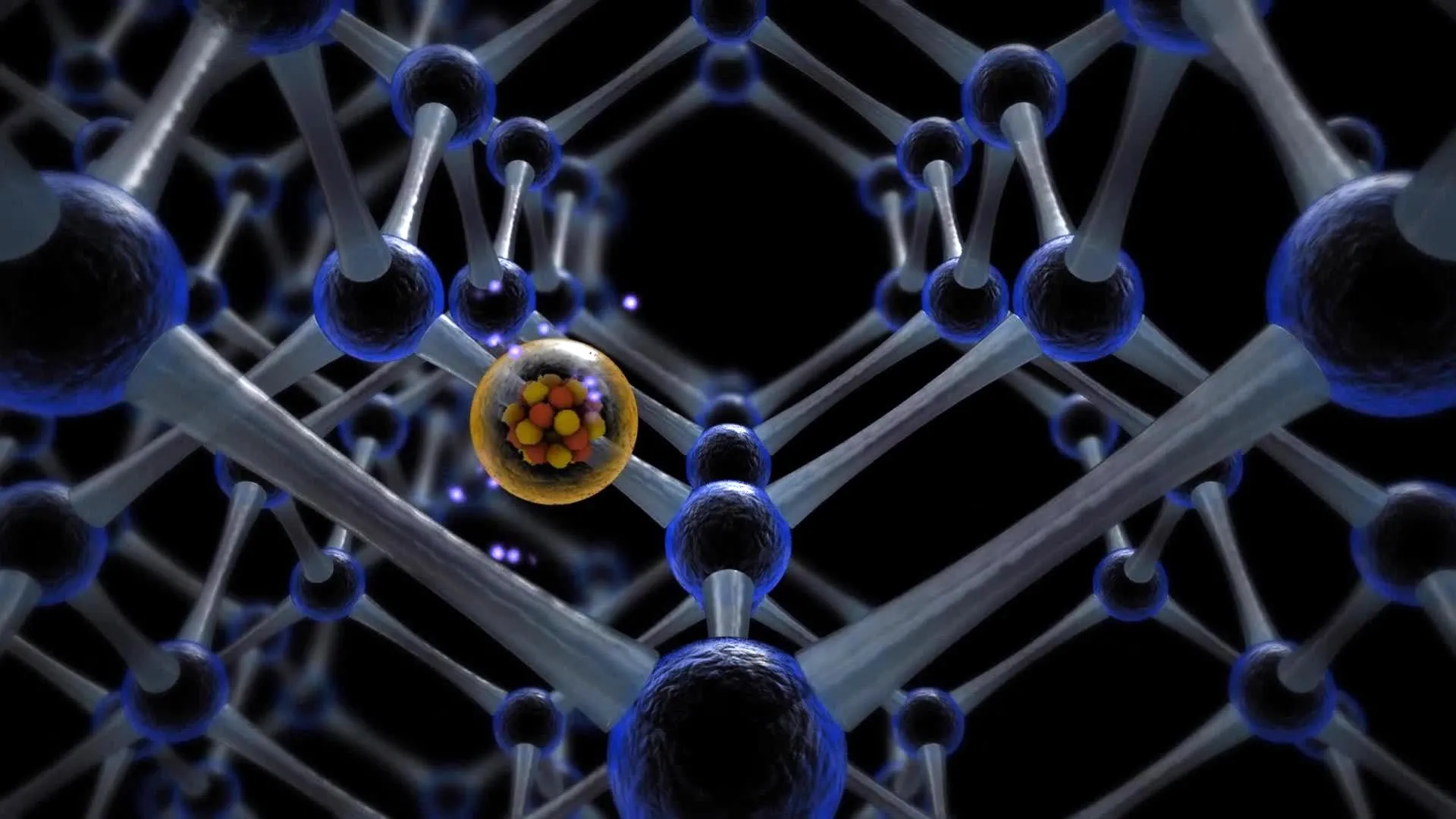
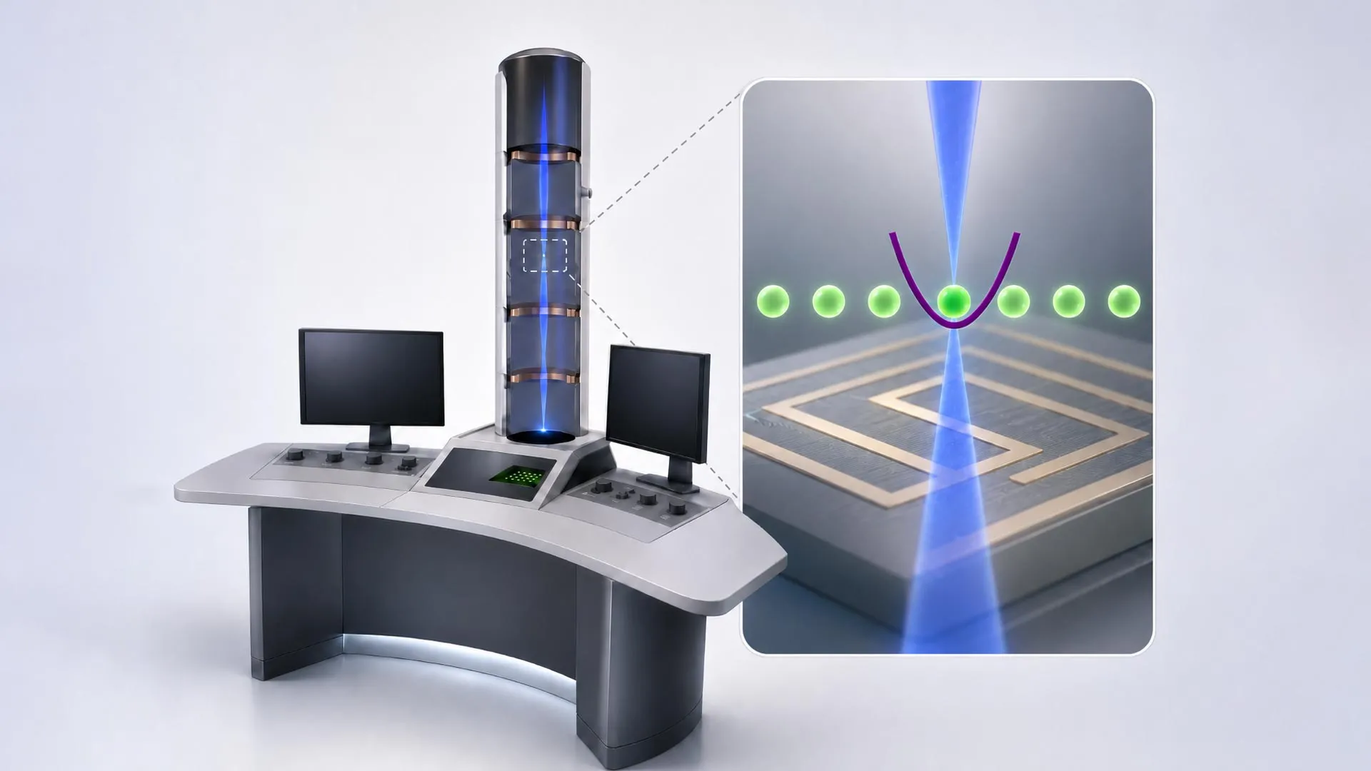
To capture this elusive data, the research team proposes routing the electron beam through a specialized interaction zone housing a trapped-ion quantum computer. According to Elias Pescoller, a doctoral student at TU Wien’s Institute for Theoretical Physics and Institute of Atomic and Subatomic Physics, the integration relies on precise physical manipulation. As the imaging electrons travel down the column, they are guided past ions that are meticulously held in place by electromagnetic fields along the beam path.
This controlled passage induces quantum entanglement between the passing electrons and the stationary ions, establishing a shared quantum state where the properties of the electron and the ion become inextricably linked. Through this mechanism, subtle perturbations experienced by the electron are instantaneously imprinted onto the quantum states of the trapped ions. Because the ion trap serves as a dynamic quantum memory register, the information is safely preserved rather than dissipated as random statistical noise.
Multiplexing and Signal Enhancement via Advanced Algorithms
Capturing information from a single entangled electron represents only the first phase of the technical challenge. The true ingenuity of the Austrian project lies in its ability to accumulate and process weak signals across multiple sequential particles.
In a traditional setup, each electron contributes an independent, often noisy data point. In the proposed quantum-enhanced microscope, the system operates iteratively. After one electron interacts with the trapped ion and deposits its quantum information, a subsequent electron passes through and becomes entangled with the same quantum computer. By executing carefully engineered quantum-computing operations between these successive events, the system can coherently aggregate data across multiple electrons.
Dennis Rätzel, a researcher at TU Wien, emphasizes the mathematical elegance of this multi-particle processing strategy. By performing specific, highly tailored quantum logic operations at every step, the system constructs a composite signal of maximum strength derived from a remarkably small number of primary electrons. This algorithmic magic transforms what would otherwise register as meaningless background clutter into a distinct, high-confidence signal.
The development of these specialized algorithms required intensive computational work, carried out in direct collaboration with Johannes Kofler’s research group at JKU Linz. By bridging theoretical computer science with experimental physics, the team successfully formulated the protocols necessary to manipulate quantum states at the speeds required for high-throughput microscopy applications. As Iva Bâezinová of TU Wien notes, this capability allows researchers to bypass the conventional statistical limits that have historically dictated the boundaries of electron detection sensitivity.
Chronology and Collaborative Development
The genesis of this unique interdisciplinary project stretches back several years, drawing momentum from Austria’s concentrated investments in quantum technologies. The formal collaboration coalesced under the quantA Cluster of Excellence framework, a national funding initiative designed to break down institutional silos and foster synergy between quantum information theorists and hardware engineers.
The chronological roadmap of the development phase highlights a methodical transition from abstract mathematical formulation to physical hardware integration:
- Phase One (Theoretical Conception): Researchers at TU Wien and JKU Linz formulated the foundational mathematics demonstrating that quantum entanglement could theoretically bridge the gap between electron scattering data and quantum memory registers.
- Phase Two (Algorithm Design): Theoretical physicists developed robust quantum-processing algorithms capable of extracting weak signals from noisy multi-electron interactions without degrading the primary imaging data.
- Phase Three (Hardware Procurement and Customization): The consortium leveraged existing hardware expertise, incorporating a state-of-the-art trapped-ion quantum computing module developed by Philipp Schindler’s team at the University of Innsbruck.
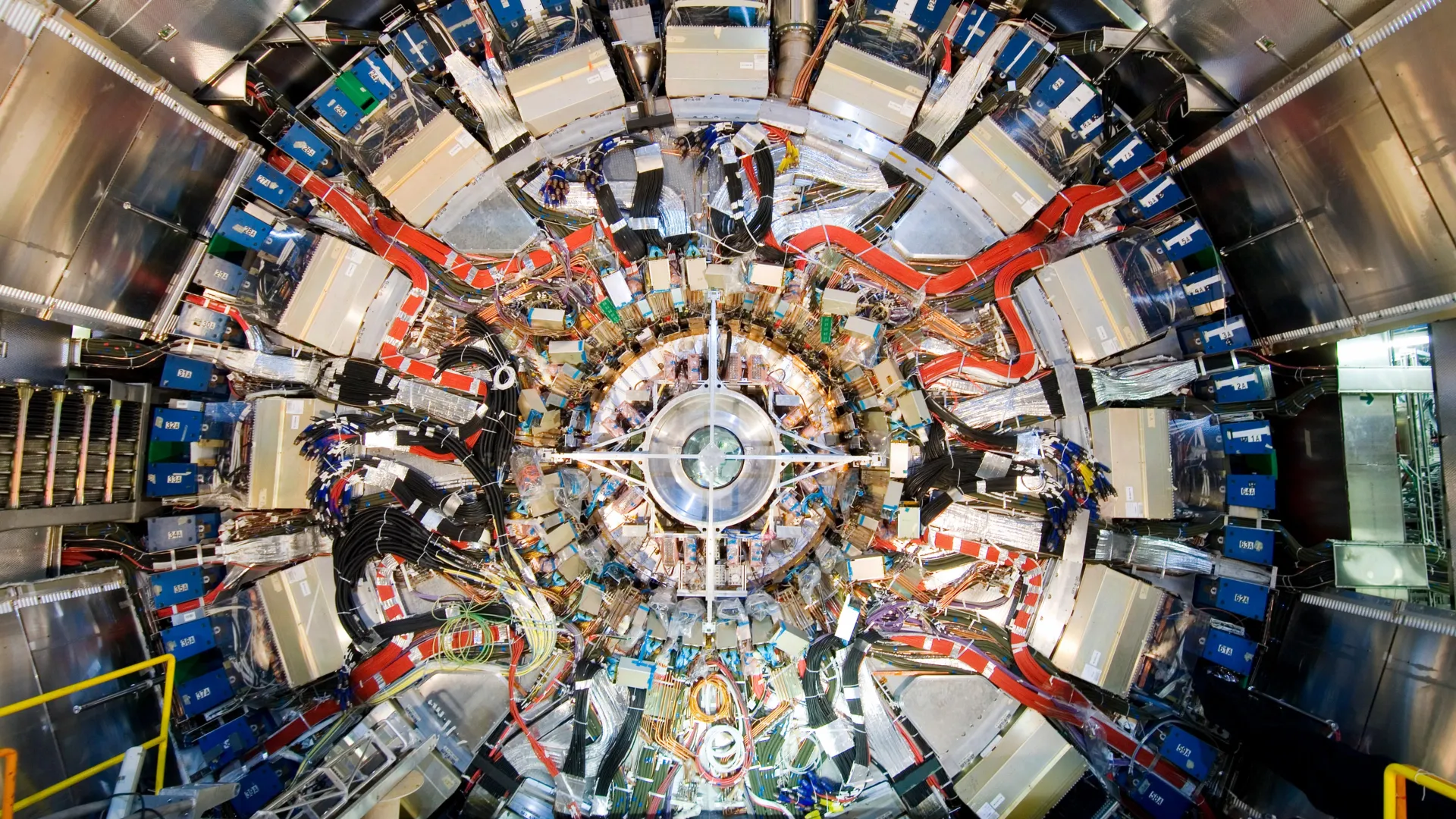
- Phase Four (Microscope Integration): Currently underway at TU Wien’s University Service Center for Transmission Electron Microscopy (USTEM), engineers are physically merging the specialized ion-trap apparatus with an advanced electron microscope column.
- Phase Five (Empirical Validation): The upcoming experimental phase will test the integrated prototype against biological and material science samples to empirically verify theoretical predictions regarding dose reduction and signal enhancement.
Thomas Juffmann of the University of Vienna underscores the rare collaborative synergy driving the project forward. By uniting distinct Austrian academic centers—each contributing specialized expertise in quantum information theory, trapped-ion hardware, and advanced microscopy—the consortium has bypassed the limitations typically faced by single-institution laboratories.
Broader Implications and Future Outlook
While the project is currently transitioning from theoretical proofs to empirical hardware demonstration, the broader implications for science and industry are profound. If the prototype microscope successfully validates the research team’s hypotheses, it could initiate a paradigm shift across multiple scientific disciplines.
In structural biology, the ability to obtain high-resolution tomograms of sensitive macromolecular assemblies—such as membrane-bound receptors, viral envelopes, and intracellular protein complexes—using a fraction of the traditional electron dose would revolutionize cryo-electron microscopy (Cryo-EM). Researchers could visualize dynamic biological processes in near-native states with minimal radiation-induced artifacts, accelerating drug discovery pipelines and deepening our fundamental understanding of cellular machinery.
Beyond biology, materials science stands to benefit immensely. Investigators studying beam-sensitive polymers, porous metal-organic frameworks, and delicate battery interfaces often struggle with structural degradation during analysis. A quantum-enhanced microscope would enable non-destructive evaluation of these advanced materials at the atomic scale, paving the way for next-generation energy storage solutions and nanotechnology manufacturing.
As the physical assembly of the hybrid instrument proceeds at USTEM, the international scientific community watches with keen interest. By wedding the macroscopic precision of electron optics with the subatomic sensitivity of quantum computation, Austrian researchers are not merely upgrading an analytical tool—they are redefining the fundamental boundaries of what human ingenuity can observe.