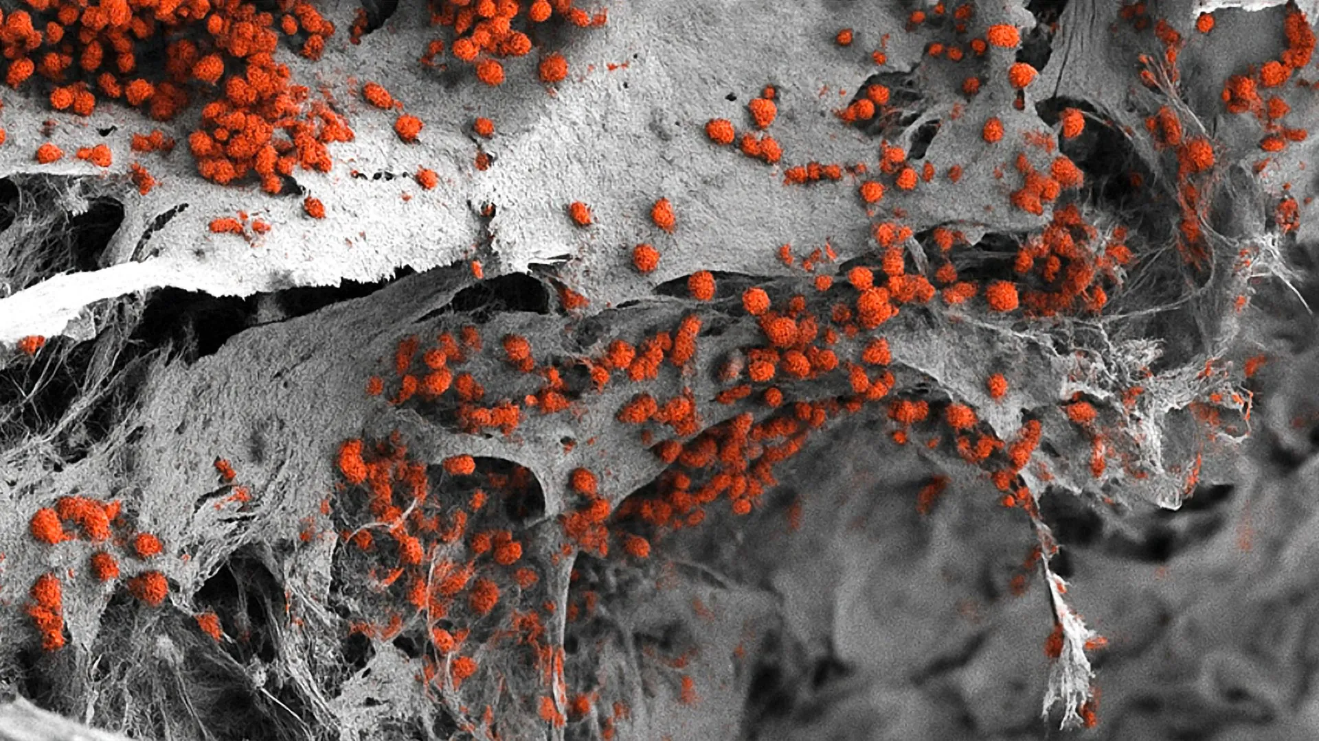


For decades, the fundamental biological understanding of glucose has remained largely unchallenged: it is the primary metabolic fuel powering nearly every living cell across the spectrum of eukaryotic life. When textbooks describe glucose, they characterize it as a molecule of thermodynamic currency, broken down through cellular respiration to release chemical energy that sustains vital physiological processes. However, groundbreaking new research led by scientists at Stanford Medicine has upended this long-held physiological dogma. According to a landmark study published in the journal Cell Stem Cell, glucose is not merely a passive energy source; it functions as a master regulator of tissue differentiation, directly controlling the complex biological trajectory through which uncommitted stem cells mature into the specialized cell types that comprise the human body.
This paradigm-shifting discovery reveals an entirely unexpected dimension of cellular biology. Instead of being catabolized for energy, intact glucose molecules physically bind to hundreds of intracellular proteins, modulating their structural conformation and directly influencing which genes are transcribed into functional proteins and when. The implications of this discovery stretch far beyond basic cell biology, offering fresh perspectives on metabolic disorders such as diabetes and illuminating novel therapeutic avenues for combating aggressive cancers characterized by failed cellular differentiation.
A Serendipitous Discovery Rooted in High-Throughput Screening
The path to this discovery was entirely unexpected. When senior author Dr. Paul Khavari, chair of dermatology and the Carl J. Herzog Professor in Dermatology at the Stanford School of Medicine, and lead author Dr. Vanessa Lopez-Pajares, a research scientist at Stanford, initiated their investigation, they were not looking for glucose. Their original objective was simply to map the biomolecular shifts that occur during human skin development.
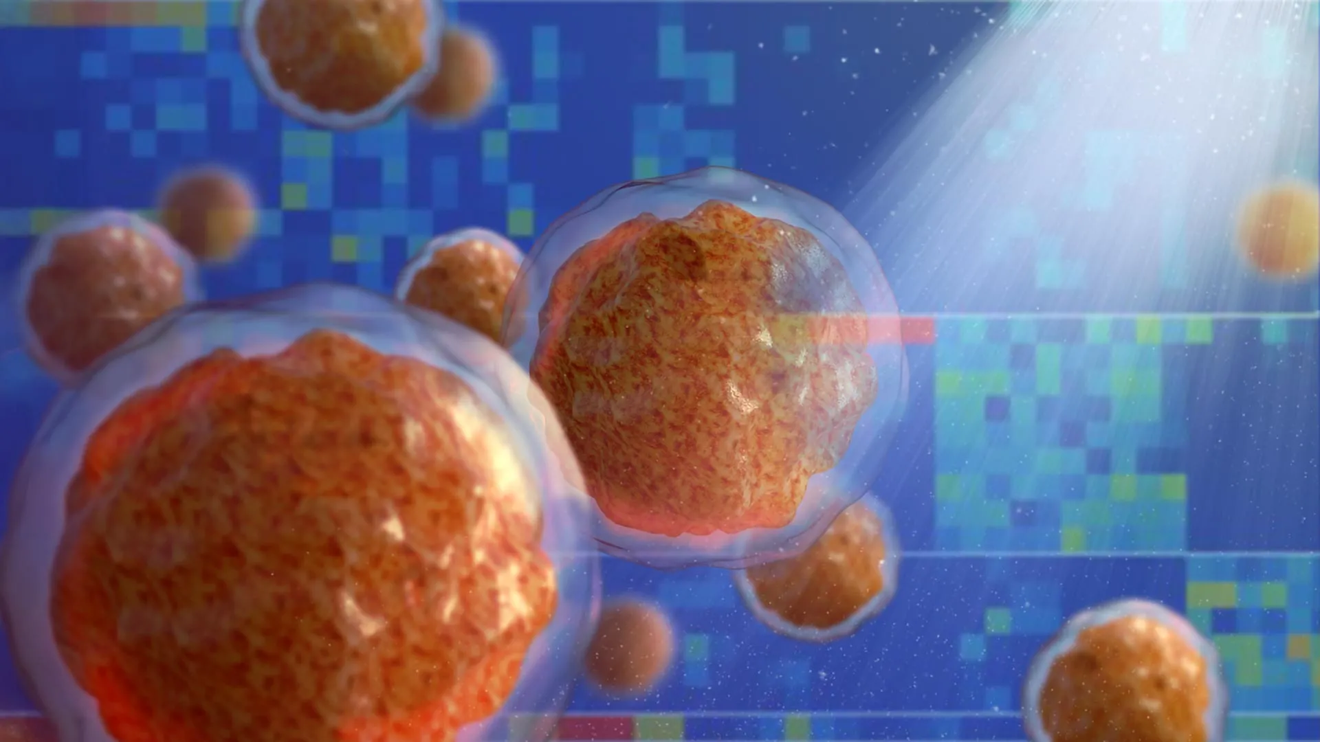
To accomplish this, the research team coupled advanced mass spectrometry with high-throughput screening methods. This technological combination allowed them to track the fluctuating abundance of thousands of distinct biomolecules within human skin stem cells as those cells underwent differentiation into mature keratinocytes—the predominant cell type making up the epidermis, the outermost layer of human skin. The core hypothesis was straightforward: molecules that significantly increase in concentration during this developmental transition likely play an active role in driving cellular maturation.
As the data compiled, the researchers identified 193 suspect molecules, many of which had established historical links to cellular differentiation. Yet, sitting comfortably as the second-most elevated molecule on the entire list was a shocking surprise: glucose.
“When we saw glucose at the top of that list, we were stunned,” Khavari recalled. Standard physiological expectation dictated that glucose levels should decline during cellular differentiation because maturing cells typically divide less rapidly, experience diminished metabolic demands, and enter a state of reduced energy expenditure as they approach senescence. Instead, the analytical data demonstrated the exact opposite. Intracellular glucose levels surged significantly as epidermal stem cells transitioned into differentiated keratinocytes.
Years of Rigorous Verification and Multi-Tissue Validation
Given the magnitude of the discovery, the Stanford research team exercised profound scientific caution. Fully recognizing that this finding contradicted decades of metabolic consensus, the investigators spent several years conducting exhaustive follow-up experiments designed to rigorously challenge and verify their initial observations before submitting their work for peer review.
“At first we just didn’t believe it,” Khavari noted. “Pero the results of extensive follow-up experiments were clear: Glucose interacts with hundreds of proteins throughout the cell and modulates their function to promote differentiation.”
To confirm the unexpected intracellular glucose accumulation, the researchers deployed multiple independent measurement techniques. They tracked cellular uptake using fluorescent and radioactive glucose analogs, and they introduced biological sensors engineered to glow green or red when exposed to physiological concentrations of glucose. As cellular differentiation progressed across various experimental models, these intracellular sensors glowed with increasing intensity.
Seeking to determine whether this phenomenon was unique to skin tissue or represented a broader biological mechanism, the team expanded their investigations. They examined diverse human cell types undergoing development, including precursors for bone, fat, and white blood cells. Furthermore, they analyzed genetically engineered murine models featuring fluorescent glucose sensors. Across every single tissue type evaluated, the pattern held true: intracellular glucose levels consistently rose during differentiation.
“In every tissue we studied, glucose levels increase as the cells differentiate,” Khavari stated. “It seems that glucose plays a global role in tissue differentiation throughout the body.”
Further mechanistic experiments revealed that this intracellular glucose accumulation is driven by a dual cellular mechanism: an active upregulation of glucose import proteins paired with a concurrent reduction in glucose export. Crucially, metabolic assays confirmed that this intracellular surge was not accompanied by an increase in glycolysis—the metabolic breakdown of glucose into energy and byproducts.
Proving the Non-Metabolic Mechanism
To definitively separate glucose’s newly discovered regulatory role from its traditional metabolic function as an energy source, the researchers conducted tissue-engineering experiments using human skin organoids. These sophisticated, three-dimensional cellular models replicate the complex composition and structural organization of native human skin when grown in specialized liquid media.
When grown in environments with abnormally low glucose levels, the skin organoids failed to differentiate properly. Genomic profiling revealed that low glucose availability altered the expression of more than 3,000 distinct genes, many of which encode proteins known to direct skin maturation.
The turning point of the study occurred when the researchers supplemented the growth medium with a specialized glucose analog—a modified molecule structurally similar to glucose but incapable of being metabolized or broken down for energy by the cells. Remarkably, these non-metabolizable glucose analogs fully rescued the differentiation defect, supporting normal skin organoid development just as effectively as regular glucose.
“That was really the biggest shock,” Khavari said, “because we were stuck in the mindset that glucose is an energy source and nothing else. But these glucose analogs support differentiation just as well as regular glucose.”
Molecular Mechanics: How Glucose Signals Cell Fate
Delving deeper into the intracellular machinery, Lopez-Pajares, Khavari, and their colleagues mapped the precise physical interactions occurring inside the cell. They discovered that the intracellular accumulation of glucose is facilitated by an enhanced production of specialized transporter proteins that shuttle glucose across the cell membrane.
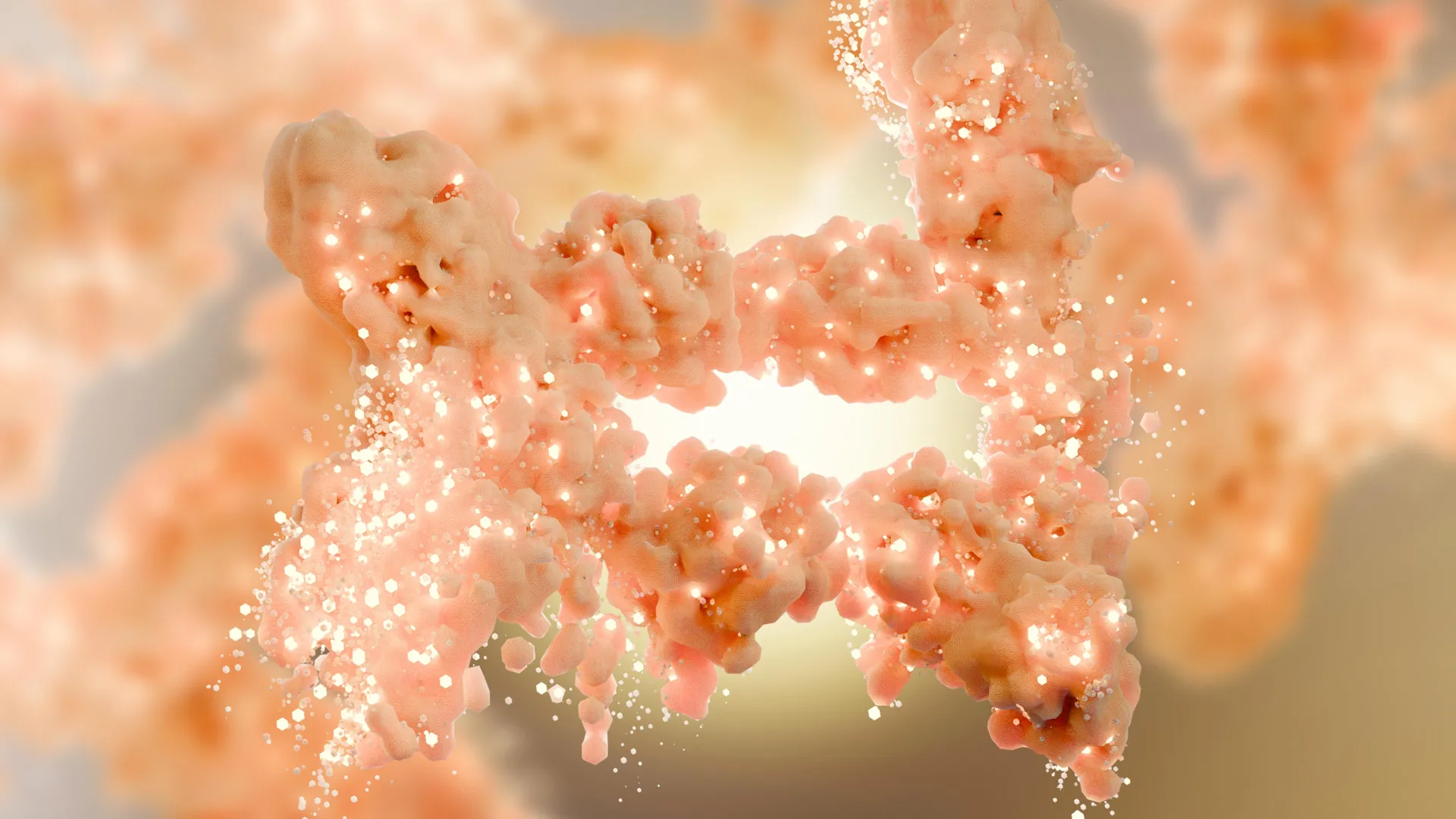
Once inside the cytoplasm, intact glucose molecules engage in direct physical binding with hundreds of distinct proteins. Among these targets is IRF6, a critical regulatory protein known to influence gene transcription. When glucose binds to IRF6, it induces a distinct conformational change in the protein’s three-dimensional structure. This structural alteration directly modifies IRF6’s ability to bind to DNA and regulate target genes involved in tissue formation.
Khavari conceptualized this sweeping physiological action with a striking analogy: “We’re seeing glucose acting like a broadcast signal in the cell, in contrast to the highly specific signaling cascades that drive many cellular functions. When glucose levels rise in a cell, they rise everywhere, all at once. It’s like a fire alarm going off in a firehouse. Everyone in the firehouse activates in response.”
Historical Context and Preclinical Implications
While this Stanford study provides the definitive molecular proof of glucose’s regulatory capacity, retrospective analysis reveals that subtle hints of this phenomenon have existed in biological literature for years. For instance, embryonic stem cells—pluripotent cells capable of giving up rise to every tissue type in the body—notoriously lose their pluripotency and differentiation capacity when cultured in environments featuring high glucose concentrations. This phenomenon is now readily explained by excess glucose prematurely driving developmental signals and stripping cells of their stemness.
Additionally, clinical observations in human medicine have long correlated systemic glucose dysregulation with impaired physiological outcomes. Patients suffering from diabetes mellitus, a chronic condition defined by chronically elevated blood glucose levels, frequently experience severe complications in wound healing, tissue regeneration, and immune response.
The findings also shed new light on experimental oncology. In recent years, certain non-metabolizable glucose analogs have advanced through preclinical and clinical trials as prospective anticancer therapies. Although originally conceptualized and developed under the assumption that they would starve malignant cells of energy by blocking metabolic pathways, the Stanford study suggests an alternative mechanism: these compounds may actually exert their therapeutic effects by forcing undifferentiated, highly proliferative cancer cells to undergo cellular differentiation, effectively halting their malignant growth.
Broader Impact and Future Research Horizons
Published online on March 21 in Cell Stem Cell, the research represents a major milestone in cellular and molecular biology. The study was supported by robust funding from the National Institutes of Health (grant numbers R01AR043799, AR045192, K01AR070895, and P30CA124435) as well as the U.S. Department of Veterans Affairs Office of Research and Development.
As the scientific community digests these findings, the Stanford research team is already looking toward the future, planning follow-up studies to investigate how this non-metabolic glucose signaling pathway operates across various disease states, including metabolic disorders and oncology. Because cancer is fundamentally a disease characterized by arrested or failed cellular differentiation, uncovering how small biomolecules direct cell fate opens an entirely new frontier in therapeutic development.
“This finding is a springboard for research on dysregulation of glucose levels, which affects hundreds of millions of people,” Khavari concluded. “Pero it’s also likely to be important in cancer development because cancer is a disease of failed differentiation. This is an entirely new and growing field. People have thought that small biomolecules like glucose were quite passive in the cell. This is another piece of evidence to pay close attention to other roles these molecules might play.”