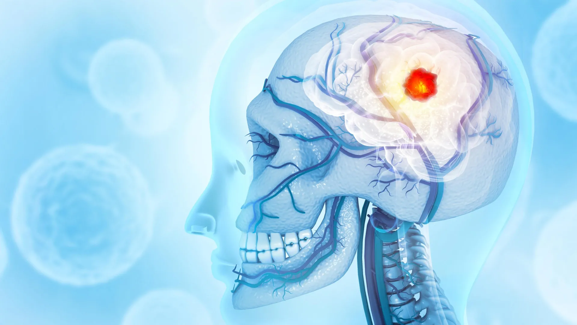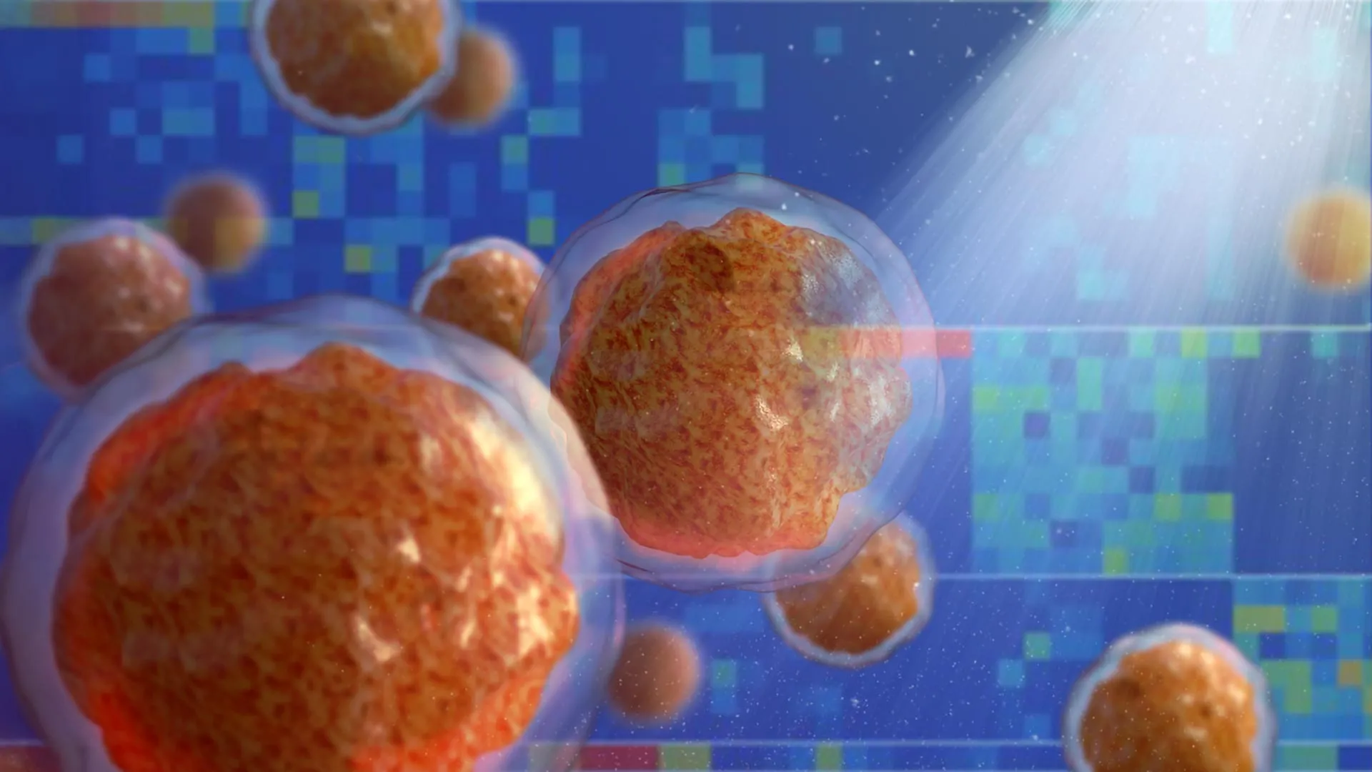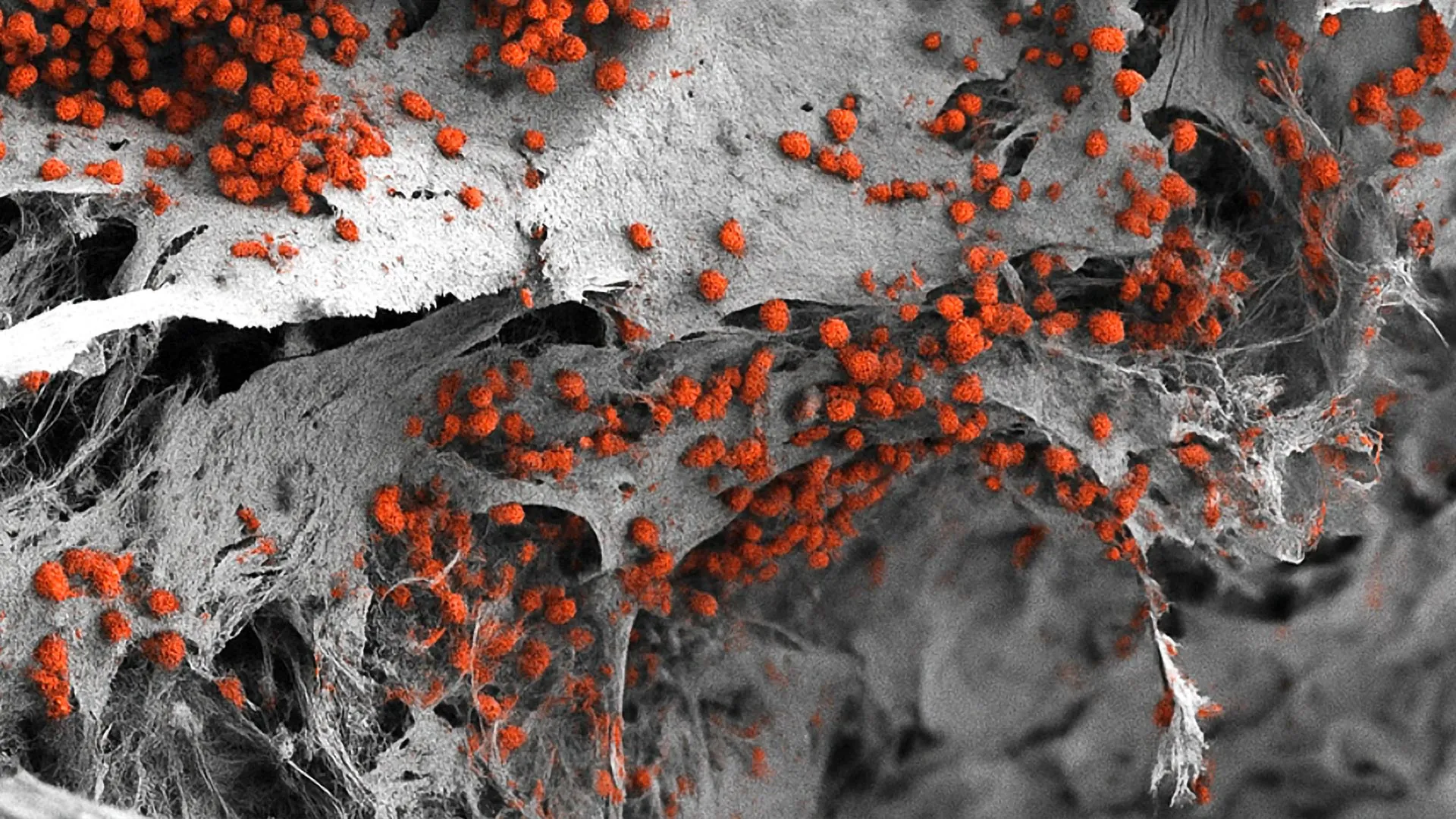


Scientists at the Indiana University School of Medicine have successfully developed a groundbreaking imaging technique designed to peer deep into the intricate microenvironment of bone marrow using mouse models. By overcoming formidable structural hurdles that have historically plagued tissue analysis, this innovative methodology promises to accelerate the pipeline for novel drug development and targeted therapies. The advancement holds profound implications for a wide spectrum of complex medical conditions, including hematological malignancies such as leukemia, various autoimmune disorders, and debilitating musculoskeletal diseases.
The research breakthrough, made possible by adapting the advanced multiplex imaging tool known as Phenocycler 2.0, allows scientists to visualize an unprecedented number of cellular markers within completely intact mouse bone marrow tissue. The details of this pioneering methodology and its initial applications were recently published in the peer-reviewed scientific journal Leukemia, marking a major milestone in cellular biology and biomedical research.
The Structural Challenges of Bone Marrow Analysis
For decades, the gelatinous consistency of bone marrow encased tightly within rigid, calcified bone has presented a formidable obstacle for researchers seeking to map its cellular landscape. Bone marrow is far from a static tissue; it is a dynamic, highly vascularized biological hub that serves as the primary site of hematopoiesis—the continuous formation of blood and immune cells. Furthermore, it houses critical populations of hematopoietic stem cells, which are responsible for replenishing the body’s supply of red blood cells, white blood cells, and platelets.
Understanding the spatial organization of these cells and how they interact with their surrounding microenvironment is vital for decoding disease progression. However, traditional analytical techniques have long forced scientists to make difficult compromises. Standard approaches such as flow cytometry require the mechanical or enzymatic digestion of tissue, destroying the native architecture and spatial relationships between cells in order to quantify populations. While flow cytometry provides valuable numerical data regarding cell types, it completely erodes the structural context of the tissue.
Conversely, conventional fluorescence microscopy preserves tissue architecture but suffers from severe technical limitations. Standard fluorescent imaging is typically constrained to detecting only three to four cellular markers simultaneously due to spectral overlap between fluorophores. This narrow window makes it nearly impossible to capture the immense cellular heterogeneity and complex signaling networks operating within the bone marrow at any given time.
The Phenocycler 2.0 Solution and Methodological Breakthrough
The research team at the Indiana University Cooperative Center of Excellence in Hematology successfully circumvented these historical barriers by harnessing the Phenocycler 2.0 platform. While the multiplex imaging tool had previously been deployed with success in structurally more accessible organs such as the spleen and kidneys, applying it to bone marrow required overcoming significant physical and chemical hurdles. The IU team is the first to successfully adapt the technology for intact mouse bone marrow tissue.
By utilizing iterative cycles of staining and gentle fluorescent barcoding, the researchers were able to simultaneously visualize and map 25 distinct cellular markers within a single, undisturbed bone marrow sample. This multiplex approach preserves the delicate spatial architecture of the tissue while providing a high-definition, panoramic view of cellular neighborhoods. Researchers can now observe how stem cells position themselves relative to immune cells, blood vessels, and structural niches, offering unprecedented clarity into normal physiological functions and pathological disruptions.
Chronology of the Research and Development
The path to this publication represents a culmination of rigorous multidisciplinary collaboration within the Indiana University School of Medicine. Although the final findings were recently unveiled in Leukemia, the initiative builds upon years of institutional expertise in hematology, orthopedic surgery, and pediatric research.
The project was spearheaded by a robust team of investigators. Sonali Karnik, PhD, assistant research professor of orthopedic surgery at the IU School of Medicine, served as co-lead author alongside a collaborative cohort of specialists. The broader research effort was guided by Reuben Kapur, PhD, co-senior author of the study, director of the Herman B Wells Center for Pediatric Research, and co-director of the IU Cooperative Center of Excellence in Hematology.
The methodology underwent extensive optimization phases to ensure reproducibility and accuracy across multiple experimental trials using mouse models. Following the successful validation of the 25-marker panel, the intellectual property potential of the technique was swiftly recognized. The IU Innovation and Commercialization Office has officially filed a provisional patent for the novel imaging methodology, securing the institution’s stake in the commercial and clinical translation of the technology.
Expanding the Horizons of Multiplex Imaging
Buoyed by their initial success, the research team is not resting on its laurels. Current efforts are already underway to significantly expand the capabilities of the imaging panel. Moving beyond the initial 25 markers, the scientists are working to integrate features that will allow for the simultaneous visualization of bone matrix, neural networks, muscular tissue, and an even broader array of immune and signaling cell types.
Expanding the marker panel will transform the tool into an even more comprehensive diagnostic and discovery platform. By mapping the cross-talk between the nervous system, skeletal structures, and hematopoiesis within the bone marrow, researchers can begin to model complex systemic diseases with a level of fidelity previously thought impossible in preclinical settings.
Perspectives from Leadership and Research Teams
The implications of this technological leap have generated considerable enthusiasm within the academic and medical research communities at Indiana University.
"Bone marrow is difficult to study because it is gelatinous and encased in hard bone," explained Dr. Sonali Karnik, co-lead author of the study, highlighting the physical constraints that have frustrated generations of pathologists and cellular biologists. "Since bone marrow plays an important role in blood and immune cell formation and houses valuable stem cells, our unique imaging approach offers a useful tool for a variety of research applications."
Because murine models serve as the foundational bedrock for preclinical trials globally, the ability to accurately image their bone marrow architecture opens up vast new avenues for translational science. Dr. Reuben Kapur emphasized the direct relevance of the technique to human pathology.
"Because mouse models are widely used to study human diseases, this technique offers a promising new method for investigating a range of conditions like autoimmune diseases, leukemia and other disorders involving bone marrow," Dr. Kapur noted. By bridging the gap between high-throughput cellular analysis and spatial biology, the tool provides investigators with a sharper lens through which to observe how diseases take root and evolve within the body’s most protected internal compartments.
Broader Impacts on Drug Development and Therapeutics
The successful deployment of the Phenocycler 2.0 platform for bone marrow analysis arrives at a critical juncture in pharmaceutical research and precision medicine. The development of effective therapeutics—particularly for blood cancers such as leukemia, multiple myeloma, and lymphomas—frequently hinges on understanding how malignant cells outcompete healthy stem cells within the bone marrow niche.
When pharmaceutical companies test novel compounds, they require robust assays that can measure not only whether a drug kills target cells, but also how it impacts the surrounding healthy tissue architecture. Traditional assays often miss subtle microenvironmental shifts that can lead to drug resistance or unexpected toxicities. By offering a 25-marker (and expanding) window into intact tissue, the IU imaging technique provides a powerful screening instrument that could streamline preclinical evaluation, reduce the attrition rate of drug candidates, and accelerate the timeline for bringing novel therapies from the laboratory bench to clinical trials.
Furthermore, the methodology holds substantial promise for regenerative medicine and immunology. Conditions characterized by chronic inflammation, immune dysregulation, and bone degeneration can now be studied with spatial precision, enabling researchers to pinpoint therapeutic targets that were previously obscured by the limitations of older technology.
Funding and Collaborative Acknowledgments
The advancement of sophisticated imaging technologies of this scale requires sustained financial investment and collaborative institutional support. The research conducted at the Indiana University School of Medicine was made possible primarily through financial backing from the National Institutes of Health (NIH), reflecting the federal agency’s ongoing commitment to fostering foundational biomedical innovations.
In addition to Dr. Karnik and Dr. Kapur, the published study credits a wide array of contributing researchers from the IU School of Medicine. The co-authors include Connor Gulbronson, Paige C. Jordan, Rahul Kanumuri, Baskar Ramdas, Ramesh Kumar, Melissa L. Hartman, Izza Khurram, Drew M. Brown, Karen E. Pollok, Pratibha Singh, and Melissa A. Kacena. Their collective expertise spanning orthopedic surgery, pediatrics, cell biology, and oncology was instrumental in validating the imaging protocol.
Outlook for Future Clinical Translation
As the Indiana University Innovation and Commercialization Office continues to navigate the provisional patent process, the research team is actively exploring pathways to scale the technology for broader academic partnerships and potential industrial applications. While the current breakthrough focuses exclusively on mouse models—a vital step for validating biological mechanisms—the underlying principles of multiplex spatial imaging hold immense potential for future adaptation in human clinical diagnostics.
If successfully translated to human tissue biopsy analysis, similar multiplex methodologies could one day revolutionize personalized medicine in oncology and hematology, allowing physicians to map the exact spatial cellular composition of a patient’s bone marrow tumor microenvironment and tailor targeted treatments accordingly. For now, the IU School of Medicine’s milestone achievement stands as a transformative leap forward, equipping the global scientific community with an unprecedented instrument to decode the complex mysteries locked within bone.