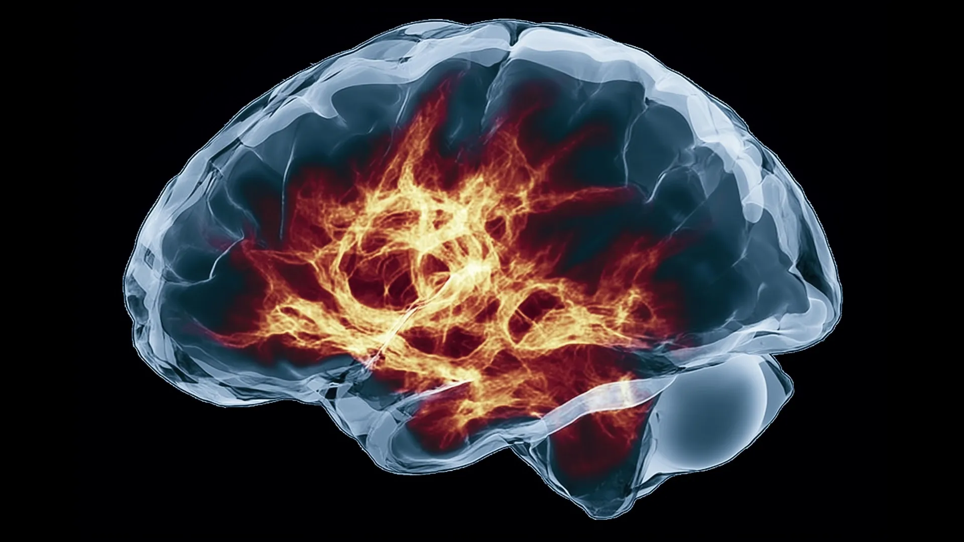

Deep within the brainstem lies a cluster of neurons known as the locus coeruleus (LC), a name derived from the Latin for "blue place" due to its distinct pigmentation. For decades, the scientific community viewed this region as a singular, monolithic "loudspeaker" for the brain, broadcasting the neurotransmitter norepinephrine (NE) in a uniform, undifferentiated manner to help regulate stress, attention, and arousal. However, groundbreaking research published in the journal Nature by scientists at the Allen Institute for Brain Science has dismantled this long-standing assumption. By utilizing high-resolution imaging and genetic mapping, researchers have revealed that the LC is not a chaotic foghorn but a highly sophisticated, targeted postal service, utilizing distinct pathways to deliver precise chemical messages to specific regions of the nervous system.
A Paradigm Shift in Neurobiology
The prevailing model of the LC—the "volume transmission" hypothesis—suggested that when the region was activated, it flooded the brain with norepinephrine, creating a blanket effect that influenced everything from wakefulness to the fight-or-flight response. This interpretation was primarily based on the fact that individual LC neurons have long, branching axons that span vast areas of the brain. While the physical reach of these neurons was known, the specificity of their communication was not.
The new study, led by researchers at the Allen Institute, employed an exhaustive multi-modal approach to challenge this. By integrating whole-brain imaging, single-cell genetic profiling of nearly 400,000 cells, and the structural reconstruction of complex neurons, the team discovered that the LC is organized into anatomically and functionally distinct sub-circuits. This evidence suggests that rather than a uniform release, the brain modulates its norepinephrine output with surgical precision.
Chronology of the Investigation
The journey to this discovery began several years ago as part of a broader effort supported by the National Institutes of Health’s Brain Research Through Advancing Innovative Neurotechnologies (BRAIN) Initiative. Scientists recognized that current behavioral models failed to account for the nuanced ways animals respond to their environments.
- Phase One (2020–2021): Researchers initiated high-resolution mapping of the mouse brain, focusing on the connectivity of the LC. Initial imaging revealed that dorsal LC neurons consistently projected to the forebrain and cerebral cortex, while ventral LC neurons showed a propensity for the brainstem and spinal cord.
- Phase Two (2022): The team performed genetic profiling to determine if these anatomical differences were matched by internal cellular differences. They found that gene expression patterns varied significantly between the dorsal and ventral populations, confirming they were distinct cell types rather than a homogeneous group.
- Phase Three (2023): Behavioral experiments were conducted to observe how these specific groups responded to environmental stimuli. The researchers observed that dorsal LC neurons were highly active during learning-based decision-making tasks, whereas ventral neurons fired specifically when animals chose to ignore environmental signals.
- Phase Four (2024): The final synthesis of data, leading to the Nature publication, provided the complete map of these pathways, confirming that the LC’s signaling is highly organized.
The Anatomy of Long-Range Signaling
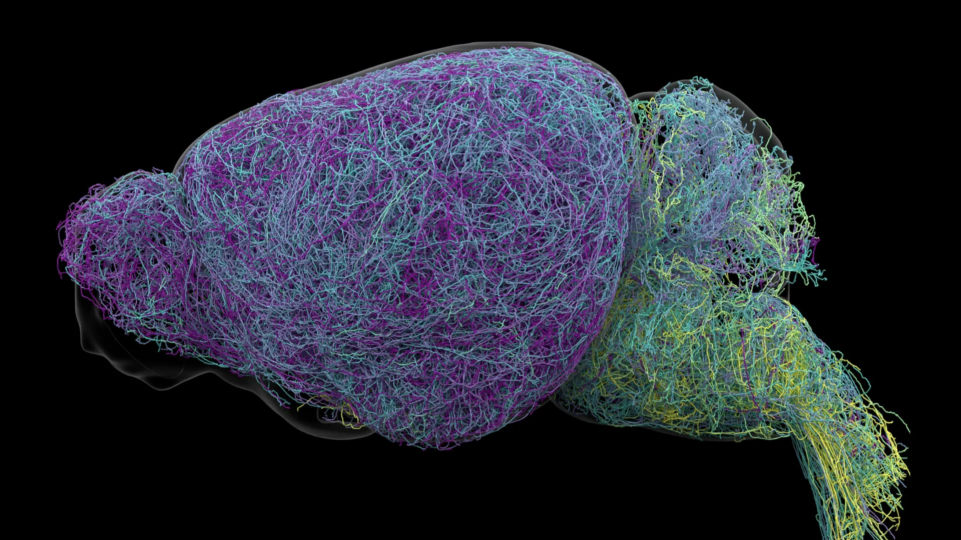
One of the most startling findings to emerge from the study is the physical scale of these neurons. In their examination of nearly 35,000 neurons, the researchers discovered that LC axons reach lengths of approximately 35 centimeters—an extraordinary distance for a single cell in a mouse.
The record-breaking neuron, reconstructed to a length of 70.32 centimeters, provides a masterclass in biological efficiency. Despite its immense reach, this neuron does not distribute norepinephrine indiscriminately. It remains highly selective, targeting specific volumes of the cerebral cortex while bypassing other regions like the cerebellum and the spinal cord. This finding effectively disproves the idea that the "length" of a neuron necessitates a "broadcast" style of signaling. Instead, the architecture of the axon is tightly controlled to ensure that chemical messengers reach only intended destinations, reinforcing the concept of a targeted neural network.
Official Perspectives and Implications
Karel Svoboda, director of Neural Dynamics at the Allen Institute and a co-author of the study, emphasized the gravity of the shift in perspective. "The findings suggest the brain’s norepinephrine system is far more like a targeted postal network than a foghorn," Svoboda noted. By comparing the system to a postal service, researchers are highlighting the ability of the brain to differentiate between types of information, whether it be a stimulus for learning or a signal to ignore irrelevant environmental noise.
Jeremiah Cohen, another co-author of the study, noted that the specificity of these neurons is highly unusual for the brain. "Most neurons are more specific in their targets," Cohen explained. "But this neuron doesn’t release NE everywhere. It ignores the cerebellum, brainstem, and spinal cord." This discovery necessitates a re-evaluation of how neuroscientists interpret brain-wide signaling systems.
Clinical Potential: Toward Precision Medicine
The implications for mental health and neurology are profound. Currently, medications that target the norepinephrine system—often prescribed for ADHD, depression, and anxiety—are systemic in nature. They act on the entire brain, which is precisely why patients often report "beneficial effects in one region while causing unwanted effects elsewhere."
For instance, a patient taking an antidepressant might see improvements in mood but experience sleep disturbances or cardiovascular side effects. Because the LC is one of the first areas to deteriorate in patients with Alzheimer’s disease, understanding its modularity could prove revolutionary. If scientists can map which specific sub-circuits are affected by disease, they may be able to develop therapeutic interventions that target only those circuits, minimizing systemic side effects.
A Synergy of Chemical Messengers
The study also opens a new dialogue regarding the interaction between norepinephrine and dopamine. The researchers observed that the LC’s signaling pathways share architectural similarities with the brain’s dopamine systems, particularly those that communicate with the basal ganglia. It is now hypothesized that these two systems—norepinephrine and dopamine—work in tandem to manage the acquisition of complex, abstract concepts.
By providing a clear map of how the LC communicates, the research suggests that learning is not a generalized brain state but a specific activation of the dorsal LC pathway. This could lead to new cognitive training therapies that specifically engage these pathways, potentially aiding those with learning disabilities or cognitive decline.
Conclusion: Future Directions
While these findings are derived from the mouse model, the fundamental anatomy of the brainstem is highly conserved across mammalian species, including humans. This suggests that the "postal network" model is likely a universal feature of the mammalian brain.
The next phase of research will likely focus on confirming these pathways in human post-mortem tissues and developing refined pharmacological agents that can selectively stimulate or inhibit specific LC sub-circuits. As we move closer to a granular understanding of the brain’s wiring, the "blue place" deep in the brainstem is proving to be far more than just a source of chemical arousal; it is the control center for a complex, organized, and remarkably precise communication system that dictates how we learn, react, and engage with the world around us. This research marks a definitive departure from the simplistic models of the past and sets the stage for a new era of neurobiology defined by precision and targeted intervention.