
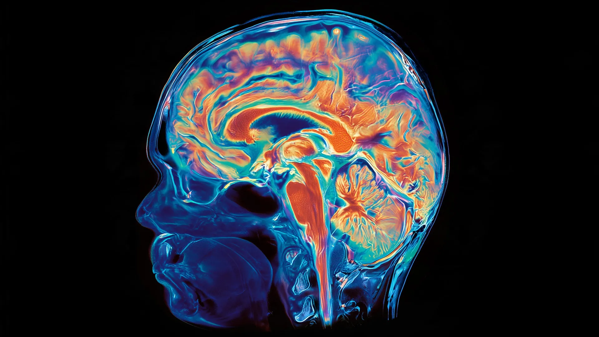
In a landmark case study that has sent ripples through the neurological community, researchers from the Perelman School of Medicine at the University of Pennsylvania have uncovered rare, high-resolution evidence that the successful removal of amyloid plaques—a hallmark of Alzheimer’s disease—may effectively stall the subsequent development of tau protein tangles and neurodegeneration. Presented at the 2026 Alzheimer’s Association International Conference and published simultaneously in the journal JAMA, this investigation provides perhaps the most compelling human evidence to date that modern anti-amyloid therapies do more than just clear “gunk” from the brain; they may fundamentally alter the disease’s trajectory.
The study centers on a unique, post-mortem examination of a patient who participated in a long-term clinical trial for an anti-amyloid monoclonal antibody. By comparing neighboring regions of the same brain—some of which were cleared of amyloid by the medication and others that remained heavily burdened—researchers observed a striking "Goldilocks" effect: areas free of amyloid exhibited significantly lower levels of tau and healthier cellular architecture, while adjacent, amyloid-rich regions showed advanced signs of cell death and disease progression.
The Mechanism of Neurodegeneration: A Protein-Driven Crisis
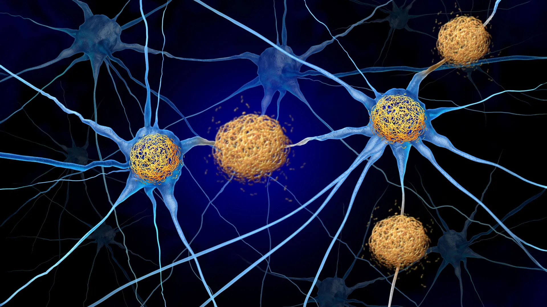
To understand the gravity of these findings, one must consider the two primary biological villains in Alzheimer’s pathology. Beta-amyloid is a protein that aggregates into extracellular plaques, effectively choking the space between neurons and disrupting neural signaling. Tau, by contrast, is an intracellular protein that, under the stress of amyloid presence, becomes hyperphosphorylated and forms neurofibrillary tangles. These tangles destabilize the internal transport systems of neurons, eventually leading to the catastrophic cell death that characterizes the clinical decline of Alzheimer’s patients.
For decades, the “Amyloid Cascade Hypothesis” has posited that amyloid deposition is the initiating event in the disease process, acting as a catalyst that eventually triggers the more lethal tau-related damage. While clinical trials for drugs like lecanemab (Leqembi) and donanemab (Kisunla) have successfully demonstrated the clearance of plaques, demonstrating a direct, causal link between this clearance and a reduction in tau pathology in the living human brain has been notoriously difficult. The Penn study bridges this gap, providing a visual and molecular map of exactly what happens when the amyloid cascade is interrupted.
Chronology of a Unique Clinical Case
The patient at the center of this study was diagnosed with mild cognitive impairment (MCI) in his fifties, a relatively early age for the onset of Alzheimer’s symptoms. He was subsequently enrolled in a clinical trial for aducanumab, a pioneering anti-amyloid therapy. His treatment regimen was rigorous: he received 30 doses of the medication over a span of four and a half years.
Following his last dose, the patient lived for approximately four years, providing researchers with a long-term look at the lasting effects of the therapy. Before his passing, he elected to donate his brain to the Center for Neurodegenerative Disease Research, a decision that has now yielded one of the most significant insights in the history of neurodegenerative pathology. By the time of his death, he had lived more than a decade since his initial diagnosis, yet the state of his brain tissue remained remarkably heterogeneous.
Mapping the Patchwork: The Gyri vs. Sulci Phenomenon
The most compelling aspect of the study lies in the anatomical comparison of the brain’s topography. The brain’s surface is composed of folds (gyri) and the grooves between them (sulci). In this specific patient, the researchers observed a clear dichotomy: the gyri, which had successfully shed their amyloid burden due to the therapeutic intervention, showed a significant reduction in tau tangles and inflammatory markers.
Conversely, the sulci—the deeper grooves—retained high levels of amyloid. In these precise, localized areas, the presence of tau was rampant, and the tissue showed advanced neurodegeneration. This "patchwork" pattern mirrored the longitudinal PET scans taken while the patient was still alive, confirming that the drug’s efficacy was not uniform across the entire brain. This anatomical variability provided the research team with a rare internal control, allowing them to compare two different disease states within the exact same biological environment.
Implications for Modern Alzheimer’s Therapeutics
The implications for the current standard of care are profound. Drugs such as donanemab and lecanemab rely on similar mechanisms of action as the treatment received by the patient in this study. The findings suggest that the clinical benefits of these medications—which are currently measured in 18-month trial windows—might represent only the tip of the iceberg in terms of long-term neuroprotection.
"Seeing both disease patterns side-by-side in the same brain gave us a rare opportunity to understand how amyloid removal affects other proteins that contribute to Alzheimer’s disease," said Dr. David Wolk, co-senior author and director of the Penn Alzheimer’s Disease Research Center. "The findings provide some of the clearest human evidence to date that anti-amyloid therapies may limit the accumulation of tau and slow the brain changes that lead to memory loss and cognitive decline."
However, the medical community remains cautious. While the evidence is compelling, researchers admit that a single case study cannot determine the exact threshold of amyloid clearance required to achieve permanent neuroprotection. Nor can it fully account for why certain brain regions responded better to the therapy than others. The question of “regional vulnerability”—why some areas of the brain are more resistant to drug delivery or plaque clearance—is now a primary focus for future pharmaceutical research.
The Case for Early Intervention
The study adds significant weight to the growing movement toward earlier detection and intervention. Because amyloid can accumulate in the brain for up to two decades before a patient exhibits even minor memory lapses, the medical community is shifting its focus toward pre-symptomatic treatment.
Dr. Christopher A. Brown, the study’s first author, noted that the timing of the intervention is likely the most critical variable in patient outcomes. "Patients may accumulate amyloid for years before symptoms appear," Brown stated. "This case suggests that removing amyloid early may help limit the changes that ultimately damage brain cells. Ongoing trials will help determine whether starting treatment before symptoms begin can provide even greater benefit."
If these results are validated in broader, longitudinal population studies, the paradigm for Alzheimer’s management will likely shift from reactive symptom management to proactive "pathology management." This would involve aggressive monitoring of amyloid and tau levels in high-risk individuals, with the goal of neutralizing the amyloid threat long before the "tau cascade" causes irreversible cognitive damage.
Scientific Context and Future Directions
The study, supported by a significant consortium of National Institutes of Health grants, underscores the necessity of brain donation programs for advancing our understanding of neurodegenerative diseases. As we enter an era where Alzheimer’s is no longer viewed as an inevitable decline but as a treatable medical condition, the ability to study the human brain in the post-treatment era is invaluable.
Looking forward, the research team at the Perelman School of Medicine intends to investigate the specific biological mechanisms that allow some brain regions to remain resilient while others succumb to plaque buildup. By identifying these factors, scientists hope to develop "next-generation" therapies that are more uniform in their distribution and more effective at penetrating the blood-brain barrier in vulnerable areas.
While the Alzheimer’s community is far from declaring victory, this autopsy report provides a clear, documented path forward. It confirms that the amyloid plaques are not merely bystanders in the Alzheimer’s process, but active drivers of a lethal pathology. By systematically dismantling these plaques, medicine now has a tangible, observable, and scientifically verified method for slowing the biological clock of the disease, offering new hope to the millions of families worldwide affected by this devastating condition.