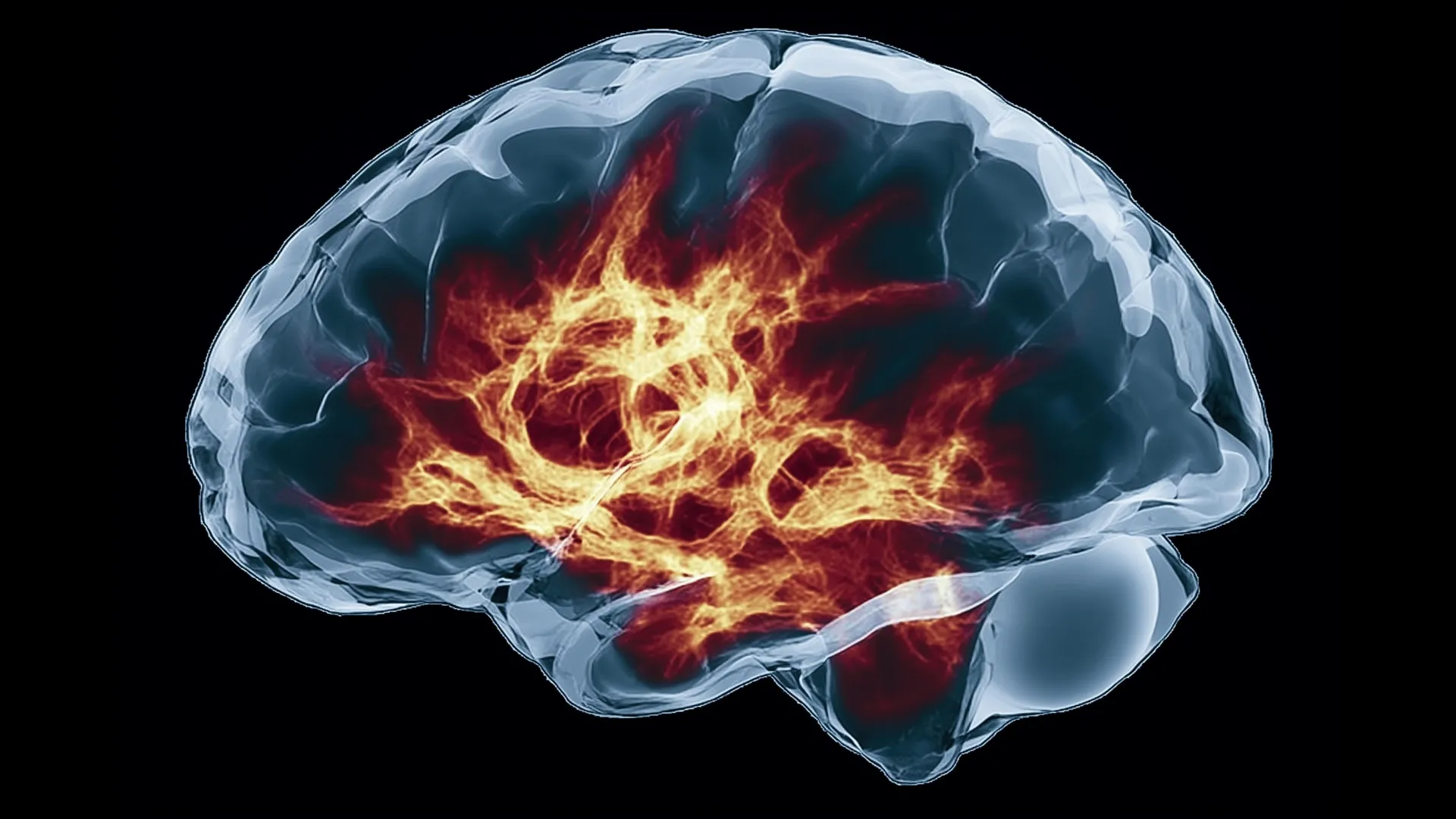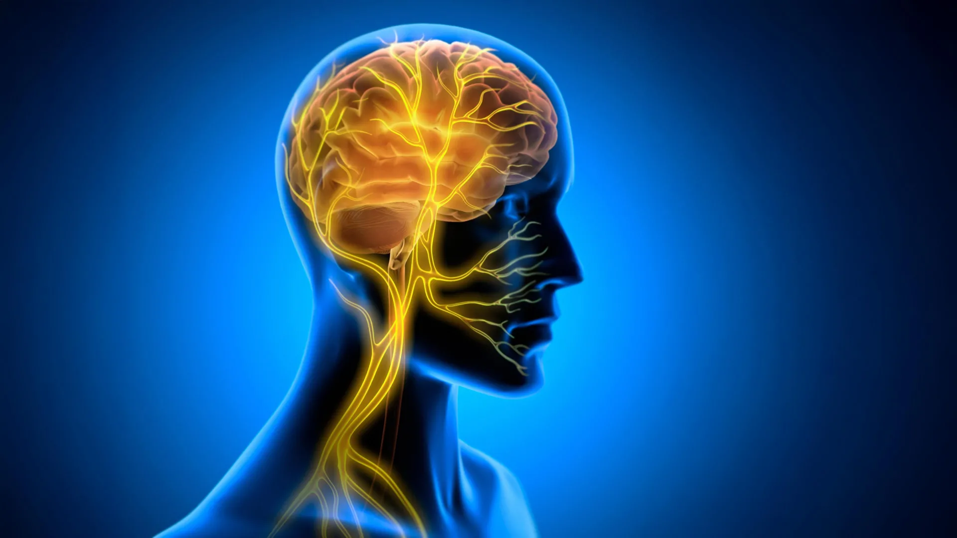
The human brain is far more industrious during general anesthesia than previously understood, maintaining sophisticated linguistic and predictive capabilities even in the absence of conscious awareness. Recent findings published in the journal Nature by a multidisciplinary team at Baylor College of Medicine reveal that the hippocampus—a critical region for memory and learning—remains active, capable of distinguishing patterns and anticipating upcoming linguistic structures while a patient is rendered unconscious. This discovery challenges the long-standing medical assumption that anesthesia effectively "shuts down" high-level cognitive functions, suggesting instead that the brain remains a vigilant, analytical engine even when the mind is "offline."
The Methodology of Deep-Brain Observation
To achieve this level of granular insight, researchers led by Dr. Sameer Sheth, professor and Cullen Foundation Endowed Chair of Neurosurgery at Baylor, utilized high-density recording technology during essential surgical procedures. The study subjects were patients undergoing epilepsy surgery, a clinical setting that permits the temporary placement of recording devices to map brain activity.
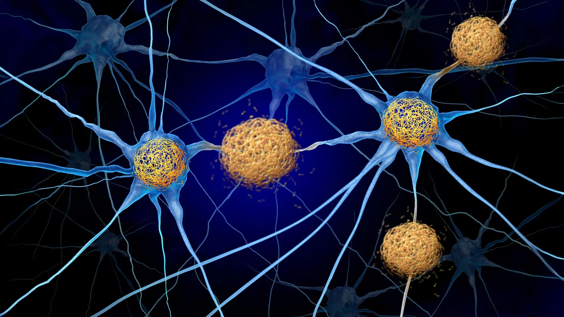
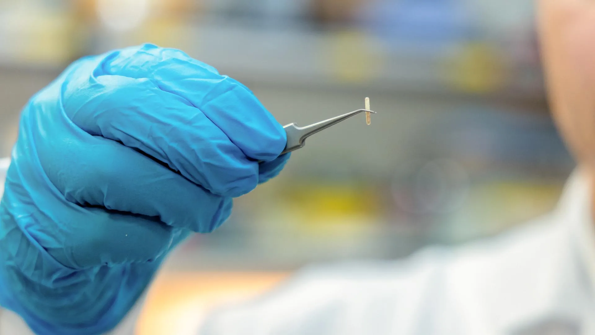
The team employed Neuropixels probes, state-of-the-art silicon devices that are significantly smaller than traditional electrodes. These probes allow researchers to monitor the firing patterns of hundreds of individual neurons simultaneously. By placing these probes directly into the hippocampus, the researchers captured the electrical "chatter" of the brain during various stages of anesthesia. This technique provided a high-resolution window into the temporal lobe, an area known for its role in organizing information and anchoring memories.
Chronology of the Discovery
The research took place over several months, involving a rigorous, multi-stage experimental design. Initially, the team established a baseline by observing how neurons responded to simple, repetitive auditory stimuli. They played a series of monotonous tones, periodically interjected with an "oddball" or unexpected sound.
The data revealed that even under deep anesthesia, the hippocampus registered the irregularity. Furthermore, as the experiment progressed, the hippocampal neurons demonstrated a capacity for adaptation—a hallmark of neural plasticity. The cells became increasingly adept at distinguishing between the standard tones and the deviant ones, effectively "learning" the environment despite the patient being incapable of conscious perception.
Following this baseline, the team advanced to more complex linguistic testing. They played short, coherent narratives to the patients while continuing to record neural activity. The resulting data showed that the hippocampus was not merely recording sound waves; it was categorizing them. Specific patterns of neuronal firing corresponded to different parts of speech, such as nouns, verbs, and adjectives. Most remarkably, the brain exhibited predictive coding, a mechanism that anticipates the next logical word in a sentence based on the preceding context.
Data-Driven Insights and Predictive Coding
Predictive coding is a fundamental theory in neuroscience which posits that the brain is not a passive receiver of sensory input but an active, anticipatory processor. In a conscious state, this allows us to finish each other’s sentences and navigate complex environments with ease.
The Baylor study provides the first robust evidence that this process persists under general anesthesia. Data analysis showed that the neural firing patterns encoded information about upcoming lexical items. This suggests that the brain’s "internal model" of the world remains online and continues to run simulations of future possibilities even when the patient is completely unresponsive.
For clinicians and neuroscientists, this suggests that the "loss of consciousness" induced by anesthetics like propofol or sevoflurane may be a disruption of global communication networks rather than a cessation of localized processing. The hippocampus continues its work, but that information is no longer being broadcast to the prefrontal cortex or other areas responsible for the subjective experience of "self" or "awareness."
Official Perspectives and Expert Analysis
Dr. Sameer Sheth, who serves as the Director of The Gordon and Mary Cain Pediatric Neurology Research Foundation Laboratories, noted that these findings necessitate a shift in how the medical community views the unconscious state. "Our findings show that the brain is far more active and capable during unconsciousness than previously thought," Sheth stated. "Even when patients are fully anesthetized, their brains continue to analyze the world around them."
Dr. Benjamin Hayden, a professor of neurosurgery at Baylor and a collaborator on the study, highlighted the discrepancy between the behavior of the unconscious brain and traditional medical definitions. "This kind of predictive coding is something we associate with being awake and attentive, yet it’s happening here in an unconscious state," Hayden observed.
The implications for this research are significant, particularly in the realm of neurology and anesthesiology. By understanding the boundaries of neural activity under anesthesia, doctors may one day be able to better monitor the depth of sedation, moving away from subjective scales toward objective, neuro-electrical markers.
Parallels with Artificial Intelligence
The study also offers a unique bridge between biological neuroscience and the rapid advancements in Artificial Intelligence. Modern Large Language Models (LLMs), such as those powering contemporary chatbots, function through a process of probabilistic prediction—calculating the likelihood of the next token in a sequence based on a vast dataset.
The observation that the human hippocampus performs a similar predictive task—even without consciousness—suggests that predictive processing is a fundamental computational principle of intelligence, whether that intelligence is carbon-based or silicon-based. While the underlying mechanisms differ, the structural similarity in how both systems anticipate future information provides researchers with a new framework for studying the biological basis of language.
Broader Implications for Speech Prosthetics
Perhaps the most promising application of this research lies in the development of Brain-Computer Interfaces (BCIs). For patients who have lost the ability to speak due to stroke, neurodegenerative disease, or traumatic brain injury, the challenge has always been "decoding" the internal language of the brain.
Dr. Vigi Katlowitz, the study’s first author and a neurosurgery resident, emphasized the clinical potential: "Can we use these signals to deploy and run a speech prosthetic for some of the parts of the brain that are damaged by stroke or injury? These are questions that we can now consider in relation to this part of the brain."
If the hippocampus can encode language structures in the absence of consciousness, it implies that the "syntax" of thought exists in a readable, electrical format that could potentially be captured and translated by external hardware. This could lead to a new generation of prosthetics that rely on the brain’s natural predictive tendencies to help restore communication for the severely impaired.
Limitations and Future Research
While the findings are groundbreaking, the research team remains cautious, emphasizing that this study represents a single step in a much larger investigation. The experiments were limited to a specific type of general anesthesia and a specific brain region—the hippocampus.
Questions remain as to whether these findings would hold true for other types of anesthetics or in other states of unconsciousness, such as natural sleep or medically induced comas. Furthermore, the researchers acknowledge that finding activity in the hippocampus does not equate to the patient "understanding" the stories in any way that would be recognizable to them upon waking. The distinction between "processing" and "experiencing" remains the central enigma of consciousness studies.
Moving forward, the Baylor team plans to expand their research to observe how other regions of the brain interact with the hippocampus during anesthesia. By mapping the communication pathways that are severed during the onset of unconsciousness, they hope to identify the precise moment—and the precise neural circuit—where consciousness emerges.
Conclusion: A New Era of Neuro-Cognitive Understanding
The work conducted at Baylor College of Medicine serves as a profound reminder that the brain is a tireless machine, constantly engaged in the labor of pattern recognition and prediction. By stripping away the layer of subjective consciousness, the researchers have revealed the "background processes" that define our cognitive existence.
As science continues to peel back the layers of the unconscious mind, the boundary between being awake and being under anesthesia becomes increasingly porous. We are beginning to see that the brain’s ability to interpret the world is not merely a consequence of being awake; it is an intrinsic, fundamental property of the neural architecture itself. This discovery does not just change our understanding of anesthesia—it challenges us to rethink the very nature of consciousness and the hidden, silent work the brain performs while we sleep.