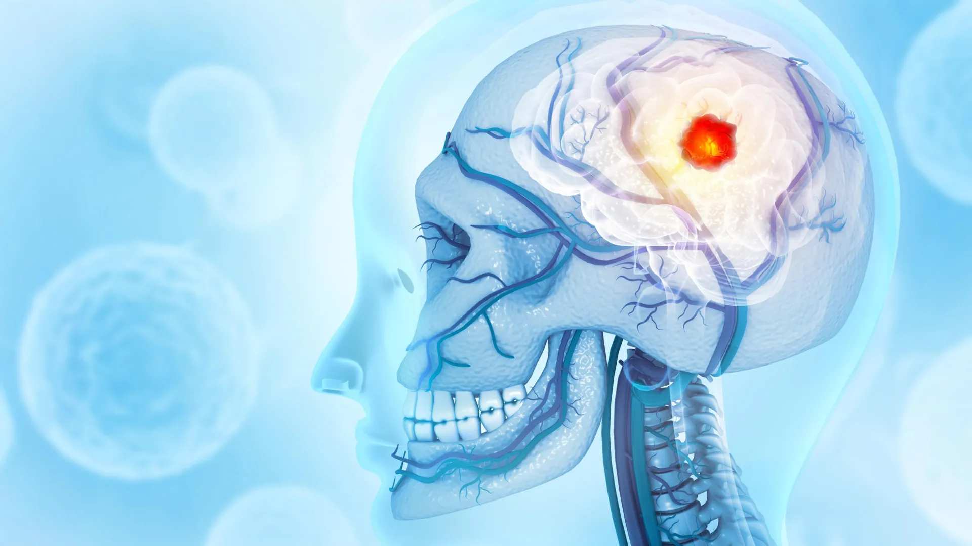
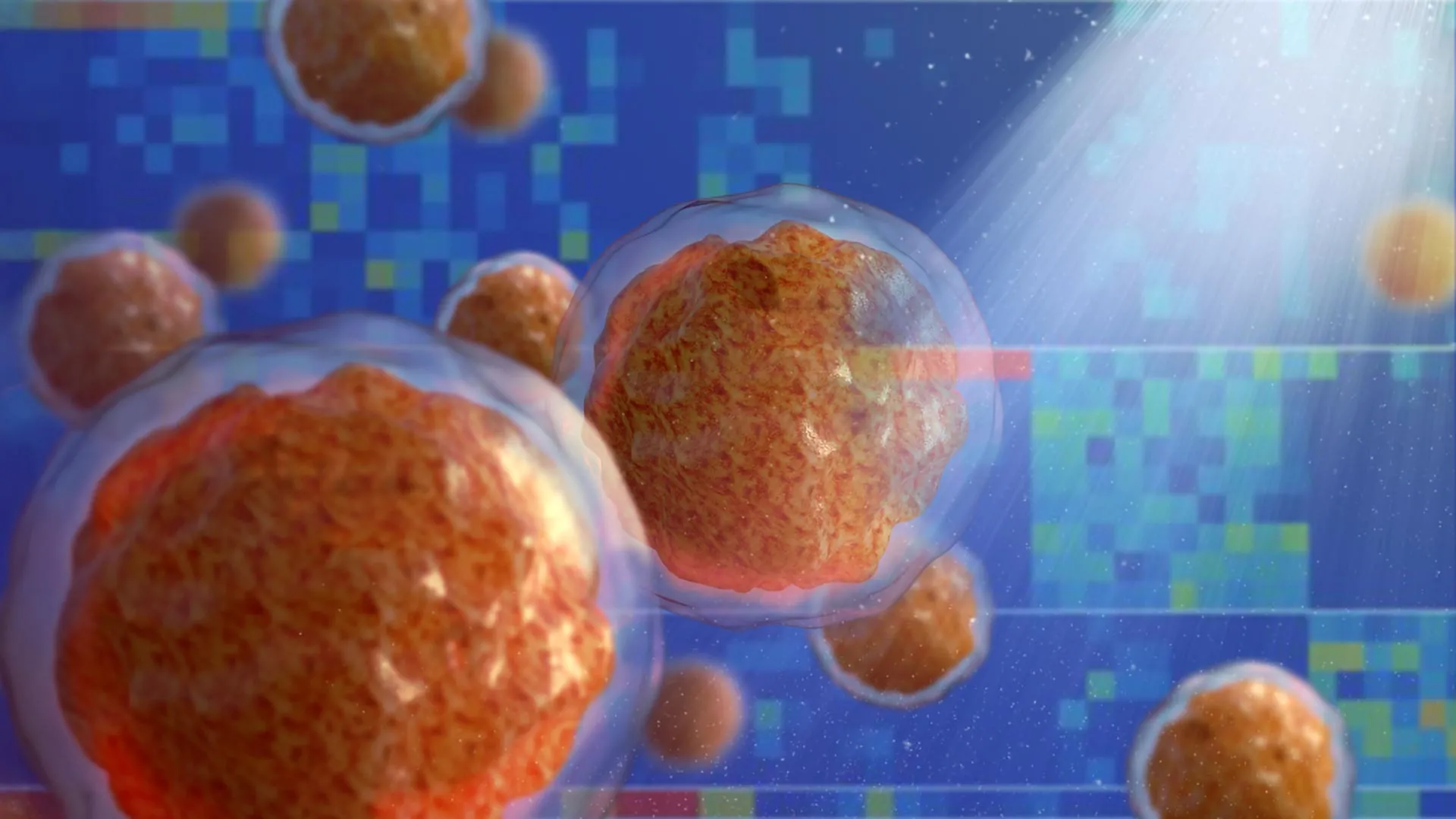
A collaborative research effort led by scientists at the Johns Hopkins Kimmel Cancer Center and the Johns Hopkins Bloomberg School of Public Health has unveiled a critical molecular mechanism driving the progression of translocation renal cell carcinoma (tRCC), a rare and often aggressive form of kidney cancer. The study, published in the journal Cell Reports on April 22, details how specific gene rearrangements lead to the formation of microscopic liquid droplets within cell nuclei. These droplets, known as biomolecular condensates, act as specialized command centers that selectively activate genes responsible for cancer growth and metastasis. By identifying the physical and chemical properties of these structures, researchers have opened a new frontier for therapeutic intervention in a disease that currently lacks a standardized treatment protocol.
The research was spearheaded by senior author Danfeng "Dani" Cai, Ph.D., an assistant professor of biochemistry and molecular biology at the Johns Hopkins Bloomberg School of Public Health, in partnership with Eneda Toska, Ph.D., an assistant professor of oncology at the Johns Hopkins Kimmel Cancer Center. Their findings suggest that the physical state of proteins—specifically their ability to transition into liquid-like phases—is as central to cancer progression as the genetic mutations themselves.
The Genetic Landscape of Translocation Renal Cell Carcinoma
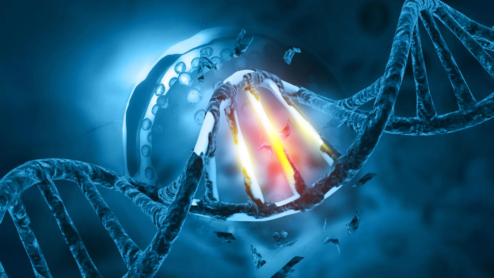
Translocation renal cell carcinoma is a distinct subtype of kidney cancer defined by the rearrangement of chromosomes. This process involves a DNA segment "swapping" places, which results in the fusion of the TFE3 gene with one of several partner genes. While tRCC accounts for approximately 1% to 5% of all renal cell carcinomas in adults, it represents a much higher proportion of kidney cancers in children and young adults, often presenting a more aggressive clinical course.
Under normal physiological conditions, the TFE3 gene codes for a protein that regulates various cellular functions. However, in tRCC, the tail end of the TFE3 gene merges with the beginning of another gene, such as PRCC, NONO, or SFPQ. This genetic "chimera" produces fusion proteins that are not found in healthy cells. While the existence of these fusion proteins has been recognized by the oncology community for years, the exact mechanism by which they reprogram a healthy cell into a malignant one remained elusive until this study.
Dr. Cai’s team focused their investigation on the two most prevalent fusion partners: NONO and SFPQ. Together, these two variants account for approximately 40% of all TFE3 fusion cases. By narrowing the focus to these common drivers, the researchers sought to find a "universal" vulnerability in the way these proteins operate.
Liquid Condensates: The Engines of Malignancy
The core discovery of the Johns Hopkins study lies in the observation that TFE3 fusion proteins do not float freely within the cell nucleus. Instead, they congregate to form "liquid condensates." These are concentrated clusters of molecules that interact in a small, demarcated space to perform specific biological tasks.
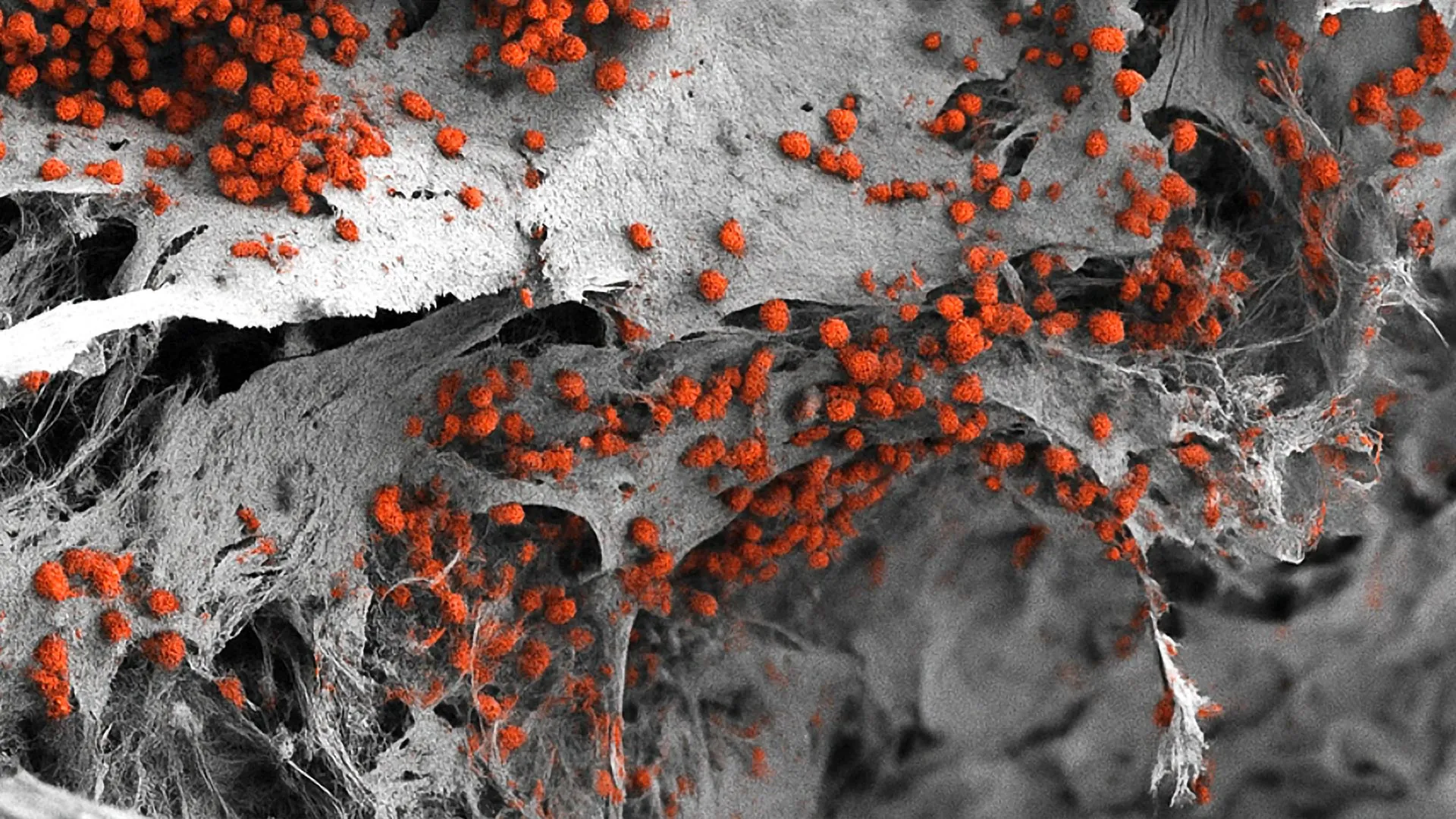
To visualize this process, the research team utilized advanced microscopy techniques, attaching fluorescent tags to TFE3 fusion proteins in cells derived from kidney cancer patients. The results were striking: the fusion proteins appeared as distinct, glowing "dots" or droplets within the nucleus. These droplets were found to be highly active hubs. Further analysis revealed the presence of marker proteins associated with active gene transcription and other proteins responsible for "turning on" genetic sequences.
The study posits that these condensates serve as a bridge between the fusion protein and the cell’s DNA. By concentrating the necessary molecular machinery in one place, the condensates allow the cancer-driving proteins to exert a disproportionate influence over the cell’s genetic output. This "phase separation" allows the cancer to bypass normal regulatory checks, effectively hijacking the cell’s internal hardware to promote rapid proliferation and survival.
Chronology of the Research and Experimental Methodology
The investigation proceeded through several distinct phases, combining molecular biology, genomics, and advanced imaging:
- Protein Mapping and Visualization: The team first established the physical presence of the condensates using patient-derived cell lines. This confirmed that the fusion proteins (specifically NONO-TFE3 and SFPQ-TFE3) naturally formed these liquid-like structures.
- Genomic Interaction Analysis: Partnering with Dr. Toska, the team investigated how these droplets interacted with chromatin—the "beads on a string" structure of DNA. Using high-throughput sequencing and chemical mapping, they identified the specific sites on the genome where the fusion proteins were binding.
- Functional Dissection: The researchers used gene-editing technology to systematically remove different segments of the TFE3 fusion proteins. They were looking for the "glue" that held the liquid droplets together.
- The Coiled-Coil Discovery: The team identified a specific structural element known as a "coiled-coil" domain—a shape where two or more protein coils wrap around each other. When this small segment was removed, the fusion proteins lost their ability to form droplets. Crucially, without the droplets, the proteins were no longer able to activate the genes that drive cancer growth.
Chromatin Remodeling and the "Landscape" of Cancer
A significant portion of the study was dedicated to understanding how these liquid droplets physically alter the cell’s DNA. In a healthy cell, DNA is tightly wound around proteins called histones, forming chromatin. Genes that are tightly wound are "off," while those in "open" or accessible areas are "on."
Dr. Toska’s analysis revealed that TFE3 fusion proteins act as master architects of this landscape. By creating chemical modifications on the chromatin, the fusion proteins force closed sections of DNA to open and open sections to close. This "redesign" of the chromosome landscape is specifically targeted toward genes that facilitate cell movement and rapid division—two hallmarks of metastatic cancer.
"Individually, all the protein components found in the TFE3 fusions… are typically involved in the cell machinery that turns on genes," Dr. Cai explained. "However, we found when in the form of these fusion proteins, they acquire an even stronger ability to control what genes get turned on." This synergy between the fusion partners creates a "super-activator" that the cell is unable to suppress through normal means.
Implications for Future Therapies and Broader Oncology
The discovery of liquid condensates as the driving force behind tRCC has profound implications for drug development. Currently, tRCC is notoriously difficult to treat; it does not respond well to standard chemotherapies, and while some targeted therapies and immunotherapies show promise, there is no "gold standard" for care.
The Johns Hopkins team suggests that disrupting the formation of these liquid droplets could be a viable therapeutic strategy. Rather than trying to inhibit the protein’s function directly—which is often difficult with transcription factors like TFE3—doctors could use "condensate-disrupting" drugs to dissolve the droplets, thereby "muting" the cancer genes.
Furthermore, the implications of this study extend far beyond kidney cancer. "Other cancers, such as Ewing sarcoma and leukemia, are caused by fusion genes as well," noted Dr. Cai. "It’s possible that these fusion genes form similar droplets… and could react to similar treatment strategies." This suggests a paradigm shift in oncology: moving away from targeting specific genetic mutations and toward targeting the physical state of the proteins those mutations produce.
Collaborative Effort and Institutional Support
The success of the study was the result of a multi-disciplinary effort involving researchers from various departments within Johns Hopkins and the National Cancer Institute (NCI). Co-authors included Choon Leng So, Ye Jin Lee, Wanlu Chen, Binglin Huang, and others from the Bloomberg School of Public Health, as well as Bujamin Vokshi from the Johns Hopkins School of Medicine and W. Marston Linehan from the NCI.
The research was heavily supported by the National Institutes of Health (NIH), specifically the National Institute of General Medical Sciences and the National Cancer Institute. Additional funding was provided by the National Human Genome Research Institute, the Department of Defense Kidney Cancer Idea Development Award, and the Johns Hopkins Provost Catalyst Award.
Dr. Toska’s contributions were also supported by her ongoing work in the field, which includes grants and consulting roles with pharmaceutical entities like AstraZeneca and Menarini, highlighting the intersection of academic research and potential industry application for new drug pipelines.
Conclusion and Next Steps
The identification of the coiled-coil domain as the linchpin of condensate formation provides a clear target for future pharmacological screening. The research team’s next phase of work will involve screening for small molecules or existing drugs that can penetrate the cell nucleus and disrupt these liquid structures without harming healthy cellular processes.
By shifting the focus from the "what" of genetic mutations to the "how" of their physical manifestation in the cell, the Johns Hopkins investigators have provided a roadmap for treating not only a rare kidney cancer but potentially a wide array of translocation-driven malignancies. As the scientific community continues to explore the role of biomolecular condensates, the findings in Cell Reports serve as a cornerstone for a new era of "physical" oncology, where the state of matter within a cell is just as important as the code within its DNA.