

In a significant departure from decades of established medical consensus, researchers at the University of Kentucky have identified a novel mechanism by which Alzheimer’s disease compromises sleep, suggesting that the condition’s most taxing symptoms may be reversible without the need to clear amyloid plaques. The findings, published in the journal Alzheimer’s & Dementia, indicate that the brain’s own immune cells—microglia—are the primary agents responsible for sleep degradation in the Alzheimer’s brain. By modulating the inflammatory response of these cells, investigators were able to restore over two hours of sleep per day in mouse models, providing a promising new therapeutic avenue that pivots away from the traditional focus on protein accumulation.
For years, the scientific community has operated under the assumption that sleep disturbances in Alzheimer’s patients were a direct byproduct of neuronal damage or the physical accumulation of amyloid plaques. However, the study led by Shannon L. Macauley, Ph.D., an associate professor of physiology at the University of Kentucky College of Medicine, and first author Nicholas J. Constantino, Ph.D., suggests that the immune system’s reaction to these plaques is the true culprit. This research introduces a "paradigm-shifting" perspective, characterizing the sleep loss as a symptom of a systemic inflammatory "overreaction" rather than a simple consequence of disease-related brain clutter.
The Mechanism of Neuro-Inflammation: A Cascade of Disruption
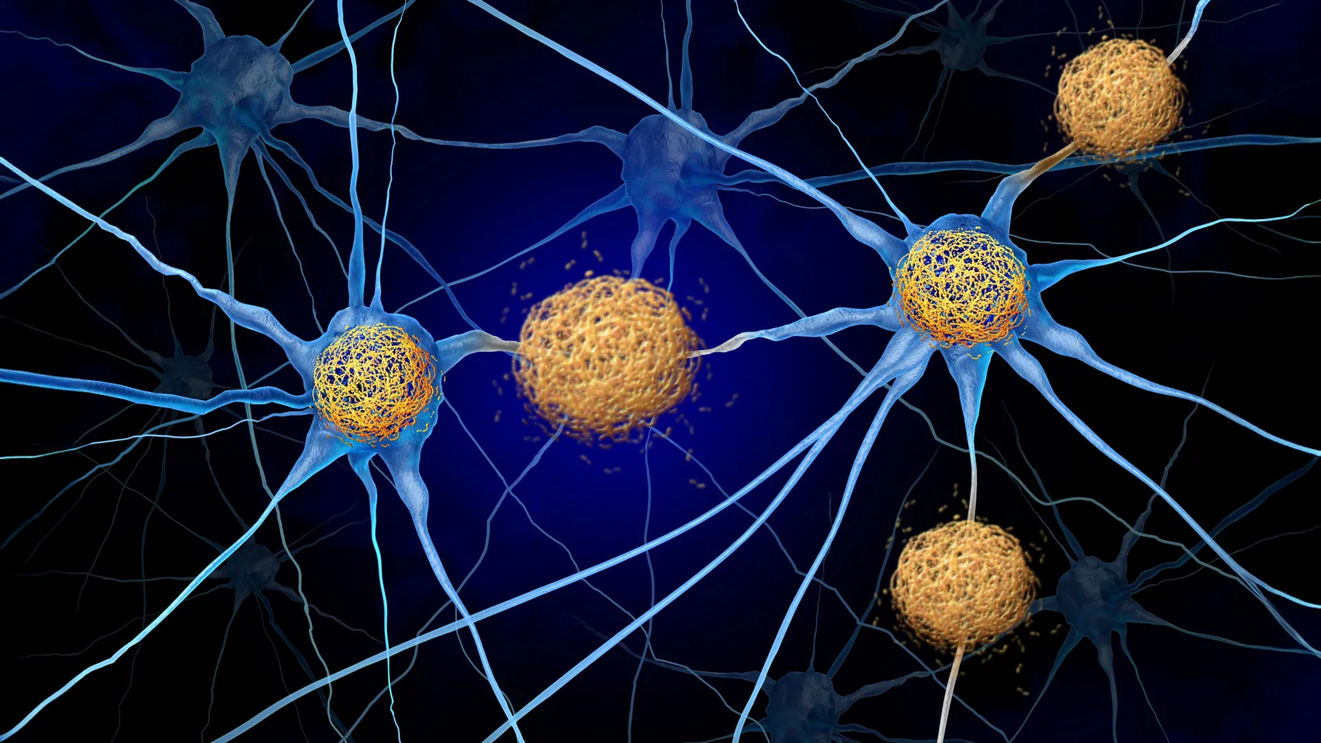
To visualize the phenomenon uncovered by the research team, one must consider the brain’s internal defense system. Microglia are essential, specialized immune cells tasked with clearing cellular debris and maintaining a stable neural environment. Under normal conditions, they are guardians of brain health. However, in the context of Alzheimer’s disease, these cells encounter amyloid plaques—sticky protein fragments that build up between nerve cells.
The study indicates that when microglia detect these plaques, they initiate an aggressive, non-specific inflammatory response. The researchers likened this to a building’s fire sprinkler system activating across an entire floor because of a small fire in a single kitchen. This excessive response releases a cascade of inflammatory cytokines, which, while intended to neutralize the plaques, ultimately destabilizes the neural circuits responsible for regulating circadian rhythms and sleep cycles. Essentially, the immune system is "partying all night," preventing the brain from transitioning into the deep, restorative stages of sleep necessary for cognitive health.
Chronology of the Investigation
The research was conducted through a multi-stage study that spanned several years, tracking mice at two critical developmental milestones: six months, when initial amyloid plaques appear, and 18 months, when the disease model shows advanced pathology. By utilizing a control group of "wild-type" mice, the team was able to differentiate between age-related sleep decline and pathology-specific disruption.
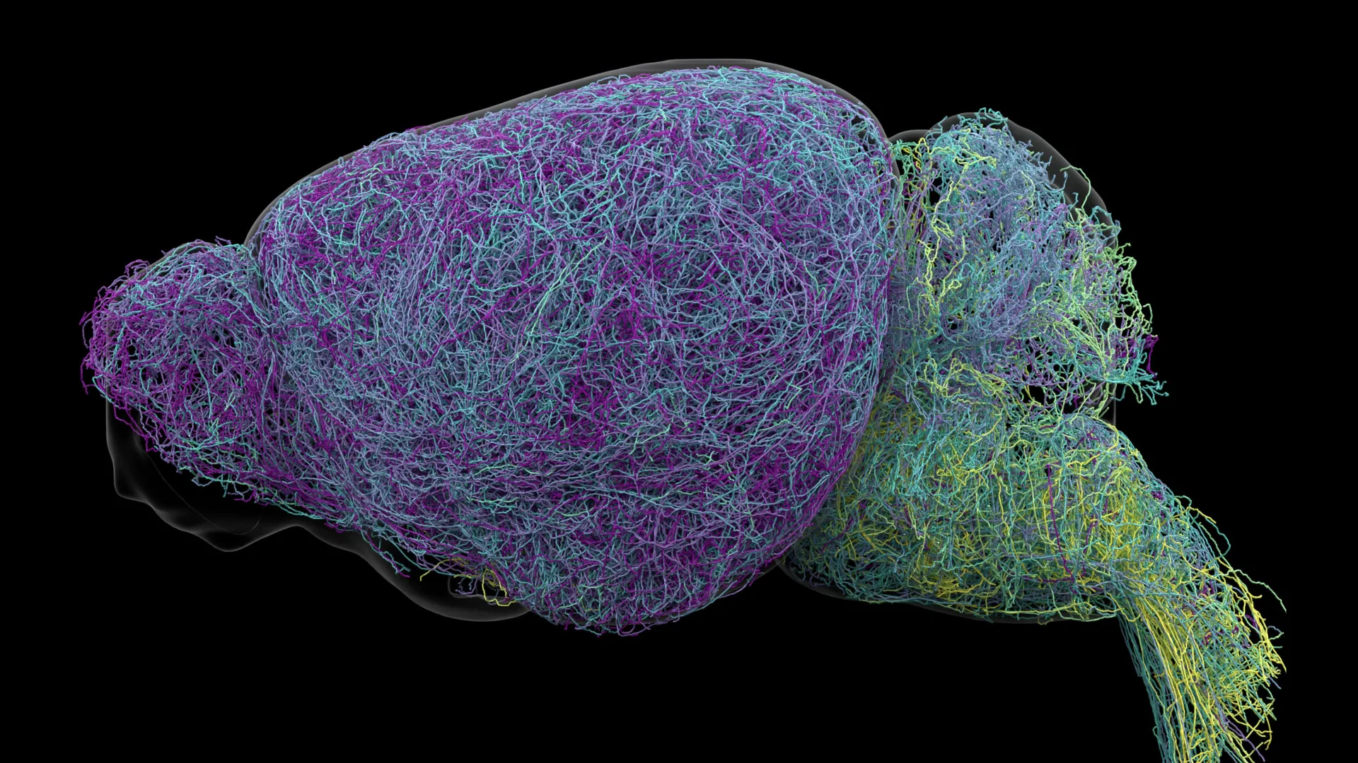
The team employed advanced technological apparatuses, including EEG (electroencephalography) and EMG (electromyography) sensors, to map the precise architecture of the mice’s sleep cycles. This was complemented by light-sheet microscopy, a sophisticated imaging technique that renders biological tissue transparent, allowing the researchers to generate detailed three-dimensional maps of how microglia and plaques were distributed throughout the brain.
A pivotal moment in the investigation occurred when the team utilized Pexidartinib (PLX3397), a pharmacological agent typically used in oncology, to temporarily deplete approximately 87% of the microglia in the mice. The results were instantaneous and statistically significant: the animals experienced a recovery of more than two hours of sleep per day, with marked improvements in the quality of NREM (non-rapid eye movement) sleep.
Analyzing the Data: The "Ceiling Effect"
One of the most counterintuitive findings of the study was the lack of correlation between the severity of plaque burden and the severity of sleep disruption. The researchers had hypothesized that as plaque accumulation increased over the 12-month interval, sleep patterns would continue to deteriorate. Instead, they observed a "ceiling effect."
"The disruptions in sleep and cortical EEG activity that occur at 6 months, when plaques first emerge, did not worsen by 18 months, despite more than double the amount of plaque burden," noted Dr. Constantino. This discovery implies that once the initial inflammatory threshold is crossed, the sleep-disrupting mechanism is fully engaged. Consequently, further accumulation of plaques does not necessarily accelerate the rate of sleep loss, suggesting that the primary window for intervention may be much earlier in the disease trajectory than previously assumed.
Furthermore, the study utilized the FOOOF (Fitting Oscillations and One Over Frequency) mathematical method to separate rhythmic brain waves from background electrical noise. This allowed the team to confirm that the brain remains in an "excited" state when it should be idle, much like a car engine left running at high RPMs despite being parked.
Clinical Implications and Future Therapeutic Strategies
The realization that sleep can be improved without clearing amyloid plaques is a massive shift in Alzheimer’s research, which has long been dominated by the "amyloid hypothesis." While the removal of plaques remains a goal for many pharmaceutical developments, the University of Kentucky study suggests that we might not need to remove the disease-causing proteins to manage its most debilitating symptoms.
The team is currently investigating existing, FDA-approved medications, such as Metformin (for diabetes) and various anti-seizure medications, to determine if they can modulate microglial metabolism. By "calming" the immune cells rather than eliminating them, researchers hope to dampen the inflammatory cascade while preserving the essential protective functions these cells provide to the nervous system.
Beyond treatment, the study highlights the potential of portable, affordable EEG technology as a diagnostic tool. Because the study successfully linked specific electrical signatures to Alzheimer’s pathology, the researchers believe that non-invasive, longitudinal monitoring via home-based EEG devices could eventually allow for earlier detection. This is particularly vital for underserved populations in rural areas, such as those in Kentucky, where access to expensive neuroimaging, like PET scans, is limited.
Perspectives from the Research Environment
The success of this study is largely attributed to the collaborative, risk-tolerant culture of the Macauley laboratory at the Sanders-Brown Center on Aging. Dr. Macauley, who fosters an environment where original hypotheses are frequently challenged, emphasizes the importance of following data where it leads, even if it contradicts the established "common knowledge" of the field.
"Some of the most interesting studies I have been a part of emerged because our original hypothesis was wrong," said Dr. Constantino. This mindset enabled the team to pivot from studying neurons to studying the broader immune environment of the brain—a shift that may well define the next generation of Alzheimer’s therapies.
Broader Impact and Expert Analysis
While the results in murine models are robust, the transition to human clinical trials remains the next great challenge. Experts in the field of neurodegeneration have noted that while the mouse model is an excellent tool for basic research, the complexity of the human brain—and the human immune system—is significantly greater. There is also the critical need to determine whether the long-term modulation of microglia will have unforeseen side effects, as these cells are integral to daily brain maintenance.
Despite these hurdles, the potential impact is profound. Poor sleep is not merely a symptom of Alzheimer’s; it is a contributor to the disease’s progression. Sleep is the time when the brain’s glymphatic system clears out metabolic waste. If sleep is chronically disrupted by immune-driven inflammation, the brain’s ability to clean itself is impaired, leading to a self-reinforcing cycle of decay. By breaking this cycle, clinicians may be able to preserve cognitive function for longer periods, significantly improving the quality of life for patients and their caregivers.
The research was supported by a series of grants from the National Institute on Aging and the National Institute of General Medical Sciences, as well as significant private funding from the Cure Alzheimer’s Fund and The CART Fund. As the team moves forward, their focus will remain on refining these drug-based interventions and validating their EEG biomarkers in human cohorts. The findings offer a rare spark of optimism in the fight against a disease that has historically proven resistant to traditional pharmacological approaches. If these results can be replicated in humans, the medical community may finally have a way to mitigate the devastating toll of Alzheimer’s by addressing the very cells that are meant to protect us.