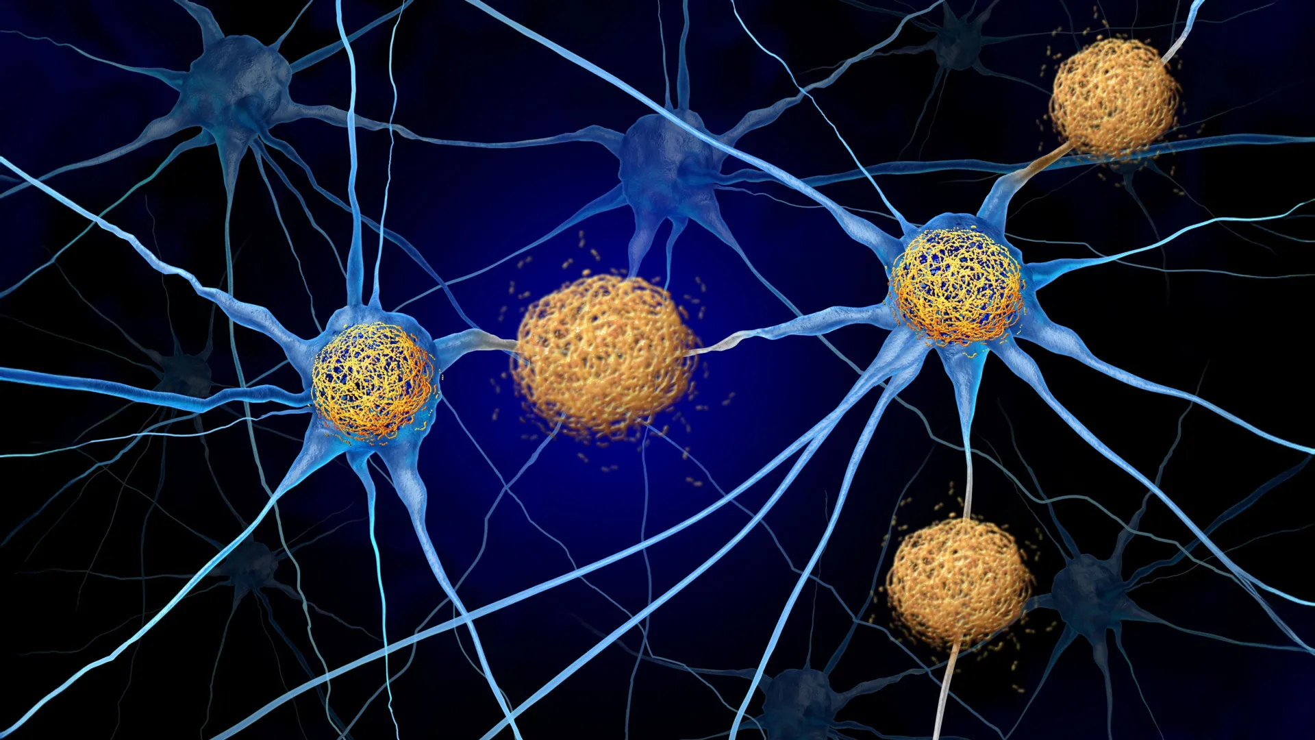

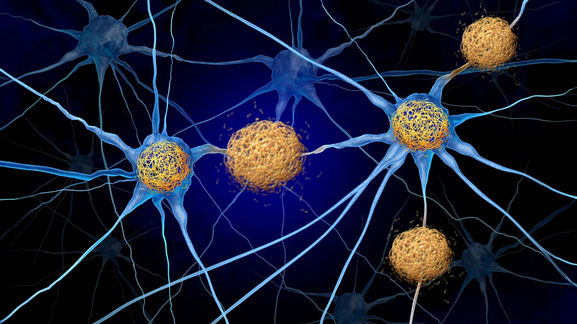
For decades, the central dogma of Alzheimer’s research has focused on the "protein-first" hypothesis. Scientists have tirelessly pursued methods to clear amyloid plaques and tau tangles, the hallmark protein aggregates that accumulate in the brains of those suffering from neurodegenerative decline. However, while recent breakthroughs like lecanemab and donanemab have demonstrated a capacity to slow cognitive decline by targeting these proteins, they have largely failed to halt the underlying death of neurons. A groundbreaking study published September 28 in the journal Neuron now suggests that the true culprit behind cognitive loss may not be the proteins themselves, but the body’s aggressive, misguided immune response to them.
A Paradigm Shift in Neuroimmunology
The research, led by Dr. David M. Holtzman, the Barbara Burton and Reuben M. Morriss III Distinguished Professor in the Department of Neurology at WashU Medicine, posits that tauopathies—a class of neurodegenerative diseases including Alzheimer’s, frontotemporal dementia, and progressive supranuclear palsy—are significantly exacerbated by the infiltration of T cells into the brain.
In a healthy state, the brain is an "immune-privileged" organ, shielded from the full brunt of the body’s systemic immune responses by the blood-brain barrier. However, in the context of tau-related neurodegeneration, this barrier appears to be compromised or bypassed. The research team identified that T cells, which are typically tasked with fighting infections, are recruited into the brain by a specific chemical "breadcrumb trail." By disrupting this trail, researchers were able to preserve memory and brain tissue in mice, even while the toxic tau tangles remained untouched.
The Chronology of Discovery
The path to these findings began several years ago, rooted in the evolving understanding of how the brain interacts with the peripheral immune system.
- 2023: A seminal study by the Holtzman lab, published in Nature, confirmed that T cells infiltrate the brains of mice with tau accumulation. These cells were found to cluster specifically in regions where tau pathology was most dense, suggesting a direct link between immune cell presence and localized tissue damage.
- Early 2024: A follow-up study published in Nature Neuroscience provided the missing piece of the puzzle, identifying that the signals directing these T cells to the brain originate in the lymph nodes located outside the cranium.
- September 2024: The current study in Neuron synthesized these findings, identifying the specific molecular pathway—the CXCL10/CXCR3 axis—responsible for guiding these cells. By blocking the CXCR3 receptor, the team effectively "blinded" the T cells to the signals drawing them into the brain, resulting in a 50% reduction of T cell infiltration and a 40% preservation of memory-related brain tissue.
The Mechanics of Immune Infiltration
The chemical signaling pathway identified by the researchers involves a protein called CXCL10, which acts as a beacon. Activated T cells express a receptor known as CXCR3, which allows them to "sense" CXCL10.
In mice models exhibiting Alzheimer’s-like tau pathology, the research team observed significantly elevated levels of CXCL10. This finding is consistent with data from human clinical studies, where high concentrations of CXCL10 have been observed in the cerebrospinal fluid of Alzheimer’s patients. When the researchers genetically removed either CXCL10 or the CXCR3 receptor, or blocked the receptor using an antibody, the T cells were unable to penetrate the brain. This remained true even when the researchers attempted to induce inflammation to trigger an immune response, proving that this specific pathway is the primary highway for T cell entry into the neurodegenerative brain.
Protecting Tissue Without Clearing Protein
Perhaps the most startling aspect of the study is the therapeutic outcome. In the trial, mice received the antibody treatment once every five days for three and a half months. At the conclusion of the study, the mice that received the therapy performed significantly better on standardized memory tests compared to the control group.
Crucially, histological analysis revealed that while the mice had better cognitive outcomes and more preserved brain tissue, the amount of tau protein in their brains remained identical to that of the untreated, cognitively impaired mice. This implies that the neurodegeneration witnessed in Alzheimer’s is largely a "bystander effect"—the neurons are dying not because the tau is inherently toxic to the cell membrane, but because the immune system is launching an inflammatory attack on cells that contain the tau.
Implications for Future Drug Development
The clinical implications of this research are substantial. One of the most significant barriers to developing CNS (central nervous system) drugs is the blood-brain barrier. Developing small molecules that can cross this selective membrane in sufficient concentrations to be therapeutic is both expensive and technically difficult.
Because the researchers found that blocking the CXCR3 receptor—which exists on T cells in the peripheral blood—prevented the cells from entering the brain, there may be no need for a drug to cross the blood-brain barrier at all. "For this therapeutic approach, if it is safe, you wouldn’t have to design the drug to get into the brain," Dr. Holtzman noted.
This opens a door to repurposing existing medications. Several drugs currently approved for autoimmune conditions like multiple sclerosis or inflammatory bowel disease target T cell migration and function. Scientists may be able to pivot these existing, well-understood medications into clinical trials for Alzheimer’s patients, potentially accelerating the timeline for a new class of treatments.
Expert Analysis and Future Outlook
The scientific community has received the findings with significant interest, as they challenge the traditional amyloid-tau-centric model. If these results can be replicated in human clinical trials, it would represent a fundamental shift in how the medical community defines Alzheimer’s. Currently, the disease is classified as a primary or secondary proteinopathy. This research suggests it should perhaps be viewed as a neuroimmunological disorder.
However, researchers urge caution. Mice models are imperfect mirrors of the human brain. The human immune system is significantly more complex, and the long-term effects of suppressing T cell entry into the brain—a process that might be a protective mechanism in other, as-yet-unknown contexts—remain to be studied.
"Tauopathies aren’t thought of as autoimmune disorders," Dr. Holtzman stated, "so they haven’t been treated the same way, but this study shows for the first time in an animal model that these diseases respond to a specific T-cell therapy."
The next steps for the WashU team involve determining the safety profiles of CXCR3-targeting antibodies in humans and identifying the specific triggers that cause the peripheral lymph nodes to start signaling to the brain in the first place. If they can confirm that the inflammatory response is indeed the primary driver of neuron loss, the focus of Alzheimer’s treatment could move away from the "uphill battle" of clearing protein deposits and toward the more manageable task of modulating the body’s systemic immune response.
This study was made possible by a wide coalition of supporters, including the National Institutes of Health, the GHR Foundation, and the Carol and Gene Ludwig Initiative in Neuroimmunology Research. While the road from a laboratory in St. Louis to a pharmacy shelf is long, the identification of a clear, targetable pathway that preserves brain tissue and memory offers a glimmer of hope for the millions of families worldwide affected by the encroaching shadows of neurodegeneration. By decoupling protein pathology from cognitive decline, researchers may have finally found the key to keeping the mind intact, even when the underlying disease processes are still present.