In a significant development for neurovascular medicine, researchers at the University of Rochester Medical Center have identified a novel therapeutic avenue for stroke recovery that hinges on the restoration of the body’s internal biological clock. Published in the Journal of Clinical Investigation, the study reveals that reinforcing circadian rhythms—the 24-hour internal cycles that govern physiological processes—can bolster the brain’s glymphatic system. By enhancing this waste-clearing network, the researchers observed improved motor function and reduced inflammation in animal models, even when interventions were introduced days after the initial neurological event. This finding challenges the traditional focus of stroke treatment, which has historically prioritized immediate, acute-phase interventions, by suggesting that the brain’s "housekeeping" mechanisms remain a viable target for recovery long after the injury has occurred.
The Chronology of Glymphatic Discovery
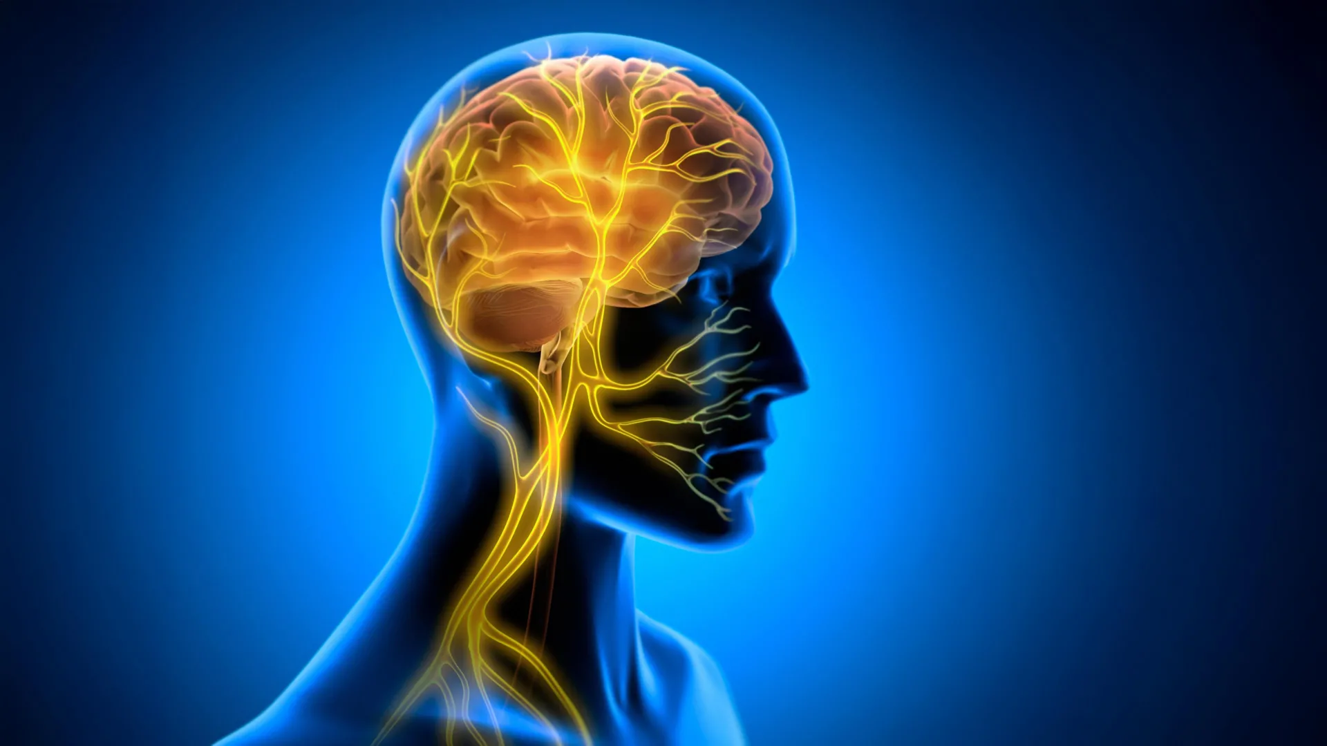
The significance of this research is rooted in a decade of pioneering work conducted by the laboratory of Maiken Nedergaard, MD, DMSc. In 2012, the Nedergaard lab fundamentally altered the scientific understanding of brain physiology by identifying the glymphatic system. This network acts as a perivascular pathway for cerebrospinal fluid (CSF), circulating through the brain to flush out metabolic waste, including proteins that would otherwise aggregate and cause neurodegeneration.
By 2015, subsequent research led by the group began to quantify the efficiency of this system, noting that glymphatic activity is most pronounced during deep sleep. The scientific narrative shifted further in 2020 when Lauren Hablitz, PhD, and her colleagues published a landmark study demonstrating that the glymphatic system is not merely a passive byproduct of sleep, but is actively gated by the circadian rhythm. This discovery provided the missing link between the body’s internal timing and the brain’s ability to maintain its own environment, establishing that the brain’s waste clearance follows a rhythmic pulse independent of sleep state alone.
Stroke as a Disorder of Biological Timing
For decades, the clinical approach to stroke has been framed as a vascular emergency, where the primary objective is the mechanical or chemical dissolution of a clot to restore blood flow. However, the University of Rochester team posits that the trauma of a stroke extends beyond vascular occlusion, functioning as a "disorder of timing."
Clinical data has long observed that stroke incidence is not random; it exhibits distinct diurnal patterns, with a higher frequency of ischemic events occurring in the early morning hours. Furthermore, the recovery period is frequently plagued by severe sleep-wake disturbances, which are statistically correlated with poorer functional outcomes, increased rates of depression, and a lower quality of life for survivors. Hablitz and her team hypothesized that if the brain’s internal clock is disrupted by the injury, then the restorative "cleanup" phase is effectively stalled.
In a healthy state, the glymphatic system serves as a conduit for clearing pro-inflammatory cytokines and metabolic debris. Following a stroke, the brain experiences an accumulation of these harmful molecules, which may sustain secondary damage and impede neuroplasticity. The team’s hypothesis suggests that the post-stroke brain is not necessarily incapable of healing, but is suffering from a failure of the waste-management system, leading to a toxic buildup that hampers recovery.
Experimental Interventions and Data Outcomes
To test whether re-synchronizing the biological clock could mitigate these effects, the researchers implemented a series of interventions designed to entrain the circadian rhythm in mice. The methodology involved several approaches: timed exposure to light, the administration of melatonin, the use of the synthetic clock-targeting drug KL001, and the implementation of time-restricted feeding.
The experimental design was particularly rigorous in its timing. While most stroke therapies, such as the administration of tissue plasminogen activator (tPA), are restricted to a narrow therapeutic window—often just a few hours post-stroke—the researchers delayed their interventions until 72 hours after the injury. Despite this delay, the results were statistically significant.
Mice that underwent time-restricted feeding or received KL001 showed a marked improvement in motor recovery compared to control groups. Diagnostic imaging revealed smaller lesion volumes, suggesting that the reduction in inflammatory markers was not merely symptomatic but protective of brain tissue. Measurements of glymphatic flow showed a measurable increase in fluid movement, which in turn correlated with a decrease in the presence of inflammatory cytokines. "All of the cytokines moved in the same direction," Dr. Hablitz noted, suggesting a systemic improvement in the brain’s clearing efficiency rather than an isolated effect on a single inflammatory pathway.
The Potential for Accessible Rehabilitation
One of the most compelling aspects of these findings is the potential for clinical translation through non-invasive, behavioral interventions. While drugs like KL001 represent a pharmaceutical future, the success of time-restricted feeding is particularly notable for its accessibility. If similar outcomes can be observed in human clinical trials, the implications for rehabilitation medicine could be transformative.
Current stroke rehabilitation often requires intensive, facility-based care. The ability to support recovery through dietary timing or light exposure therapy would provide clinicians with tools that are both low-cost and easily implemented within the home environment. This could theoretically allow for a more continuous, long-term recovery strategy that extends beyond the initial hospitalization phase.
Implications for Future Neurological Research
The implications of the Rochester study extend well beyond stroke. The glymphatic system is increasingly viewed as a central pillar of brain health, with its dysfunction linked to conditions ranging from Alzheimer’s disease and Parkinson’s to traumatic brain injury. If the brain’s waste-clearing system is a primary target for neurological recovery, understanding how to manipulate that system via circadian rhythms could unlock new protocols for treating a wide array of neurodegenerative and neuroinflammatory diseases.
However, the research team remains cautious, emphasizing that these results are currently confined to animal models. The next phase of research will focus on the precise mechanism of action: determining how the internal clock signals the glymphatic system to accelerate or decelerate, and verifying whether these findings hold true in the complex, polygenic, and often co-morbid environment of human stroke patients.
The study reflects a fundamental shift in neuroscience, moving away from a siloed view of the brain as a static organ toward a dynamic understanding of it as an integrated system dependent on temporal rhythms. By prioritizing the restoration of these rhythms, clinicians may soon be able to leverage the body’s own biological architecture to promote healing. As the scientific community looks toward future clinical trials, the goal remains clear: to transition from merely preventing damage to actively facilitating the brain’s innate ability to clear debris and reconstruct neural pathways after injury.
This research underscores that the road to recovery may be paved with better sleep, consistent schedules, and the disciplined maintenance of our internal biology. As neuroscientists continue to map the interplay between the circadian clock and the glymphatic network, the potential for non-pharmacological, time-based interventions suggests a new, more holistic chapter in modern neurology.