The fundamental challenge of molecular biology has long been the paradox of DNA interaction. Because DNA molecules possess a pervasive negative electrical charge, they inherently repel one another, much like the opposing poles of two magnets. Yet, within the crowded, bustling environment of the living cell, DNA must consistently navigate this repulsion to find its exact matching sequences. This ability to recognize and pair with specific strands is the cornerstone of genetic recombination, DNA repair, and the complex process of gene silencing. When these mechanisms fail or go awry, the resulting genomic instability is often a precursor to oncogenesis and the development of various cancers.
A Two-Decade Quest for the DNA Zipper
The scientific community has long theorized about how this "repulsion barrier" is breached. In the early 2000s, Professor Alexey Kornyshev of Imperial College London and his research partners proposed the "DNA zipper" model. This theoretical framework suggested that salt ions—electrically charged particles naturally occurring in cellular fluids—do not simply float aimlessly around DNA. Instead, they were thought to organize into alternating patterns of positive and negative charges along the grooves of the DNA helix. According to this model, these charged patterns would act as a structural bridge, allowing neighboring DNA molecules to lock together like the teeth of a zipper or two interlocking spiral staircases.
For twenty years, this remained an elegant but unverified hypothesis. The primary obstacle was the sheer scale of the interaction; the molecular dance occurs at a level of detail that traditional microscopy could not capture. However, a collaborative team of researchers from the University of York, the University of Sheffield, and Imperial College London has now successfully bridged the gap between theory and observation. By integrating cutting-edge atomic force microscopy (AFM) with high-resolution molecular dynamics simulations, the team has provided the first direct experimental evidence for the DNA zipper mechanism.
The Methodology of Molecular Visualization
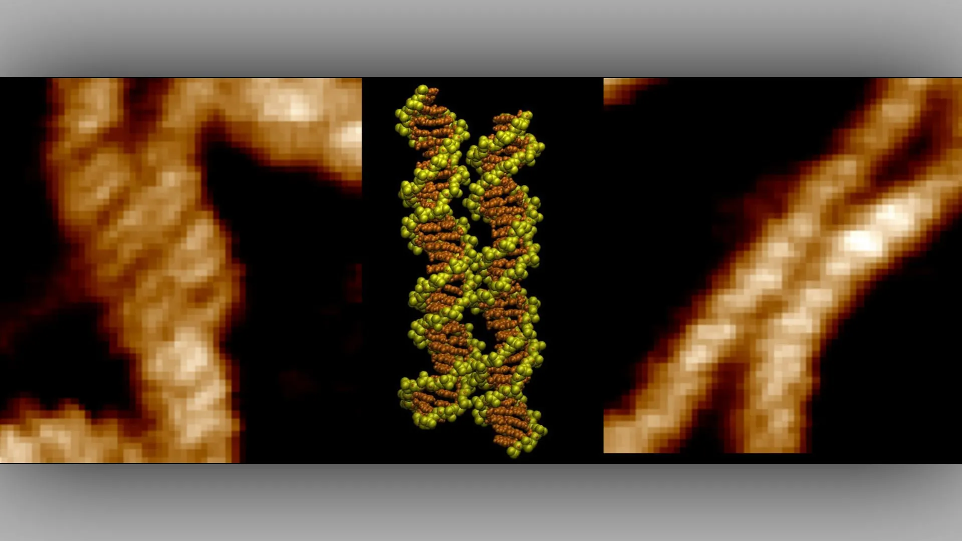
The investigation, recently published in the journal Nucleic Acids Research under the title "Imaging and mechanism of DNA-DNA recognition mediated by divalent ions," employed a dual-track approach. First, the team utilized atomic force microscopy to map the physical topography of DNA samples. AFM functions by scanning a physical probe over a sample, allowing researchers to visualize molecules at the nanometer scale. Through this process, the researchers captured high-resolution imagery of short DNA fragments aligning with extraordinary precision, essentially matching one another "groove for groove."
However, imagery alone cannot explain the forces at play. To understand the "why" behind the "what," the team turned to sophisticated computer simulations. These simulations tracked the movement of individual atoms and ions within the proximity of the DNA strands. The data revealed that divalent metal ions—ions carrying a double positive charge—act as essential mediators.
The simulations demonstrated that these metal ions settle into the grooves of the DNA, effectively serving as molecular bridges. By interacting with both DNA molecules simultaneously, these ions act like charged arms that pull the strands together and stabilize their alignment, overriding the natural repulsion that would otherwise drive them apart. This discovery transforms the DNA zipper from a theoretical construct into a physically observed biological reality.
Expert Perspectives and Scientific Implications
The significance of this study extends far beyond the resolution of a long-standing academic debate. By identifying the specific role of divalent ions in facilitating DNA recognition, the research team has opened new avenues for understanding genomic health.
Professor Agnes Noy of the University of York, who co-led the research, emphasized the clinical potential of these findings. "This discovery could help researchers identify regions of the genome specially involved in DNA pairing," Professor Noy stated. "These regions may become particularly important when mutations disrupt normal cellular processes and contribute to cancer."
The ability to map these "pairing hotspots"—sequences where DNA binds with greater affinity due to the ion-bridging effect—could revolutionize how scientists look at genetic instability. When the delicate balance of DNA recognition is disturbed, the cell may fail to repair breaks in its genetic code or may misinterpret sequences, leading to the mutations that drive tumor growth.
Dr. Thomas Catley, a co-lead author from the University of Sheffield’s School of Chemical Materials and Biological Engineering, reflected on the milestone: "It was incredible to be able to directly visualize the long-hypothesized mechanism for the first time. The advanced imaging techniques at our disposal are allowing us to uncover these key DNA interactions which have implications in many key cellular processes."
Dr. Victor Velasco-Berrelleza, also of the University of Sheffield, underscored the importance of the interdisciplinary nature of the work. "Microscopy shows us what happens, but the simulations allow us to uncover the molecular mechanism behind it," he noted. The synergy between physical imaging and computational modeling is increasingly becoming the gold standard for structural biology, allowing researchers to move from descriptive science to predictive science.
Broader Impact: From Oncology to Biotechnology
The implications of this research are broad, spanning from medical diagnostics to the emerging field of synthetic biology. In the realm of oncology, understanding the mechanics of DNA pairing could lead to the development of novel therapeutic strategies. If researchers can pinpoint sequences that are hyper-susceptible to improper pairing, they may be able to design small-molecule inhibitors that prevent cancer-promoting genetic interactions.
Furthermore, the discovery has significant potential in the field of nanotechnology and biotechnology. Because specific DNA sequences have been shown to pair with varying degrees of strength, scientists can now treat these sequences as programmable components. By engineering specific DNA structures that utilize this ion-mediated "zipper" mechanism, bioengineers could develop sophisticated molecular scaffolds, biosensors, or drug-delivery vehicles that self-assemble with unprecedented accuracy.
The study also provides a necessary foundation for future investigations into how the cellular environment—such as the concentration of different salt ions—modulates gene expression. Since the strength of the DNA interaction depends on the presence of these divalent ions, changes in the ionic composition of the cell could theoretically alter how genes are regulated, providing a new layer of epigenetic control that warrants further study.
A New Chapter in Genomic Understanding
The transition from a two-decade-old hypothesis to a confirmed molecular mechanism represents a significant victory for the collaborative scientific process. By confirming the DNA zipper model, the research team has not only satisfied a curiosity in structural biology but has also provided a functional map for exploring the most intimate interactions of the human genome.
As the scientific community digests these findings, the focus will likely shift toward the functional analysis of the identified "hotspots." If these sites of high-affinity pairing are indeed the focal points of cellular miscommunication in disease, they represent high-value targets for future drug discovery.
The successful integration of atomic force microscopy and atomic-level simulation serves as a blueprint for future studies in the life sciences. As imaging technology continues to improve, and as computational power grows, the ability to observe the microscopic world with such precision will likely continue to demystify the complex, often counter-intuitive processes that allow life to function at the molecular level. For now, the "DNA zipper" stands as a testament to the fact that even the most invisible, counter-intuitive forces of nature can be mastered once the right tools are applied.