At the fundamental level of biology, the architecture of life is governed by a persistent paradox: DNA carries a strong negative electrical charge, a property that should, by all laws of physics, force DNA molecules to repel one another. Yet, within the confined, crowded, and chaotic environment of the living cell, DNA must frequently navigate this repulsion to find its matching sequences. This precise alignment is not merely a biological curiosity; it is the cornerstone of genetic recombination, DNA repair, and gene silencing. When these processes falter, the results can be catastrophic, often leading to the genomic instability associated with cancer.
For decades, the mechanism behind this "molecular recognition" remained largely theoretical. Now, a collaborative team of researchers has successfully visualized this process, providing the first direct evidence for a phenomenon long dubbed the "DNA zipper." By combining high-resolution atomic force microscopy (AFM) with state-of-the-art computational simulations, the team has demonstrated how positively charged metal ions act as bridges, effectively neutralizing the electrostatic barrier and allowing DNA strands to interlock with extraordinary precision.
A Two-Decade Quest for the DNA Zipper
The history of this discovery dates back approximately twenty years to a hypothesis proposed by Professor Alexey Kornyshev of Imperial College London. Kornyshev and his colleagues theorized that the secret to DNA pairing lay in the surrounding salt environment. They suggested that divalent metal ions—positively charged atoms such as magnesium or calcium—could arrange themselves along the DNA backbone in specific, repeating patterns.
According to the "DNA zipper" model, these ions create a periodic distribution of electrical charges that complementary DNA sequences recognize. Much like two spiral staircases being joined by interlocking steps, these ions allow two separate helices to sit comfortably against one another despite their inherent negative charge. For nearly two decades, however, this model remained an elegant, albeit unproven, mathematical construct. The difficulty in testing the theory stemmed from the extreme scales involved; DNA helices are only about two nanometers wide, and the interactions between ions and DNA happen on the scale of picoseconds and angstroms, far beyond the reach of standard light microscopy.
Integrating Advanced Microscopy and Molecular Dynamics
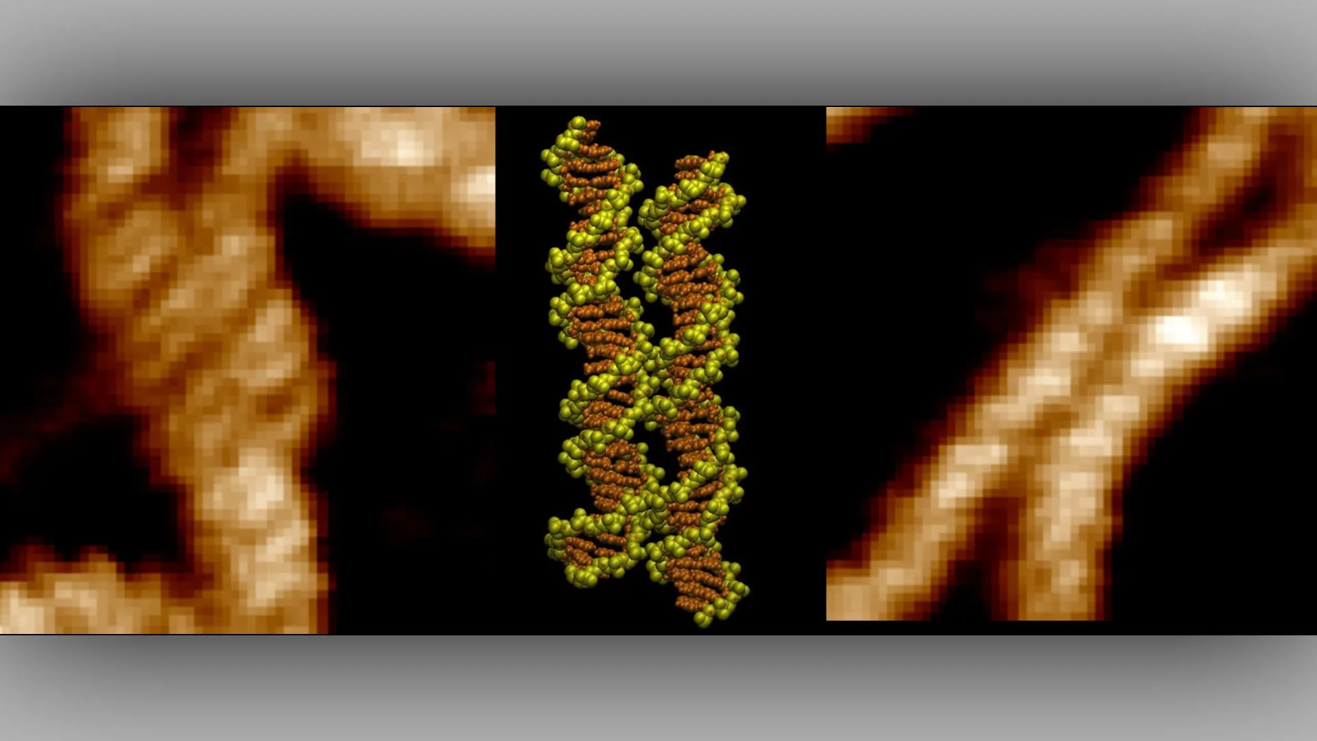
The breakthrough was achieved through a dual-pronged approach that married experimental observation with theoretical modeling. The study, published in the journal Nucleic Acids Research under the title "Imaging and mechanism of DNA-DNA recognition mediated by divalent ions," utilized atomic force microscopy to map the physical topography of DNA samples. By scanning the surface of the DNA at an atomic level, the researchers were able to capture images of the strands in close contact, revealing that they align "groove for groove."
To explain how these physical configurations were achieved, the team turned to molecular dynamics simulations. Dr. Victor Velasco-Berrelleza of the University of Sheffield, who led the simulation efforts, utilized computational power to track the movement of individual atoms and ions in real-time. The simulations revealed that divalent metal ions do not simply surround the DNA randomly. Instead, they act as active facilitators. A single, double-charged ion can reach out to both DNA molecules simultaneously, acting as a molecular "bridge" or clamp. This reduces the energy barrier that would otherwise push the strands apart, creating a stable, temporary union.
"Microscopy shows us what happens, but the simulations allow us to uncover the molecular mechanism behind it," Dr. Velasco-Berrelleza explained. This synthesis of data provided a holistic view of the interaction that neither method could achieve in isolation.
Supporting Data and the Discovery of Pairing Hotspots
One of the most significant findings of the study is that DNA pairing is not a uniform process across the entire genome. The researchers identified specific "hotspots" where DNA helices show a much higher propensity to align. This suggests that the genetic code itself, through its sequence of base pairs, influences how ions interact with the molecular backbone.
When the sequence of the DNA changes, the resulting topography of the groove changes as well. This dictates where the metal ions can settle, effectively creating a "code" for recognition. This discovery has profound implications for our understanding of genetic regulation. It suggests that the cell may have evolved to use these ionic bridges to "scan" for specific sequences, a process that is essential for the rapid identification of homologous DNA during repair cycles.
The data collected by the team indicates that when these ionic bridges are disrupted—whether through changes in salt concentration or mutations in the DNA sequence itself—the efficiency of recombination drops significantly. This quantitative insight provides a baseline for future studies looking at how environmental factors or cellular stress might derail genetic maintenance.
Official Reactions and Scientific Significance
The research team, led by Professor Agnes Noy from the University of York’s School of Physics, Engineering and Technology, alongside Dr. Thomas Catley from the University of Sheffield, emphasizes that this is a milestone in structural biology.
"It was incredible to be able to directly visualize the long-hypothesized mechanism for the first time," said Dr. Catley. "The advanced imaging techniques at our disposal are allowing us to uncover these key DNA interactions which have implications in many key cellular processes."
Professor Noy noted the broader implications for human health, stating, "This discovery could help researchers identify regions of the genome specially involved in DNA pairing. These regions may become particularly important when mutations disrupt normal cellular processes and contribute to cancer."
The scientific community has received these findings as a long-awaited validation of a foundational theory in biophysics. By proving that the "DNA zipper" is a physical reality, the study provides a new lens through which to view genome stability. Researchers in oncology and genetic medicine are now expected to investigate whether specific cancer-prone regions of the genome exhibit altered ionic binding properties, potentially opening new avenues for diagnostic markers.
Broader Implications: From Biotechnology to Medicine
The discovery that DNA can be programmed to pair through ionic interactions has utility that extends well beyond fundamental biology. In the field of DNA nanotechnology, scientists have long sought ways to create self-assembling structures. If DNA sequences can be engineered to pair preferentially in the presence of specific ionic environments, it could allow for the construction of "smart" materials—structures that only assemble or change shape when triggered by specific metal ions.
This has immediate potential for drug delivery systems, where a synthetic DNA carrier could be designed to hold its payload until it reaches an environment with a specific ionic charge, triggering a structural shift that releases the medication.
Furthermore, the work highlights the importance of the ionic environment inside the cell. While biology textbooks often focus on proteins and enzymes as the primary actors in cellular processes, this research underscores the critical, often overlooked role of ions and electrostatic forces. The "DNA zipper" is a reminder that the cell’s internal environment is a highly tuned chemical landscape where the movement of even the smallest ions can dictate the fate of the entire genome.
Future Directions
As the team moves forward, the focus will likely shift toward observing these interactions within the much more complex environment of the cell nucleus, which is crowded with histones, proteins, and other cellular machinery. While the current study successfully modeled the process in a controlled, in vitro environment, determining how these ionic bridges compete with protein-DNA interactions remains a vital frontier.
The successful integration of AFM imaging and molecular simulation has set a new standard for how researchers investigate sub-cellular phenomena. By bridging the gap between theoretical physics and experimental biology, this study not only confirms a twenty-year-old hypothesis but also provides the tools necessary to decode the complex, charge-mediated language that governs the life and health of every cell. As researchers continue to map the genome, the "zipper" mechanism may prove to be one of the most essential, if invisible, tools that life uses to maintain its integrity.