
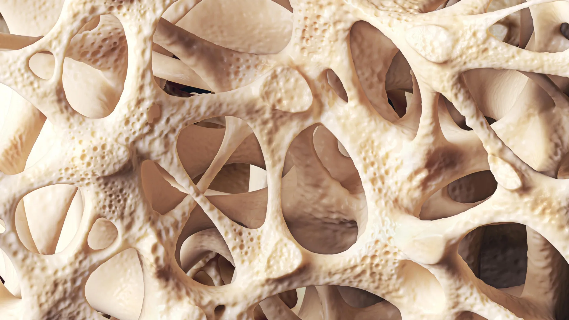
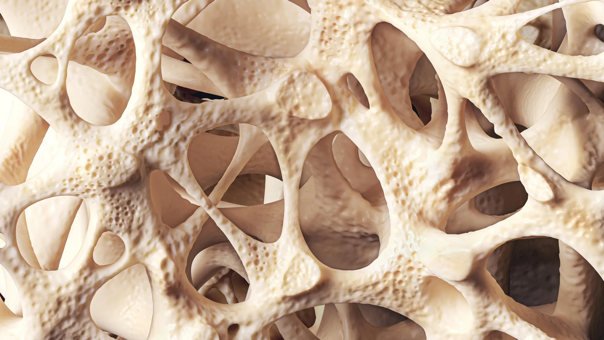
Around 40% of adults worldwide are affected by osteopenia, a prevalent and often silent medical condition defined by a reduction in bone mineral density that falls below normal levels yet has not crossed the diagnostic threshold into full-blown osteoporosis. This widespread public health issue disproportionately impacts postmenopausal women and elderly populations, laying bare the vulnerability of aging skeletal systems globally. In the United Kingdom alone, health statistics indicate that more than 500,000 fractures occur annually as a direct consequence of low bone density, creating a massive financial and operational strain on healthcare systems. Despite these striking figures, osteopenia frequently evades early detection. Because the condition develops gradually without obvious physical symptoms, millions of individuals remain entirely unaware of their compromised bone architecture until they suffer an unexpected fracture or undergo a specialized diagnostic scan triggered by age or menopausal status.
The Lifespan Chronology of Skeletal Mass
To understand how osteopenia develops, medical researchers examine the biological timeline of human bone tissue. Bone is not an inert structural frame; rather, it is a dynamic, living tissue that undergoes continuous renewal through a finely tuned metabolic process known as bone remodeling. Throughout this cycle, specialized cells break down old or damaged bone tissue—a phase called resorption—while other cells synthesize new bone matrix to replace it.
During childhood, adolescence, and early adulthood, the bone remodeling process is heavily skewed toward formation. Bone mass accumulates rapidly, reaching its highest level—known medically as peak bone mass—typically during a person’s mid-20s to early-30s. This peak serves as the structural savings account for the remainder of life. Once this biological milestone is passed, however, the physiological balance shifts. Gradual bone loss begins to outpace bone formation year after year. While this age-related decline is a natural facet of human physiology, various secondary catalysts can dramatically accelerate the process, transforming a normal aging trajectory into a pathological vulnerability like osteopenia.
Primary Risk Factors and Accelerants of Bone Breakdown
Aging remains the single most influential risk factor for bone mineral density loss, but a constellation of hormonal, genetic, and lifestyle factors can drastically worsen the condition. Foremost among these is the hormonal shift accompanying menopause. The sharp decline in estrogen levels significantly impairs the body’s ability to protect skeletal integrity. Estrogen acts as a natural shield for bones by suppressing the activity of osteoclasts, the cells responsible for bone resorption. When estrogen levels plummet, this protective brake is released, accelerating bone breakdown. Consequently, medical data shows that approximately one in two women over the age of 50 will experience a debilitating fragility fracture in their remaining lifetime.
Beyond biology and endocrinology, lifestyle choices exert a profound influence over bone strength. Chronic habits such as cigarette smoking, excessive alcohol consumption, and prolonged physical inactivity deprive the skeleton of the mechanical stress and chemical balance it needs to maintain density. Nutritional deficiencies compound these risks. Insufficient dietary calcium intake and widespread vitamin D deficiencies starve the body of the raw materials required to mineralize and maintain strong bone architecture.
Furthermore, secondary medical conditions and specific therapeutic interventions can severely disrupt skeletal health. Long-term use of corticosteroid medications is well-documented to impair bone formation and increase resorption. Similarly, chronic gastrointestinal and autoimmune disorders that hinder nutrient absorption—such as Crohn’s disease and celiac disease—frequently place patients at an elevated risk of premature bone thinning.
Diagnostic Frameworks and Clinical Detection
Because osteopenia produces no overt clinical symptoms in its early stages, identification relies heavily on proactive medical screening and quantitative imaging. Healthcare professionals typically evaluate patients using a dual-energy X-ray absorptiometry (DXA) scan. This low-dose radiological examination measures bone mineral density at key weight-bearing sites, such as the hip and spine.
The output of a DXA scan is expressed as a T-score, a statistical measure comparing the patient’s bone density to the normative data of a healthy young adult of the same sex. According to World Health Organization criteria, a T-score falling between −1.0 and −2.5 indicates osteopenia. A score dipping below −2.5 crosses the threshold into osteoporosis.
To refine clinical decision-making, physicians rarely rely on T-scores in isolation. Instead, they utilize advanced fracture risk assessment tools, such as the FRAX algorithm, which calculates a patient’s ten-year probability of suffering a major osteoporotic fracture. This calculation integrates multiple variables, including age, body mass index, personal and family fracture history, smoking status, glucocorticoid usage, and secondary clinical disorders. This multi-factorial approach ensures that interventions are tailored precisely to individual risk profiles rather than blanket diagnostic cutoffs.
Modern Management Strategies: Lifestyle, Nutrition, and Pharmacotherapy
When osteopenia is identified, the primary clinical objective is to arrest or significantly slow further bone loss while minimizing the lifetime risk of fractures. Management strategies are multifaceted, combining targeted lifestyle modifications, nutritional interventions, and, in select high-risk cases, pharmaceutical treatments.
Physical activity stands as a cornerstone of non-pharmacological management. Weight-bearing exercises—such as brisk walking, dancing, and light jogging—subject the skeleton to controlled mechanical loading, which stimulates bone-forming cells to deposit new matrix. Resistance training further fortifies both bone and surrounding musculature. Moreover, specialized movement practices like Tai Chi have been shown in clinical studies to enhance dynamic balance and lower-limb strength, directly reducing the incidence of falls that often precipitate severe fractures in older adults.
Nutritional optimization is equally critical. Adequate dietary calcium intake provides the foundational mineral for bone structure, while vitamin D acts as an essential hormonal regulator that enables the intestinal tract to absorb calcium efficiently. Dairy products, dark leafy greens, and fortified foodstuffs are standard dietary recommendations. However, in regions with limited natural sunlight—such as the United Kingdom—public health authorities frequently recommend daily vitamin D supplementation across entire demographic segments to combat widespread deficiency.
When lifestyle measures prove insufficient or when a patient’s calculated fracture risk is exceptionally high, clinicians may turn to pharmacological interventions. Antiresorptive medications, most notably bisphosphonates, are frequently deployed to inhibit osteoclast activity, thereby preserving existing bone density. While these pharmaceutical agents are traditionally prescribed for established osteoporosis, growing clinical consensus supports their selective use in high-risk osteopenic patients who exhibit rapid bone loss or additional compounding vulnerability factors.
Societal Implications and the Paradigm Shift in Bone Health
Public health experts and medical societies increasingly emphasize that osteopenia should not be dismissed as an inevitable, benign consequence of growing older. Rather, it must be reframed as a critical window of clinical opportunity—a physiological early-warning system that allows patients and healthcare providers to intervene well before severe structural damage occurs.
The progression from osteopenia to osteoporosis is far from guaranteed. Emerging clinical research underscores that early detection, coupled with persistent lifestyle adjustments and proactive medical management, can successfully stabilize bone mineral density. In some favorable cases, patients even achieve measurable improvements in skeletal strength.
Ultimately, safeguarding lifelong bone health requires a paradigm shift toward a lifelong perspective. Skeletal strength at age 70 is the cumulative product of nutrition, physical activity, hormonal milestones, and health behaviors maintained across decades. By recognizing osteopenia as a manageable public health priority rather than an unavoidable destiny, modern medicine can drastically reduce the personal and societal toll of fragility fractures in an aging global population.