Around 40% of adults worldwide are affected by osteopenia, a prevalent medical condition characterized by a measurable loss of bone mineral density that falls short of the diagnostic threshold for osteoporosis. While it frequently flies under the radar of public health awareness, its societal and personal implications are profound. In the United Kingdom alone, health authorities estimate that more than 500,000 fractures occur annually due to low bone density, placing a heavy and sustained burden on emergency departments, orthopedic services, and broader healthcare systems. Despite these striking statistics, osteopenia remains widely misunderstood, often dismissed as an inevitable consequence of aging rather than a critical window for medical intervention.
Unlike acute conditions that announce themselves with sudden pain or overt distress, osteopenia develops silently and progressively over many years. The vast majority of individuals who live with the condition experience no physical symptoms whatsoever. Consequently, many remain entirely unaware of their compromised skeletal health until they suffer a fragility fracture from a minor fall or undergo a routine bone density screening. These screenings are typically recommended only when clinicians identify baseline risk factors, such as advanced age or the onset of menopause. This asymptomatic nature transforms osteopenia into an insidious public health challenge, requiring proactive medical vigilance to catch and manage before severe complications arise.
To comprehend how osteopenia develops, one must examine the dynamic nature of human bone tissue. Far from being an inert, static structure, bone is a living matrix that undergoes continuous, lifelong renewal through a biological mechanism known as bone remodeling. This finely tuned cycle involves two opposing cellular processes: bone resorption, wherein specialized cells break down and remove old or damaged bone tissue, and bone formation, wherein other cells synthesize new bone matrix to replace it.
The Chronology of Skeletal Development: From Peak Mass to Gradual Decline
The human skeletal system follows a predictable chronological trajectory across the lifespan, dictating our vulnerability to bone loss. During childhood and adolescence, bone formation significantly outpaces resorption, allowing the skeleton to grow rapidly in both size and density. This physiological momentum continues into early adulthood, a critical phase when bone mass reaches its absolute peak—a milestone scientifically referred to as peak bone mass.
For most healthy adults, peak bone mass is achieved somewhere between their mid-20s and early-30s. This period represents the pinnacle of skeletal strength, acting as a biological savings account that must sustain an individual for the remainder of their life. Once this peak is attained, the delicate equilibrium of the bone remodeling cycle shifts. Gradually, the rate of bone resorption begins to outpace bone formation.
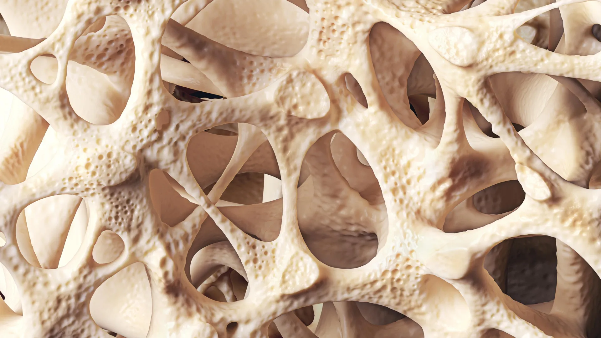
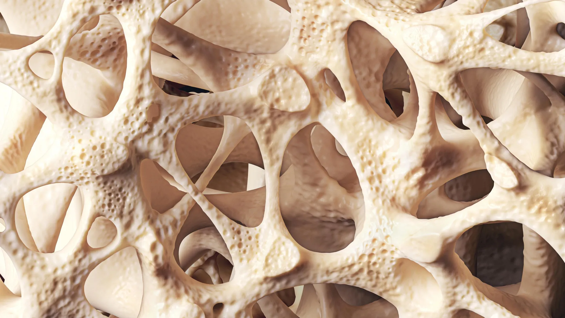
As individuals transition into middle age and beyond, this micro-architectural imbalance accumulates. Year by year, the microscopic cavities within the trabecular (spongy) bone widen, and the outer cortical bone thins. When the net loss of bone mineral density becomes pronounced enough to weaken the skeleton—yielding a T-score between −1.0 and −2.5 on a standard diagnostic scan—clinicians classify the condition as osteopenia. If this gradual erosion continues unchecked, bone density crosses the threshold of −2.5, transitioning into osteoporosis, where the risk of debilitating fractures multiplies exponentially.
The Multi-Factorial Etiology: Why Bone Density Declines
While chronological aging serves as the primary driver of bone density reduction, a complex web of biological, hormonal, and environmental factors can severely accelerate the process. Medical researchers and endocrinologists emphasize that bone health is a cumulative record of an individual’s lifetime exposures, nutritional habits, and genetic predispositions.
Hormonal shifts represent one of the most powerful catalysts for accelerated bone breakdown. This is most vividly demonstrated during the menopausal transition in women. Estrogen plays a vital, protective role in skeletal health by regulating the lifespan and activity of osteoclasts—the cells responsible for bone resorption. As estrogen levels drop sharply during and after menopause, this protective brake is removed, triggering a rapid phase of bone loss. Epidemiological data indicates that roughly one in two women over the age of 50 will experience a fragility fracture in their remaining lifetime, heavily driven by this postmenopausal surge in bone resorption.
Beyond hormonal dynamics, lifestyle choices exert a profound influence over long-term bone strength. Chronic physical inactivity robs the skeleton of the mechanical loading it requires to stimulate bone-forming cells. Conversely, habits such as cigarette smoking and excessive alcohol consumption directly impair osteoblast function, poisoning the bone-building machinery and reducing overall mineral density.
Nutritional adequacy is equally fundamental. Calcium serves as the primary structural mineral deposited within the bone matrix, while vitamin D acts as the vital biochemical key that unlocks the intestines’ ability to absorb dietary calcium. Insufficient intake of either nutrient cripples the body’s capacity to maintain robust skeletal architecture. In northern latitudes like the UK, where sunlight exposure is seasonal and vitamin D deficiency is relatively common, public health bodies frequently advise routine supplementation to bridge the nutritional gap.
Furthermore, secondary factors can complicate skeletal health. Certain pharmacological interventions, most notably long-term systemic corticosteroid therapy used to treat inflammatory and autoimmune diseases, are notorious for degrading bone density. Concurrently, underlying gastrointestinal and metabolic conditions that impair nutrient absorption—such as Crohn’s disease, ulcerative colitis, and coeliac disease—frequently place patients at an elevated risk of premature bone loss, regardless of their age or biological sex.
Diagnostic Standards and Clinical Assessment
Detecting osteopenia before it culminates in a fracture requires standardized medical diagnostics. The gold standard for assessing skeletal integrity is the dual-energy X-ray absorptiometry (DXA) scan. This non-invasive, low-dose radiological test measures the bone mineral density of key skeletal sites, typically the hip, spine, and forearm.
The results of a DXA scan are interpreted using a metric known as a T-score. A T-score represents the standard deviations by which a patient’s bone density deviates from the mean bone density of a healthy young adult reference population. By international consensus:
- A T-score of −1.0 or higher is considered normal.
- A T-score between −1.0 and −2.5 indicates osteopenia.
- A T-score of −2.5 or lower diagnoses osteoporosis.
However, a DXA scan alone does not paint a complete clinical picture. Medical professionals increasingly rely on sophisticated fracture risk assessment tools, such as FRAX, to evaluate a patient’s ten-year probability of sustaining a major osteoporotic fracture. These algorithms factor in age, bone mineral density measurements, body mass index, personal and family fracture history, smoking status, and secondary medical conditions like rheumatoid arthritis or long-term steroid use. This holistic evaluation ensures that interventions are tailored precisely to the individual’s true clinical risk rather than relying on a single numerical threshold.
Modern Strategies for Managing Osteopenia
The overarching goal of managing osteopenia is twofold: to arrest or slow the progression of bone loss and to minimize the absolute risk of accidental fractures. Because the condition spans a broad spectrum of severity, management strategies are highly individualized, ranging from simple lifestyle modifications to targeted pharmacological interventions.
Non-pharmacological approaches form the cornerstone of early management. Exercise prescription, in particular, has emerged as a powerful therapeutic modality. Weight-bearing aerobic exercises—such as brisk walking, jogging, dancing, and stair climbing—subject the skeletal system to controlled gravitational and muscular forces. These forces create micro-strains within the bone tissue, sending biochemical signals that stimulate osteoblasts to lay down new matrix.
In addition to impact activities, progressive resistance training has been shown to yield significant benefits, simultaneously increasing muscle mass and strengthening bone mineral density. Specialized movement practices like Tai Chi have also gained clinical endorsement; by challenging dynamic stability and lower-limb coordination, Tai Chi significantly reduces the incidence of falls among older adults, thereby preventing the mechanical triggers of fractures.
Nutritional optimization runs parallel to physical activity. Clinical guidelines emphasize a balanced diet rich in dairy products, fortified plant-based alternatives, leafy green vegetables, and oily fish, supported by targeted supplementation of calcium and vitamin D where dietary intake falls short.
When lifestyle measures prove insufficient—particularly in patients with high ten-year fracture risk scores or those who have already suffered a fragility fracture—clinicians may consider pharmacological therapies. Although medications such as bisphosphonates and other antiresorptive agents are traditionally prescribed for established osteoporosis, growing clinical consensus supports their selective use in high-risk osteopenia patients. These drugs work by inhibiting the overactive resorption of bone, effectively locking in current density and safeguarding the skeleton against further deterioration.
Implications for Public Health and Future Outlook
The evolving medical consensus surrounding osteopenia challenges the historical tendency to view the condition as an unavoidable, untreatable aspect of growing older. Health authorities and clinical researchers increasingly frame osteopenia not as a disease in its own right, but as a critical warning sign and an invaluable window of opportunity.
Progression from osteopenia to full-scale osteoporosis is not a biological inevitability. Emerging evidence underscores that early detection, coupled with persistent lifestyle adjustments and timely medical guidance, can stabilize bone health, significantly decelerate mineral loss, and, in some favorable cases, foster measurable improvements in bone density.
Nevertheless, safeguarding skeletal integrity demands a long-term, preventative mindset. Because bone health reflects the cumulative biological ledger of a person’s entire lifetime—encompassing decades of dietary habits, physical activity levels, and hormonal milestones—protecting the skeleton requires sustained, lifelong diligence. By elevating public awareness and integrating routine risk assessments into primary healthcare, modern medicine can successfully intercept this silent epidemic, preserving mobility, independence, and quality of life for millions of aging adults worldwide.