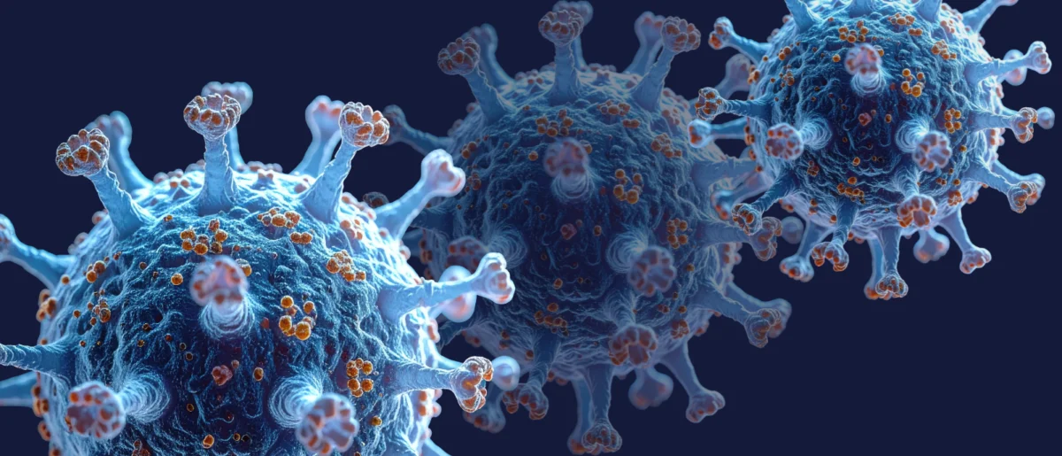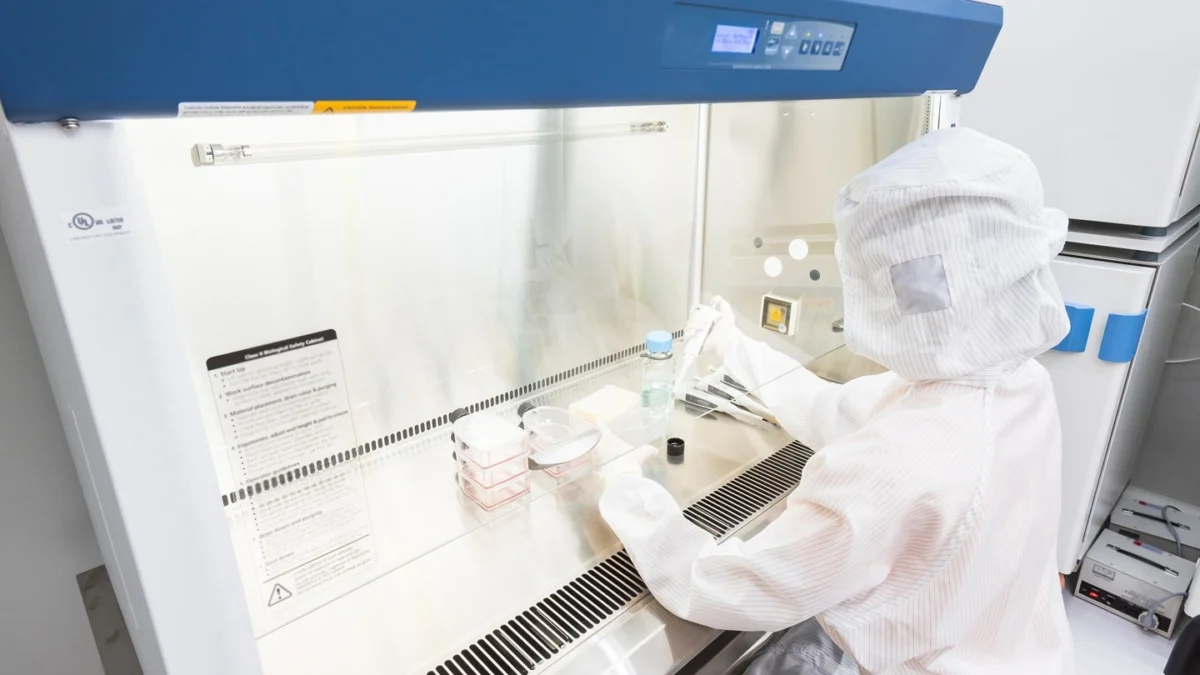
Deep within the skeletal architecture of the human body, bone marrow operates as the primary engine of life. Every single day, this spongy, vascular tissue generates hundreds of billions of vital cellular components, ranging from oxygen-transporting erythrocytes to infection-fighting leukocytes and platelets essential for coagulation. However, this delicate biological machinery is exceptionally fragile, frequently suffering catastrophic damage during intensive medical interventions such as chemotherapy and radiation therapy administered to cancer patients. This treatment-induced marrow suppression often results in severe neutropenia—dangerously depleted white blood cell counts—leaving vulnerable patients highly susceptible to opportunistic infections, systemic sepsis, and potentially fatal complications that can force clinicians to delay or discontinue life-saving cancer therapies.
To address this persistent clinical challenge, an interdisciplinary collaborative team of bioengineers, medical researchers, and pediatric specialists from the University of Pennsylvania School of Engineering and Applied Science (Penn Engineering), the Perelman School of Medicine (PSOM), and the Children’s Hospital of Philadelphia (CHOP) has successfully engineered an advanced microfluidic platform. This groundbreaking technology meticulously emulates the native microenvironment, complex architecture, and dynamic functionality of human bone marrow in vitro. Published in the peer-reviewed scientific journal Cell Stem Cell, this breakthrough bridges a critical translational gap in modern medicine, overcoming the profound limitations of traditional animal models that historically fail to faithfully replicate the intricate physiological and immunological nuances of human hematopoiesis.
The Architecture of Artificial Marrow: Mimicking Nature in Microfluidics
The newly developed device consists of a compact polymer microfluidic chip featuring specially sculpted chambers designed to house human hematopoietic stem and progenitor cells alongside the stromal and supportive cell populations with which they naturally interact. Encased within a precisely formulated hydrogel matrix, these cellular components are cultured to replicate the intricate morphogenetic processes characteristic of bone development during human embryonic growth.
Unlike static cell cultures, this biologically inspired platform creates a living, functional human marrow tissue capable of independently generating mature, healthy blood cells. These newly formed cells are actively released into a continuous stream of culture media flowing through engineered microcapillary blood vessels embedded within the chip. This dynamic fluid flow successfully mirrors the sinusoidal blood vessels found in natural bone marrow, allowing researchers to observe and quantify the real-time intravasation and release of blood cells into the circulatory network.
By incorporating these sophisticated fluidic and cellular elements, the bone marrow-on-a-chip enables scientists to simulate the toxic side effects of aggressive therapeutic regimens, including localized radiotherapy and systemic chemotherapy. Furthermore, when physically or digitally coupled with complementary organ-on-a-chip platforms—such as microfluidic models of human lung tissue—the system can successfully demonstrate inter-organ biochemical communication. This capability allows researchers to observe how bone marrow coordinates defensive responses with peripheral tissues, tracking the mobilization and trafficking of immune cells as they migrate from the marrow compartment into infected pulmonary airways to neutralize bacterial pathogens via phagocytosis.
From Extraterrestrial Ambitions to Terrestrial Breakthroughs
The genesis of this transformative technology dates back nearly a decade, born out of an unconventional scientific inquiry into the physiological impacts of space exploration on human physiology. Recognizing the accumulating clinical data that indicated an elevated risk of immune dysregulation and severe infections among astronauts deployed on prolonged spaceflights, Dr. Dan Huh, Professor in Bioengineering at Penn Engineering, and Dr. G. Scott Worthen, an attending physician at CHOP and Professor Emeritus in Pediatrics at PSOM, initiated a collaborative project to study human bone marrow under conditions of weightlessness.
The initial research timeline encountered significant operational hurdles. Nearly ten years ago, the research teams proposed sending a miniaturized model of human bone marrow to the International Space Station (ISS) to conduct comparative analyses between terrestrial controls and samples subjected to microgravity. However, logistical and technical setbacks plagued the initiative. During the primary attempted launch, the specialized fluidic control system required to maintain the viability of the engineered tissue models suffered an electrical short-circuit during atmospheric ascent. Subsequent plans to execute a rescheduled payload delivery were indefinitely postponed due to the global disruptions caused by the COVID-19 pandemic.
Despite these initial disappointments, the foundational research required to prepare the hardware for spaceflight yielded unexpected dividends. The engineering solutions developed to sustain living marrow tissues in automated, closed-loop microfluidic systems laid the groundwork for the terrestrial iteration of the platform. Reflecting on the trajectory of the project, Dr. Huh noted that while the space-based experiments were never realized, the rigorous engineering process ultimately produced one of the most sophisticated bioengineered tissue models developed to date.
Biological Self-Assembly: Borrowing Nature’s Recipe
Replicating human bone marrow outside the living body has long been considered one of the formidable challenges in tissue engineering. Natural bone marrow is a heterogeneous composite tissue primarily driven by hematopoietic stem cells (HSCs), which retain the capacity for multilineage differentiation; endothelial cells, which construct the inner lining of vascular channels; and mesenchymal stromal cells, which synthesize and maintain the extracellular matrix and structural integrity of the niche.
Historically, attempts to combine these distinct cell types in laboratory settings frequently resulted in disorganized cellular aggregates that failed to mimic the functional hierarchy or structural organization of native marrow. The anatomical seclusion of human bone marrow deep within rigid skeletal structures has historically restricted access, making in vitro physiological study exceptionally difficult.
The critical breakthrough achieved by the Penn-CHOP research team stemmed from a shift in methodology. Rather than attempting to manually assemble tissues cell by cell, the investigators focused on mimicking the developmental biology of embryonic bone marrow formation. During human gestation, bone marrow development is governed by self-organizing cellular phenomena driven by reciprocal signaling cascades among a core group of progenitor cells.
Andrei Georgescu, a former doctoral student in the Huh laboratory who served as a co-author on the study and is now the CEO of Vivodyne—a biotechnology startup co-founded to commercialize organ-on-a-chip platforms—emphasized the novelty of this design principle. According to Georgescu, the system leverages the intrinsic capacity of stem and progenitor cells to self-organize and self-assemble into complex, highly structured tissues when provided with a precisely tuned biochemical and biophysical microenvironment. Identifying the exact environmental parameters required to facilitate this spontaneous tissue morphogenesis demanded extensive empirical optimization.
Broader Implications for Medicine, Pharmacology, and Space Science
The validation of this automated, large-scale production-compatible bone marrow model carries profound implications across multiple scientific and industrial sectors. In the realm of pharmaceutical drug development, the platform offers a powerful tool for automated, high-throughput preclinical screening. By accurately predicting the marrow toxicity of novel anticancer compounds and experimental therapeutics early in the drug discovery pipeline, pharmaceutical companies can significantly reduce development costs, refine candidate selection, and mitigate the risks associated with clinical trials.
Moreover, the technology holds substantial promise for advancing the field of regenerative medicine and cell therapy. A significant finding of the study was that the marrow-on-a-chip environment successfully supports the long-term maintenance and viability of hematopoietic stem and progenitor cells without inducing spontaneous differentiation or senescence. This capability suggests that future iterations of the platform could provide the optimized biochemical signals required to maintain, expand, or rejuvenate human hematopoietic stem cells harvested from donors—a process currently constrained by the rapid loss of stemness when cells are removed from their native niche. Given the critical clinical reliance on stem cell transplantation for treating leukemias, lymphomas, and other hematologic disorders, exploring the utility of this technology for clinical cell therapy represents a primary objective for future investigations.
Even as terrestrial applications take precedence, the original impetus for the research—understanding the vulnerabilities of the human immune system during space travel—remains relevant. As commercial spaceflight and long-duration interplanetary missions transition from theoretical concepts to operational realities, platforms capable of modeling radiation exposure and microgravity effects on human immunity will become indispensable for safeguarding astronaut health.
The study’s successful execution was made possible through extensive institutional and financial backing, including support from the National Institutes of Health, the National Science Foundation, the Paul G. Allen Foundation, the National Research Foundation of Korea, the Ministry of Trade, Industry, and Energy, and the National Center for Advancing Translational Sciences. The collaborative effort brought together investigators from diverse disciplines, including Samira Mehta, Pouria Fattahi, Anni Wang, Sezin Aday Aydin, and Jeongyun Seo of Penn Engineering; Joseph Hai Oved of CHOP and PSOM; Jonathan H. Galarraga and Thomas Cantrell of Vivodyne; Brian M. Dulmovits and Timothy S. Olson of CHOP; Pelin L. Candarlioglu, Asli Muvaffak, and Anthony Lynch of GlaxoSmithKline; and Michele M. Kim and Eric S. Diffenderfer of PSOM.
As this technology advances toward broader commercial and clinical adoption, it stands to redefine the boundaries of human tissue modeling, offering unprecedented visibility into the microscopic systems that sustain human life and defend against disease.