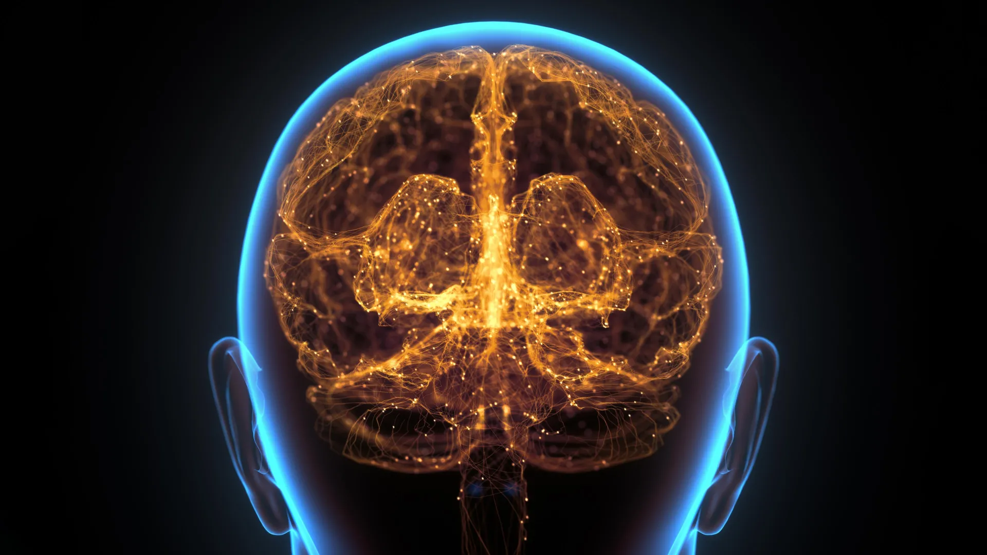
Researchers at the Washington University School of Medicine in St. Louis have fundamentally reshaped modern neuroimmunology with the discovery of previously unknown, lymph node-like immune structures nestled within the skull bone marrow of mice. Published in the prestigious journal Nature, this landmark study challenges decades of textbook dogma regarding the absolute isolation of the central nervous system from the body’s wider defensive networks. By identifying these localized "security stations," scientists have revealed that the brain possesses a rapid-response defensive vanguard positioned mere millimeters from the tissue it protects—a biological architecture that becomes active during aggressive neurological conditions like brain cancer long before traditional, distant lymph nodes receive distress signals.
The implications of this discovery extend far beyond murine models. The investigative team has already unearthed parallel evidence of comparable immune cells residing within human skull bone marrow, signaling that this localized protective apparatus is likely an evolutionarily conserved feature of mammalian anatomy. As biomedical researchers race to understand the intersection of neurology and immunology, this breakthrough provides a conceptual bridge that could soon transform therapeutic strategies for glioblastoma, Alzheimer’s disease, Parkinson’s disease, schizophrenia, and long COVID.
A Historical Paradigm Shift in Neuroimmunology
For the better part of a century, medical science operated under the assumption of central nervous system immune privilege. The prevailing theory held that the brain and spinal cord were strictly partitioned from the systemic immune system by the blood-brain barrier and the skull, kept in a state of quiet isolation to prevent potentially destructive inflammatory responses from damaging delicate neural circuitry. While textbooks taught that the brain lacked conventional lymphatic drainage, clinical observations frequently hinted at a more complex reality. Patients with neuroinflammatory disorders often exhibited immune-mediated pathology, yet the precise pathways of communication between the brain and the immune system remained shrouded in mystery.
Over the past decade, however, this long-standing paradigm has systematically crumbled, largely driven by pioneering work originating from the laboratory of Dr. Jonathan Kipnis, the Alan A. and Edith L. Wolff Distinguished Professor of Pathology & Immunology and a BJC Investigator at WashU Medicine. Several years ago, Kipnis and his research team upended conventional wisdom by discovering functional lymphatic vessels weaving through the dura mater—the protective, fibrous outer layer of tissue enveloping the brain directly beneath the structural vault of the skull.
Following that discovery, the Kipnis lab continued to map the intricate physical topography connecting the central nervous system to the skeletal framework encasing it. In subsequent studies, the team identified microscopic physical channels bridging the skull, the dura mater, and the underlying brain tissue. These microscopic tunnels serve as vital logistical corridors, providing a direct, unobstructed conduit through which immune cells, signaling molecules, and cellular waste products can freely transit between the active parenchyma of the brain and the neighboring bone marrow of the cranium.
The Genesis of the Discovery: Tracing Molecular Highways
Building upon the foundation of these physical channels, the research team initiated a series of sophisticated tracking experiments designed to monitor the exact trajectory of proteins exiting the brain. By following these molecular markers, scientists observed that the proteins did not merely dissipate into the general circulation; instead, they traveled purposefully through the micro-channels and accumulated directly within the skull bone marrow.
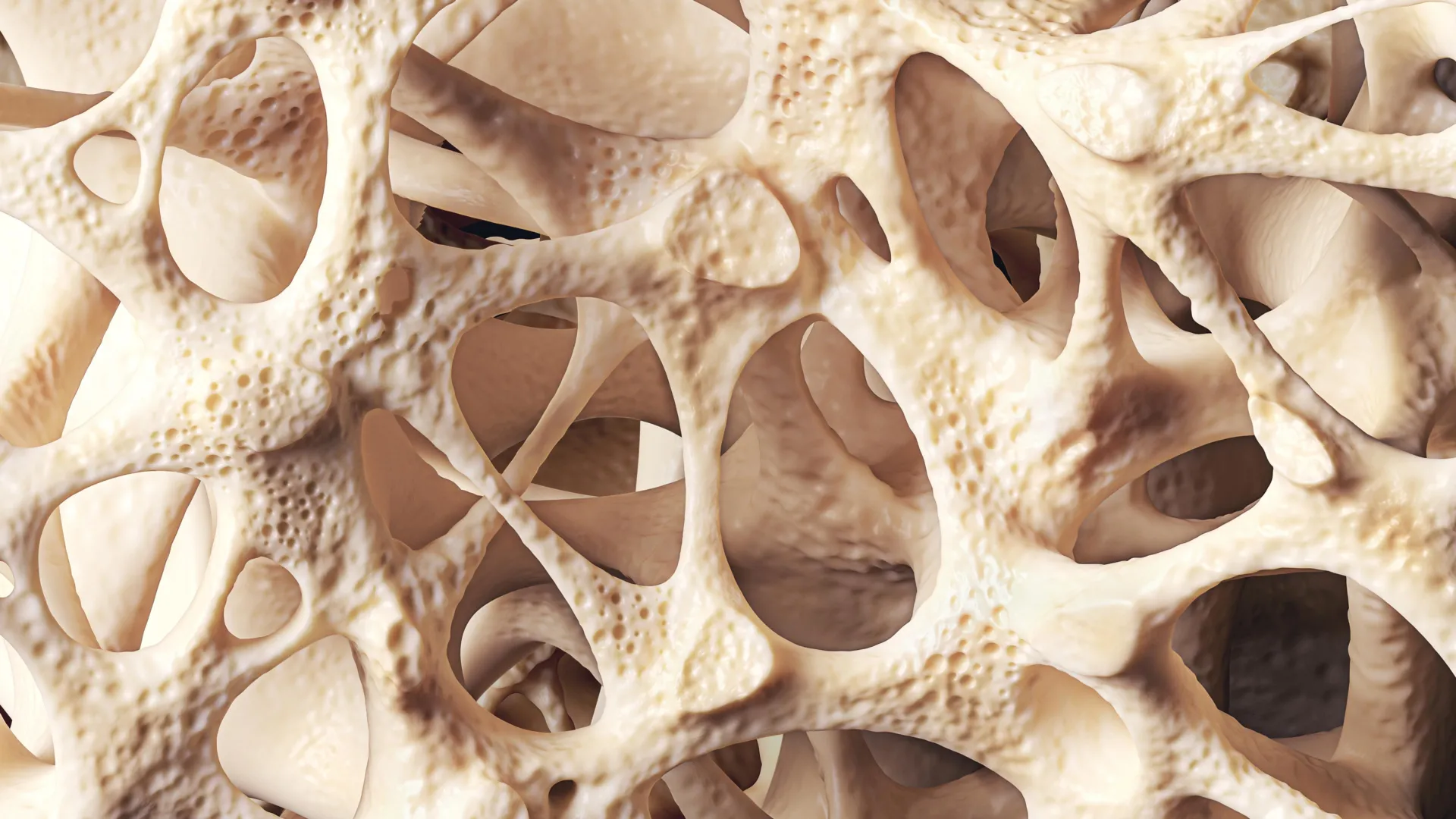
Upon closer inspection of the cranial bone marrow in healthy mice, the researchers stumbled upon an entirely unexpected biological architecture. Nestled within the skeletal tissue were highly organized immune structures bearing a striking morphological and functional resemblance to peripheral lymph nodes—the specialized coordination centers where adaptive immune responses are meticulously orchestrated. Within these newly mapped cranial niches, T follicular helper cells were observed interacting dynamically with B cells, establishing the foundational machinery required to manufacture massive quantities of disease-fighting antibodies.
"We have never seen such structures in healthy bone marrow before," remarked Dr. Jang Hyun Park, the study’s first author and a former postdoctoral research fellow in the Kipnis lab who is establishing his own independent research group at the Korea Advanced Institute of Science and Technology. "It is an exciting discovery that points out that a complex brain requires its own specialized immune structures to defend it."
Unlike standard bone marrow—which primarily functions as a hematopoietic factory generating red blood cells, platelets, and general leukocyte populations—the skull bone marrow acts as a specialized forward operating base. Because the skull encases the most complex and energy-demanding organ in the body, evolutionary pressures apparently favored the establishment of dedicated, proximal immune hubs capable of mounting an immediate, tailored counter-offensive against localized insults without waiting for systemic mobilization.
Evaluating the Frontline Defense Against Glioblastoma
To determine whether these newly cataloged immune structures served an active physiological purpose or merely represented biological relics, the research team turned to experimental models of glioblastoma. Glioblastoma is notoriously one of the most aggressive, treatment-resistant forms of human brain cancer, characterized by rapid cellular proliferation and a notorious ability to evade or suppress systemic immune surveillance.
The investigators utilized a murine glioblastoma model and systematically disrupted the function of the skull immune hubs using targeted pharmacological intervention. The results were both immediate and pronounced: tumors expanded at a significantly accelerated rate in mice whose cranial immune hubs had been experimentally compromised compared to control animals with intact cranial defenses. Furthermore, the mice subjected to impaired skull immune structures suffered from markedly reduced overall survival rates.
This empirical evidence demonstrated conclusively that the skull bone marrow does not merely observe brain pathology from a distance; it actively engages in the containment and suppression of intracranial malignancies. When these local sentinel stations are incapacitated, the brain’s ability to defend itself against aggressive neoplastic growth is severely compromised.
Therapeutic Potentiation: Engineering a Localized Immune Surge
Encouraged by the discovery that the skull’s immune hubs actively combat brain tumors, the research team sought to determine whether these specialized niches could be artificially stimulated to enhance therapeutic outcomes. The goal was to develop a localized intervention that could boost antibody production and cellular activity within the skull bone marrow without triggering systemic toxicity—a common and debilitating side effect of conventional immunotherapy regimens.
The researchers engineered a novel, targeted treatment protocol combining three distinct immune-boosting proteins suspended within a specialized biocompatible gel. This therapeutic matrix was placed directly beneath the scalp, in immediate proximity to the outer table of the cranium.
The application of the protein-loaded gel produced a dramatic, localized surge of immune activity directed squarely against the intracranial tumors. Temporal tracking of the immune response revealed a distinct sequence of activation: the defensive surge erupted first within the immune hubs nestled inside the skull bone marrow, and only manifested later in secondary, more distant lymph nodes located outside the skull. This chronological sequence confirmed that the skull bone marrow acts as the primary responder, orchestrating the early phases of the adaptive immune cascade before systemic networks are fully engaged.
When evaluated for clinical efficacy, the therapeutic strategy yielded highly promising results. Mice treated with the protein-releasing gel rejected glioblastoma tumors significantly more effectively and demonstrated extended overall survival compared to untreated control cohorts.
Broad Implications Across Neurological and Psychiatric Medicine
The identification of specialized immune hubs within the cranium fundamentally expands the burgeoning field of neuroimmunology, offering a paradigm-shifting lens through which to examine a vast spectrum of neurological, neurodegenerative, and psychiatric disorders. Because these immune niches sit in such immediate anatomical proximity to the central nervous system, they represent an ideal therapeutic gateway.
"The finding fundamentally changes our current understanding of neuroimmunology," Dr. Kipnis emphasized. "Knowing that the brain relies on first responders in the surrounding skull for defense has the potential to change how we think about developing therapies for many neurological conditions, including Alzheimer’s disease, Parkinson’s disease, schizophrenia, long COVID, and many others that have an immune component to them. Such therapies could access these immune hubs directly through the skull, without major peripheral side effects."
In conditions such as Alzheimer’s disease and Parkinson’s disease, chronic neuroinflammation and the accumulation of misfolded proteins—such as amyloid-beta, tau, and alpha-synuclein—are central hallmarks of pathology. Historically, attempts to harness the immune system to clear these toxic aggregates have encountered severe clinical hurdles, primarily due to the difficulty of crossing the blood-brain barrier effectively or avoiding widespread, dangerous systemic inflammation, such as autoimmune meningoencephalitis.
By designing therapies targeted specifically to the skull bone marrow hubs, future clinicians may be able to modulate central nervous system immunity with high precision.药物 (medicines) or biological agents applied topically to the scalp or delivered via minimally invasive cranial procedures could potentially stimulate microglial clearance mechanisms, enhance antibody-mediated clearance of pathological proteins, and restore immune homeostasis within the brain parenchyma while leaving peripheral organ systems entirely undisturbed.
Future Directions and Clinical Translation
Following the publication of these findings in Nature, independent research groups and neuroimmunology laboratories worldwide are shifting focus to validate the existence and functional capacity of human cranial immune hubs. While preliminary histological examinations of human skull bone marrow have yielded encouraging similarities to the murine models, comprehensive clinical mapping is required to understand the variability of these structures across different age groups, genetic backgrounds, and disease states.
The transition from murine models to human clinical trials will undoubtedly present complex regulatory and technical challenges. However, the prospect of non-invasive or minimally invasive scalp-applied treatments that harness the skull’s native immune machinery represents a highly attractive frontier in translational medicine. As pharmaceutical developers begin to design next-generation biologics tailored for cranial delivery, the traditional boundaries separating neurology, immunology, and oncology continue to dissolve.
Ultimately, the discovery at Washington University School of Medicine in St. Louis serves as a powerful reminder of the human body’s intricate design. The brain, long viewed as a solitary fortress operating in biological isolation, is in fact closely guarded by a specialized security apparatus built directly into the bone that protects it—a localized defensive network poised to rewrite the future of neurotherapeutic medicine.