In a significant advancement for the field of oncology, investigators at the Johns Hopkins Kimmel Cancer Center and the Johns Hopkins Bloomberg School of Public Health have identified the precise molecular mechanism by which specific gene rearrangements drive the progression of translocation renal cell carcinoma, a rare and aggressive form of kidney cancer. The study, published on April 22 in the journal Cell Reports, reveals that proteins produced by these "fusion genes" aggregate into tiny liquid droplets within the cell nucleus. These droplets, known as condensates, act as command centers that activate genes responsible for cancer growth and metastasis. By demonstrating that the disruption of these droplets can halt the activation of oncogenic pathways, the research team has opened a promising new avenue for therapeutic intervention in a disease that currently lacks a standardized treatment protocol.
The Clinical Challenge of Translocation Renal Cell Carcinoma
Translocation renal cell carcinoma (tRCC) represents a distinct subtype of kidney cancer characterized by chromosomal translocations involving the TFE3 (transcription factor E3) gene. While it accounts for a small percentage of adult kidney cancer cases—estimated at approximately 1% to 5%—it is significantly more prevalent in pediatric and young adult populations, where it may comprise up to 50% of renal cell carcinoma diagnoses.
Historically, tRCC has been difficult to treat because it does not respond predictably to the tyrosine kinase inhibitors or immunotherapies commonly used for the more prevalent clear cell renal cell carcinoma. The rarity of the disease has also hampered large-scale clinical trials, leaving clinicians with few evidence-based options for patients whose cancer has spread. The discovery made by the Johns Hopkins team provides a foundational understanding of the disease’s biology, shifting the focus from simply identifying the presence of fusion genes to understanding the physical state of the proteins they produce.
The Mechanics of Fusion Genes and Chromosomal Rearrangement
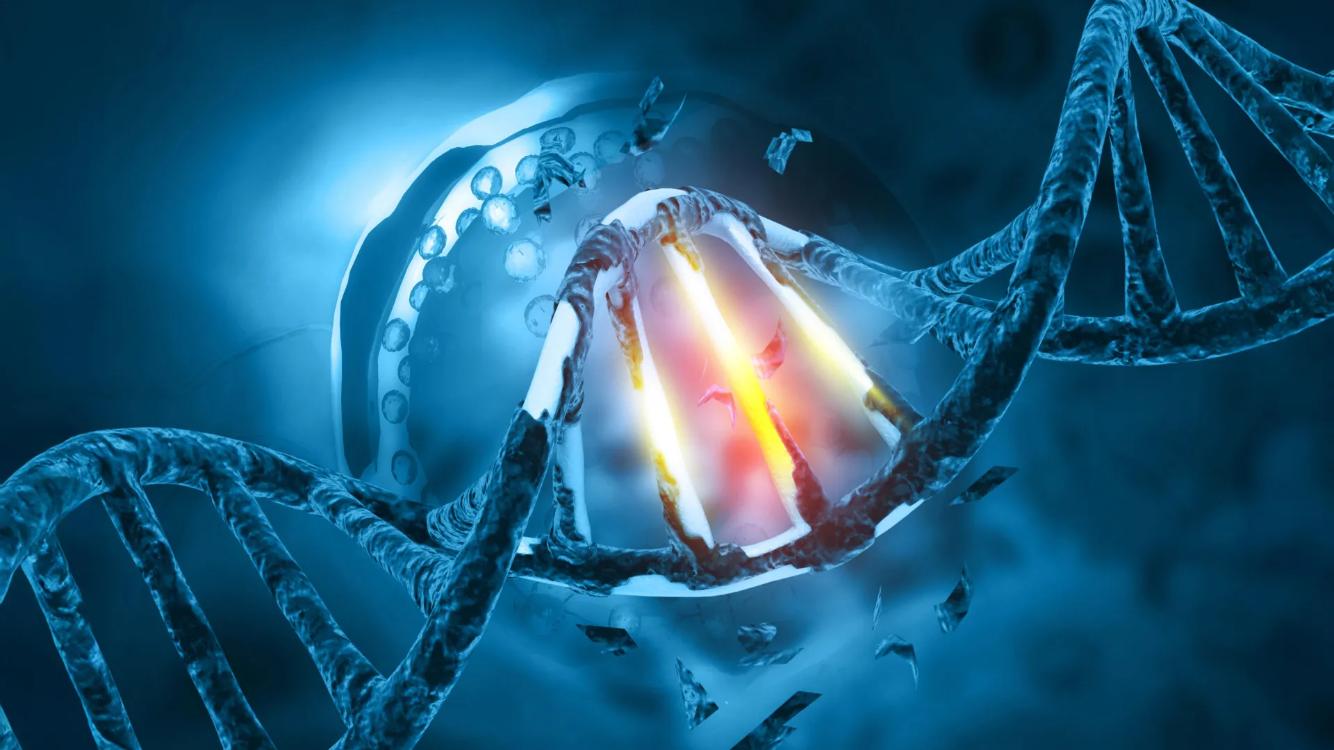
At the heart of tRCC is a genomic "swapping" event. During cell division or as a result of DNA damage, a segment of a chromosome breaks off and attaches to another. In the case of tRCC, the tail end of the TFE3 gene combines with the beginning of various "partner" genes. Common partners include PRCC, NONO, and SFPQ. This fusion creates a hybrid gene that produces a chimeric protein—one that contains elements of two different molecules.
While scientists have recognized the presence of these TFE3 fusions for years, the exact "how" of their pathogenicity remained elusive. The Johns Hopkins study focused specifically on the NONO and SFPQ fusion partners, which together account for approximately 40% of all TFE3 fusions in patients. The researchers sought to determine why these specific combinations were so effective at transforming healthy cells into malignant ones.
Visualizing Liquid Condensates: The "Oil in Water" Phenomenon
The research, led by senior author Danfeng "Dani" Cai, Ph.D., an assistant professor of biochemistry and molecular biology at the Johns Hopkins Bloomberg School of Public Health, utilized advanced imaging techniques to track the behavior of these fusion proteins. By attaching fluorescent tags to the TFE3 fusion proteins in patient-derived kidney cancer cells, the team was able to observe their localization under a microscope.
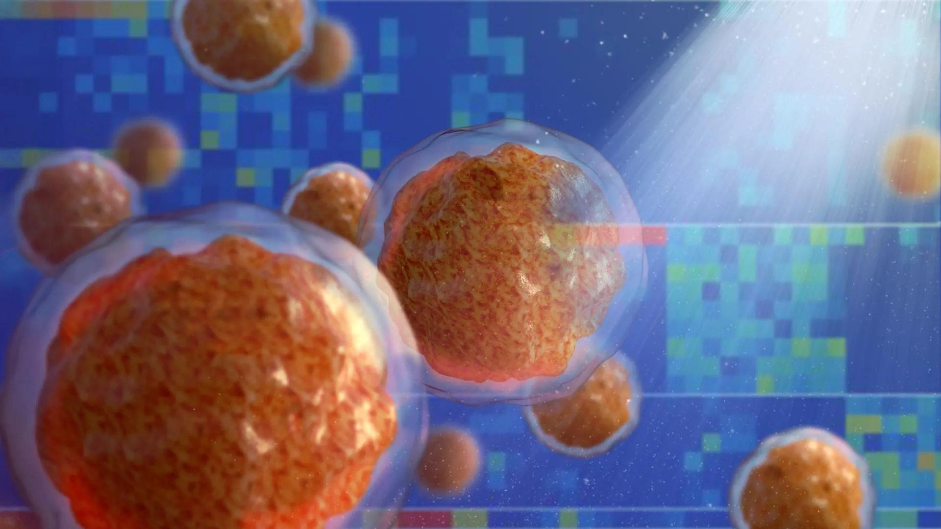
The results were striking: the fusion proteins did not distribute evenly throughout the nucleus. Instead, they congregated into discrete, glowing dots. Dr. Cai’s laboratory specializes in "condensates"—concentrated assemblies of molecules that form through a process known as liquid-liquid phase separation. This phenomenon is analogous to oil droplets forming in water; the proteins spontaneously separate from the surrounding nuclear fluid to create a high-concentration micro-environment.
The study found that these droplets were not merely passive clumps. Instead, they were active hubs of genetic regulation. The researchers observed that marker proteins associated with gene activation and the machinery required to "read" DNA were being pulled into these droplets. This suggested that the condensates were acting as "transcriptional factories," specifically designed to churn out the messages required for cancer cells to proliferate and migrate.
Redesigning the Chromosomal Landscape
To understand how these droplets interact with the genetic code, Dr. Cai collaborated with Eneda Toska, Ph.D., an assistant professor of oncology at the Johns Hopkins Kimmel Cancer Center. Together, they examined the "chromatin landscape"—the way DNA is packaged inside the cell.
DNA is often described using the "beads on a string" analogy. The DNA (the string) wraps around histone proteins (the beads) to form chromatin. When the string is tightly wound, the genes are inaccessible and "turned off." When the string is loose, the genes are "turned on."
The research team discovered that the TFE3 fusion proteins possess a unique ability to physically remodel this structure. "We found that these fusion proteins open and close different sites on the chromatin by making chemical modifications," Dr. Toska explained. By binding to specific sites and redesigning the landscape, the fusion proteins ensure that the genes necessary for cell movement and rapid division are permanently accessible. This epigenetic remodeling effectively rewires the cell’s identity, forcing it into a state of perpetual growth.
Identifying the Structural Trigger: The Coiled-Coil Segment
A critical phase of the study involved "deconstructing" the fusion protein to identify which part was responsible for forming the liquid droplets. Through precise molecular editing, the researchers removed various segments of the TFE3 fusion proteins.
They identified a specific structural motif known as a "coiled-coil"—a shape where protein helices wrap around each other like the strands of a rope. When this small coiled-coil segment, located in the region connecting the TFE3 tail to its partner protein, was removed, the results were definitive: the proteins could no longer form liquid condensates. More importantly, without the formation of these droplets, the proteins lost their ability to activate cancer-promoting genes.
This finding is of paramount importance because it identifies a specific structural target. If a drug could be developed to interfere with the coiled-coil interaction or otherwise dissolve these droplets, it could potentially "turn off" the cancer’s driving force without the need for traditional, often toxic, chemotherapy.
Broader Implications for Ewing Sarcoma and Leukemia
The implications of this study extend far beyond the rare confines of translocation renal cell carcinoma. Many other forms of cancer are driven by similar fusion genes. Dr. Cai noted that Ewing sarcoma—a bone cancer primarily affecting children—and certain types of leukemia are also characterized by fusion proteins.
"It’s possible that these fusion genes form similar droplets, or condensates, that regulate genes in these cancers and could react to similar treatment strategies," Cai stated. The realization that the physical state of a protein (its ability to form a liquid droplet) is as important as its chemical composition represents a paradigm shift in oncology. It suggests that "undruggable" proteins—those that lack a traditional binding pocket for a drug molecule—might be targeted by disrupting their ability to phase-separate.
Chronology of Research and Future Directions
The study represents a multi-year effort involving a diverse team of specialists in biochemistry, oncology, and genomics. Following the initial observation of the "dots" in the nucleus, the team spent months mapping the chromatin modifications and validating their findings across different fusion partners.
The next phase of the research, already underway, involves high-throughput screening. The team hopes to identify other proteins and molecules that reside within these TFE3 condensates. By understanding the full "inventory" of the droplet, they can look for existing drugs or develop new small molecules that can break these structures apart.
Furthermore, the team plans to investigate whether these condensates can be used as a diagnostic marker. If the presence of these droplets can be easily detected in biopsy samples, it could lead to faster and more accurate diagnosis of tRCC, which is currently often misidentified as other forms of kidney cancer.
Institutional Support and Collaborative Effort
The research was a collaborative effort involving several departments within Johns Hopkins and external partners. Co-authors included Choon Leng So, Ye Jin Lee, Wanlu Chen, Binglin Huang, Emily De Sousa, Yangzhengyu Gao, Marie Elena Portuallo, Sumaiya Begum, Kasturee Jagirdar, Vito Rebecca, and Hongkai Ji of the Bloomberg School of Public Health; Bujamin Vokshi of the Johns Hopkins University School of Medicine; and W. Marston Linehan of the National Cancer Institute.
The work was supported by extensive funding from the federal government and private foundations, reflecting the high priority placed on finding treatments for rare cancers. Support came from the National Institutes of Health (NIH) National Institute of General Medical Sciences, the National Cancer Institute (NCI), and the National Human Genome Research Institute. Additional funding was provided by a Department of Defense Kidney Cancer Idea Development Award, a Jayne Koskinas Ted Giovanis grant, and a Johns Hopkins Provost Catalyst Award.
Dr. Toska’s involvement also highlights the intersection of academic research and the pharmaceutical industry, as she has received grants and consulting fees from AstraZeneca and Menarini, though the study itself was independently conducted and peer-reviewed.
Analysis: A New Frontier in Precision Medicine
The Johns Hopkins study is a landmark in the study of "phase separation" in disease. For decades, cancer research focused on the "lock and key" model, where a drug (the key) fits into a specific protein (the lock). However, fusion proteins have often lacked a clear "lock," making them notoriously difficult to target.
By framing the problem as a matter of biophysics—specifically the formation of liquid droplets—Cai and Toska have provided a new "surface" to attack. This approach aligns with the growing field of precision medicine, where treatments are tailored not just to the organ where the cancer originated, but to the specific genetic and physical drivers of the tumor. As the medical community moves toward "condensate-modifying" drugs, the lessons learned from this rare kidney cancer may eventually provide the blueprint for treating some of the most challenging malignancies in modern medicine.