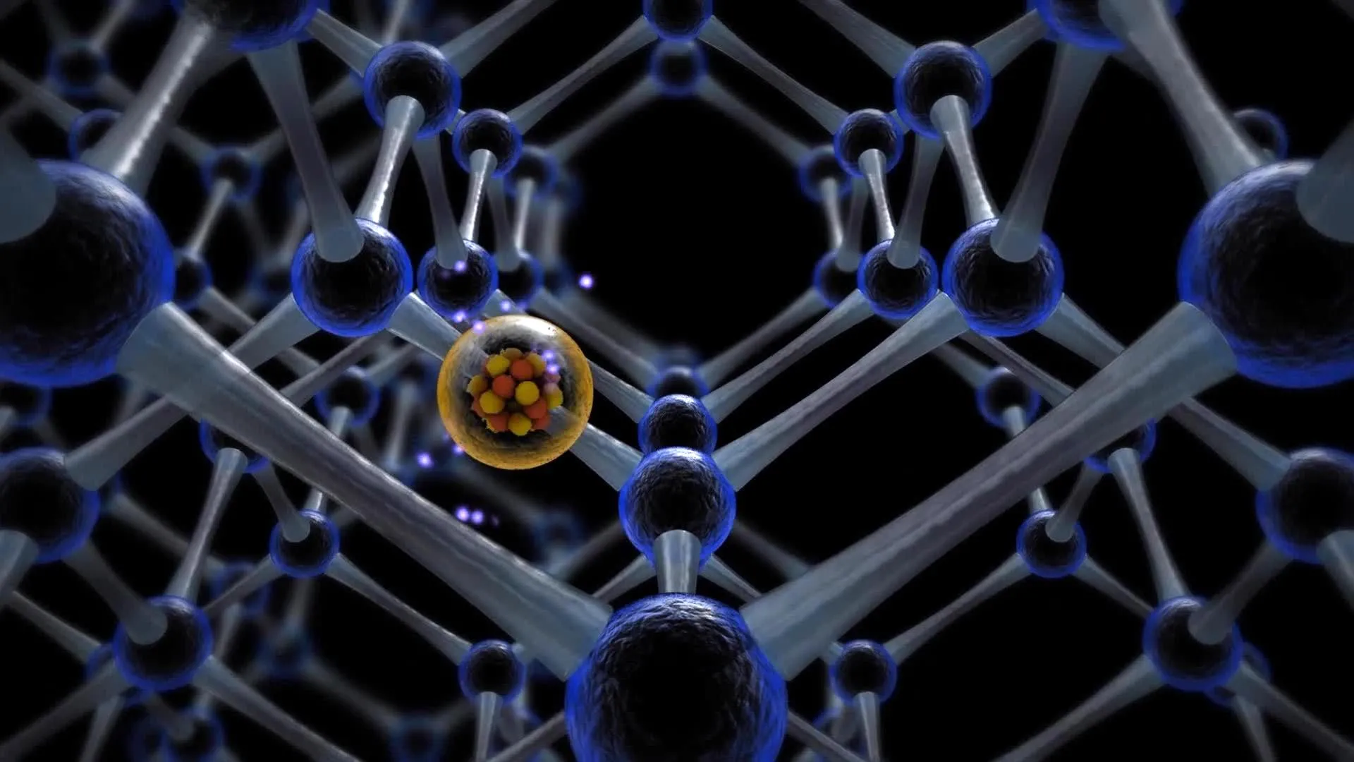
A breakthrough preclinical study led by investigators at Weill Cornell Medicine has identified a singular molecular switch, known as FLI-1, that is essential for blood stem cells to transition from a dormant state into an active, regenerative phase. This discovery, published in the journal Nature Immunology, represents a significant advancement in regenerative medicine, offering the potential to drastically improve the efficacy of bone marrow transplants and the delivery of life-saving gene therapies. By understanding the mechanism that governs how these cells multiply and integrate into the body, researchers have opened a new door for treating a wide array of hematologic disorders and cancers.
The Biological Foundation of Hematopoietic Stem Cells
Stem cells serve as the fundamental building blocks of the human body’s regenerative system. In the context of the blood and immune system, these are known as hematopoietic stem cells (HSCs). For the majority of an individual’s life, these cells reside in a state of quiescence—a form of cellular "hibernation" within the bone marrow. In this state, they divide very slowly, preserving their long-term viability and protecting their genetic integrity from the wear and tear of rapid replication.
However, when the body experiences an injury, severe infection, or the depletion of blood cells due to medical treatments like chemotherapy, these stem cells must "wake up." This transition to an activated state allows them to multiply rapidly and differentiate into various mature, functional cells, such as oxygen-carrying red blood cells, clot-forming platelets, and infection-fighting white blood cells. The ability to control this switch has long been a "holy grail" for hematologists, as the primary challenge in many blood-related treatments is the difficulty of getting harvested stem cells to expand and successfully "take" or engraft in a patient’s body.
Identifying FLI-1: The Master Regulator
The research team, led by Dr. Shahin Rafii, director of the Hartman Institute for Therapeutic Organ Regeneration and the Ansary Stem Cell Institute at Weill Cornell Medicine, utilized advanced single-cell profiling and computational modeling to compare the genetic signatures of quiescent and activated blood stem cells. Their analysis led them to FLI-1, a DNA transcription-regulating protein.
Transcription factors like FLI-1 act as master controllers, capable of turning thousands of genes on or off simultaneously. The study revealed that FLI-1 is the primary driver behind the regenerative process. When FLI-1 is absent or inactive, blood stem cells remain locked in their quiescent state. In this dormant phase, they are largely isolated from their surroundings. Conversely, the activation of FLI-1 restores the stem cells’ ability to interact with their microenvironment—specifically the "vascular niche."
The Critical Importance of the Vascular Niche
One of the most significant findings of the study involves the relationship between blood stem cells and the specialized endothelial cells that line the blood vessels within the bone marrow. This environment, known as the vascular niche, provides the necessary signals for stem cells to survive and function.
The research demonstrated that FLI-1 activity is what allows stem cells to "talk" to these endothelial cells. This co-adaptability is essential for the cells to expand their numbers and eventually migrate into the bloodstream to replenish the body’s blood supply. The study clarifies that stem cell activity is not an autonomous process; rather, it is a symbiotic relationship where the stem cell and the vascular niche must be in constant communication. This finding shifts the understanding of stem cell biology from a cell-centric view to a systems-based view of the marrow environment.
Overcoming the Limitations of Adult Stem Cell Transplants
Bone marrow transplants are a cornerstone of treatment for many leukemias, lymphomas, and other blood disorders. These procedures rely on the replenishment of the recipient’s immune and blood cells using donor stem cells. However, the success of these transplants is often limited by the quantity and quality of the stem cells available.
"The approach we outlined in this study could substantially improve the efficiency of marrow transplants and marrow-cell-targeted gene therapies, especially in cases where the donor has a very limited supply of viable blood stem cells," noted Dr. Shahin Rafii.
For patients who serve as their own donors (autologous transplants), the challenge is even greater if their stem cells have been damaged by previous rounds of chemotherapy or radiation. Such cells are often sluggish and difficult to activate. By transiently boosting FLI-1 levels, doctors could potentially "prime" these exhausted cells, making them more robust and increasing the likelihood of a successful engraftment in the patient.
Innovation via mRNA Technology
While FLI-1 is essential for regeneration, its overactivity is a known risk factor for certain types of leukemia. Therefore, a permanent increase in FLI-1 would be dangerous. To navigate this, the Weill Cornell team developed a sophisticated method to stimulate the cells only temporarily.
Borrowing from the technology used in modified mRNA-based vaccines, the researchers introduced FLI-1 into the stem cells for a brief window of only a few days. This "pulse" of activity was sufficient to wake the cells from hibernation and initiate the expansion process without altering their long-term genetic profile or increasing the risk of malignancy.
Dr. Tomer Itkin, study co-first author and current director of Tel Aviv University’s Neufeld Cardiovascular Research Institute, emphasized the safety of this method: "The stem cells we prime with FLI-1 modified mRNA in this way wake up from hibernation, expand and functionally and durably engraft in the recipient host, without any evidence of cancer."
Solving the Umbilical Cord Blood Mystery
For decades, clinicians have observed that blood stem cells derived from umbilical cords have a much higher regenerative potential than those harvested from adult bone marrow. However, the biological reason for this disparity remained a mystery until now.
The team’s research showed that umbilical cord-derived stem cells naturally possess higher levels of FLI-1 activity. This higher baseline of FLI-1 allows them to interact more effectively with the vascular niche, explaining their superior potency. By identifying this mechanism, the researchers have provided a blueprint for how adult stem cells might be "upgraded" to match the regenerative power of cord blood, potentially expanding the pool of viable transplant options for adult patients.
Implications for Gene Therapy and Blood Disorders
The discovery has immediate implications for the burgeoning field of gene therapy. For conditions like beta-thalassemia and sickle cell anemia, patients’ own stem cells are harvested, genetically modified in a lab to correct a defect, and then re-infused.
During the laboratory phase, these cells are highly vulnerable and often struggle to survive and multiply. Using FLI-1 to safely expand these cells outside the body could ensure that a larger, healthier population of corrected cells is available for re-infusion, significantly increasing the chances of a "cure" for these chronic and debilitating genetic conditions.
Chronology of the Research and Computational Analysis
The path to this discovery involved years of multidisciplinary collaboration. The timeline began with the observation of how endothelial cells in the bone marrow influence stem cell behavior. From there, the team moved into deep-tissue profiling, using bioinformatics to filter through the noise of thousands of genetic expressions to find the single "switch" that mattered most.
Sean Houghton, a bioinformatics analyst and co-first author, played a pivotal role in deciphering these complex signaling pathways. "We showed that stem cell activity is not autonomous but also is not fully determined by endothelial cell vascular niche signals—it depends instead on signaling and adaptability between the two," Houghton explained. This computational heavy lifting allowed the team to integrate FLI-1 into the known signaling pathways that drive stem cell self-renewal and survival, providing a comprehensive map of the activation process.
Institutional Support and Future Outlook
The research was a massive undertaking supported by multiple branches of the National Institutes of Health (NIH), including the National Heart, Lung, and Blood Institute; the National Institute of Diabetes and Digestive and Kidney Diseases; and the National Institute of Allergy and Infectious Diseases. Additional funding came from the Hartman Institute for Therapeutic Organ Regeneration and the Ansary Stem Cell Institute.
Looking forward, the researchers are planning to scale up their modified mRNA-based method for further preclinical development. The ultimate goal is to transition into human clinical trials, where the transient introduction of FLI-1 could become a standard part of the transplant and gene therapy protocol.
If successful in humans, this approach could set the stage for treating a wide range of blood disorders with long-term stable and safe blood production. It represents a shift toward "precision regeneration," where molecular switches are flicked with surgical accuracy to heal the body from the inside out, reducing the reliance on massive donor volumes and improving the survival rates of some of the most vulnerable patients in the healthcare system.