Researchers at the University of Kentucky have uncovered a pivotal mechanism in the progression of Alzheimer’s disease, identifying the brain’s resident immune cells—microglia—as the primary cause of sleep disruption associated with the condition. The study, published in the peer-reviewed journal Alzheimer’s & Dementia, challenges the long-held scientific assumption that sleep loss is caused primarily by the physical presence of amyloid-beta plaques or the direct degradation of neurons. Instead, the research suggests that an overactive immune response, triggered by early-stage plaque accumulation, creates a state of chronic neurological "noise" that prevents the brain from entering restorative sleep cycles.
Lead researcher Shannon L. Macauley, Ph.D., an associate professor of physiology at the University of Kentucky College of Medicine, described the phenomenon using a domestic analogy: while amyloid plaques represent a localized fire in a kitchen, the microglial response acts as a malfunctioning sprinkler system that floods the entire house. This "whole-house" immune reaction persists long after the initial trigger, effectively keeping the brain in a state of heightened arousal. The findings represent a significant paradigm shift in neurodegenerative research, suggesting that targeting inflammation rather than just plaque removal may be the key to restoring sleep and potentially slowing the disease’s progression.
The Role of Microglia in the Neurodegenerative Landscape
Microglia are specialized immune cells that serve as the first line of defense in the central nervous system. Under healthy conditions, these cells act as "scavengers," clearing cellular debris, pruning synapses, and monitoring the brain for pathogens. However, in the context of Alzheimer’s disease, microglia become chronically activated by the presence of amyloid-beta plaques—misfolded protein clumps that are a hallmark of the pathology.
The University of Kentucky team, including first author Nicholas J. Constantino, Ph.D., utilized an animal model to isolate the specific impact of these cells. By using a pharmacological agent called Pexidartinib (PLX3397) to temporarily deplete approximately 87% of the microglia in the brain, the researchers observed a dramatic reversal in sleep deficits. Mice with advanced Alzheimer’s pathology regained more than two hours of sleep per day following the treatment. Crucially, this improvement occurred without any change in the volume or density of the amyloid plaques themselves, indicating that the immune cells’ behavior, rather than the physical plaques, was the driver of wakefulness.
Experimental Methodology and Advanced Neuro-Monitoring
To distinguish between the effects of normal aging and Alzheimer’s-specific pathology, the researchers conducted a comparative study involving two distinct groups of mice. The first group was genetically predisposed to develop amyloid plaques, while the "wild-type" control group aged naturally. Observations were recorded at two critical intervals: six months, representing the onset of plaque formation, and 18 months, representing late-stage disease progression.
The team employed a multi-modal approach to monitor brain activity. Small, head-mounted devices recorded electroencephalography (EEG) and electromyography (EMG) data, providing a continuous "electrical fingerprint" of the brain. These tools allowed the researchers to categorize sleep into two primary phases: Rapid Eye Movement (REM) sleep, associated with dreaming and memory consolidation, and Non-Rapid Eye Movement (NREM) sleep, the deeply restorative stage essential for physical repair and toxin clearance.
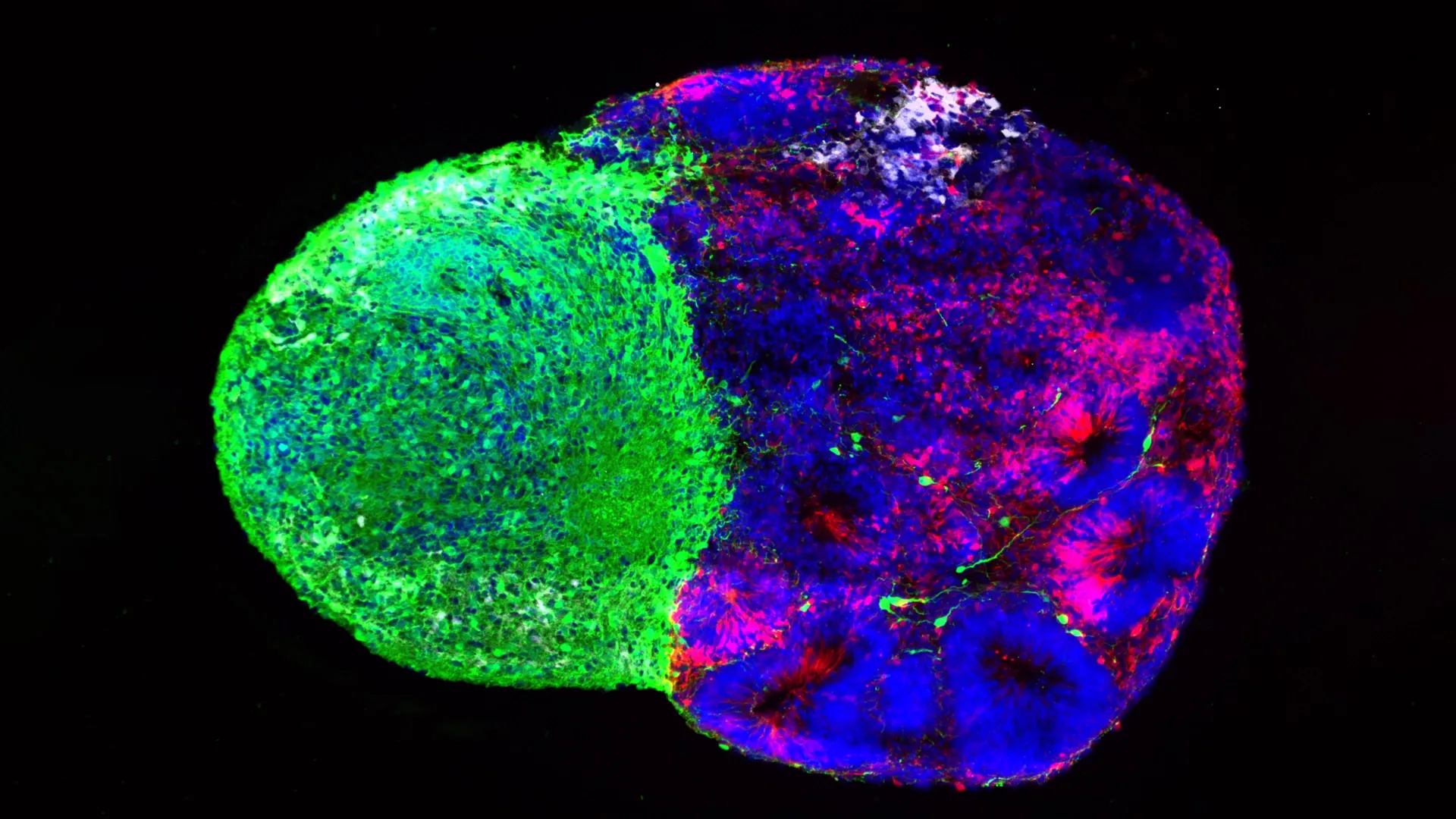
Furthermore, the researchers utilized light sheet microscopy, a sophisticated imaging technique that renders brain tissue transparent. By applying a thin plane of laser light, the team created high-resolution 3D digital maps of the brain, allowing them to visualize the spatial relationship between amyloid plaques and activated microglia across the entire organ.
The "Ceiling Effect" and Early-Stage Intervention
One of the most unexpected findings of the study was the non-linear relationship between plaque accumulation and sleep loss. Traditionally, scientists expected that as the "plaque burden" increased, the severity of sleep disruption would follow a similar upward trajectory. However, the data revealed what the researchers termed a "ceiling effect."
The disruptions in sleep and cortical EEG activity observed at the six-month mark—when plaques first emerge—did not worsen significantly by the 18-month mark, despite a doubling of the plaque burden. This suggests that the initial wave of immune activity triggered by early amyloid deposits is sufficient to maximize the sleep deficit. From a clinical perspective, this finding underscores the importance of early intervention. If the immune response becomes "locked" in an overactive state early in the disease process, therapeutic efforts to calm the microglia may need to begin well before significant memory loss or cognitive decline becomes apparent.
The Vicious Cycle: NREM Sleep and Toxin Clearance
The study highlighted a specific vulnerability in NREM sleep, which was selectively reduced by Alzheimer’s pathology. In contrast, normal aging was found to primarily impact REM sleep. The loss of NREM sleep is particularly damaging because of its role in the "glymphatic system"—the brain’s waste-clearance mechanism.
During deep NREM sleep, the space between brain cells increases, allowing cerebrospinal fluid to wash away metabolic waste, including the very amyloid-beta proteins that characterize Alzheimer’s. Macauley noted that the loss of this "cleaning cycle" creates a dangerous feed-forward loop: plaques trigger microglial inflammation; inflammation prevents restorative NREM sleep; the lack of sleep prevents the clearance of plaques; and the accumulating plaques further drive the inflammatory response. By breaking this cycle through microglial modulation, researchers hope to restore the brain’s natural ability to maintain its own health.
Data Analysis: Aperiodic Activity and the "Engine" Metaphor
To delve deeper into the electrical disruptions caused by microglia, the team applied a mathematical method known as "Fitting Oscillations and One Over Frequency" (FOOOF). This allowed them to separate periodic brain waves (the rhythmic oscillations usually studied in EEG) from aperiodic activity (the background "noise" or "offset" of the brain’s electrical state).
The researchers compared the brain’s electrical state to a car engine. In a healthy brain, the engine idles quietly during rest. In the Alzheimer’s model, however, the "aperiodic offset" remained high, suggesting that the brain’s "engine" was running at a high speed even when the animal was attempting to sleep. This background noise, driven by the "partying" microglia, essentially makes the brain too "loud" for the animal to remain in a deep, stable sleep state.
Future Clinical Applications: Portable EEG and Drug Repurposing
The implications of this research extend beyond laboratory models to potential diagnostic and therapeutic tools for human patients. One of the study’s long-term goals is the development of affordable, non-invasive screening methods. Macauley envisions a future where portable EEG systems could monitor patients in their homes, identifying the specific electrical signatures of Alzheimer’s-related sleep disruption years before clinical symptoms emerge.
"Portable EEG systems could allow us to monitor people in their home environments and potentially screen for changes associated with Alzheimer’s disease without the initial need for expensive or invasive tests," Macauley stated. This would be particularly beneficial for rural populations, such as those in Kentucky, who may lack immediate access to major specialized medical centers.
On the therapeutic front, the Macauley lab is investigating ways to "calm" microglia without the drastic step of removing them entirely, as microglia still perform essential protective functions. The team is currently testing existing, FDA-approved medications to see if they can modulate microglial metabolism. Among the candidates are Metformin, a widely used diabetes medication, and Stiripentol, an anti-seizure drug. The goal is to determine if these drugs can shift the way microglia process energy, thereby reducing their inflammatory output and allowing the brain to return to a restful state.
Collaborative Research and Institutional Support
The success of the study has been attributed to the highly collaborative environment at the University of Kentucky’s Sanders-Brown Center on Aging. Dr. Macauley emphasized a lab culture that encourages "calculated risk-taking" and the investigation of data that contradicts original hypotheses. Dr. Constantino noted that this atmosphere was crucial in moving the focus away from traditional neuron-centric models toward the role of the immune system.
The research was supported by substantial federal and private funding, reflecting the scientific community’s interest in new Alzheimer’s targets. Grants were provided by the National Institute on Aging (NIA) and the National Institute of General Medical Sciences (NIGMS), both part of the National Institutes of Health (NIH). Additional support came from a $287,236 award from the Cure Alzheimer’s Fund and a $250,000 award from The CART Fund (Coins for Alzheimer’s Research Trust).
As the global population ages, the prevalence of Alzheimer’s disease is expected to rise, placing an immense burden on healthcare systems. By identifying microglia as a reversible cause of sleep loss, the University of Kentucky team has provided a new roadmap for treatments that could improve the quality of life for millions, targeting the disease not just as a collection of plaques, but as a manageable immune-driven condition.