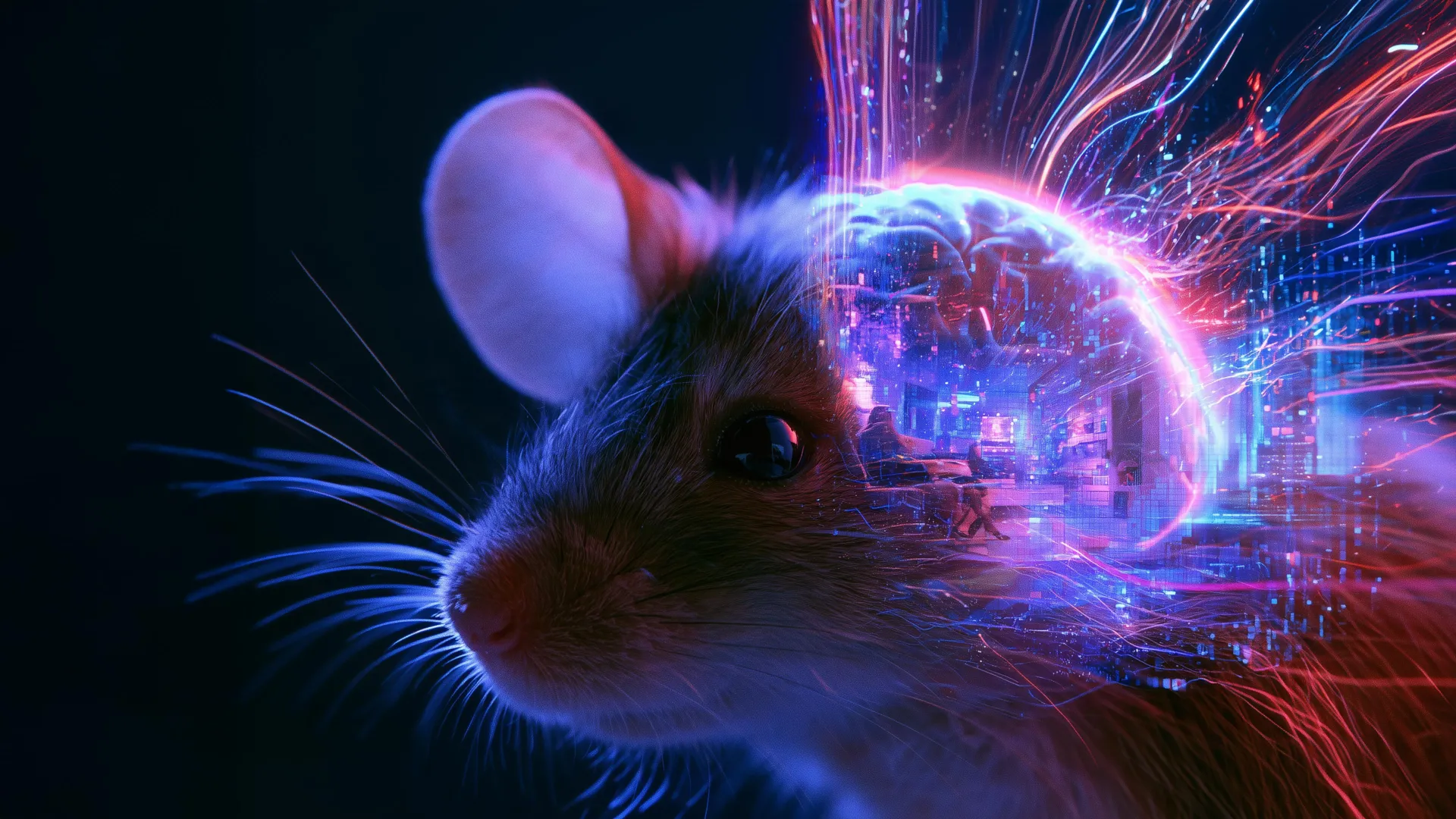
The research, conducted at the Sainsbury Wellcome Centre at UCL, moves beyond the limitations of macro-level brain imaging by focusing on the granular activity of individual neurons. While previous studies have utilized functional Magnetic Resonance Imaging (fMRI) to decode human visual perception, the spatial resolution of such methods is often too coarse to capture the nuances of neural computation. By shifting the focus to the visual cortex of mice, the UCL team has demonstrated that the brain’s "internal movie" can be reconstructed with startling fidelity, provided the underlying neural data is sufficiently comprehensive.
The Evolution of Neural Decoding: A Chronology
The quest to decode the brain has been a decades-long endeavor. In the early 2000s, researchers first began exploring the feasibility of reconstructing simple geometric shapes from the visual cortex of cats. These early experiments relied on linear decoders that were limited by low computational power and a lack of sophisticated machine learning models.
By the 2010s, the field saw a transition toward more complex visual stimuli. Studies involving human subjects inside fMRI machines allowed for the reconstruction of basic silhouettes and patterns. However, the "pixel-level" accuracy remained elusive, and the models were largely confined to static images. The year 2023 served as a pivotal point for the discipline with the launch of the Sensorium Competition—a global challenge focused on predicting neural responses to naturalistic stimuli. It was within this environment of rapid innovation that Dr. Joel Bauer and his colleagues found the inspiration for the dynamic neural encoding model utilized in their latest work. By late 2023 and early 2024, the UCL team had refined this architecture, moving from mere prediction of neuron activity to the generative reconstruction of dynamic video sequences.
Methodological Innovations in Brain Mapping
To achieve this reconstruction, the research team employed a sophisticated calcium imaging technique. As neurons fire, they experience a localized influx of calcium ions; the researchers used microscopic sensors to detect these fluorescent pulses, allowing them to map the activity of thousands of individual neurons in real-time.
The core of the methodology relies on a "difference-based" algorithm. The model begins with an arbitrary, blank visual canvas. It then calculates the discrepancy between the expected neural response to that blank screen and the actual, measured neural response of the mouse as it watches a high-definition video. Through an iterative feedback loop, the algorithm adjusts the pixels on the screen to minimize the error between the simulated neural activity and the biological reality. As the reconstructed pixels converge with the actual visual input, the "hidden" video emerges.
Crucially, the model does not operate in a vacuum. It integrates auxiliary data streams, including the animal’s physical orientation, velocity of movement, and subtle changes in pupil diameter. This holistic approach recognizes that the brain does not process vision as an isolated feed, but as an integrated component of sensory-motor experience.
Quantitative Accuracy and Statistical Validation
In scientific terms, the success of the reconstruction is measured via pixel correlation. In the recent UCL trials, the team observed that the correlation between the original and reconstructed footage remained high, even when the model was presented with novel, unseen videos.
Data from the study indicates that the fidelity of the reconstruction is directly proportional to the number of neurons sampled. When the model was fed data from a small cluster of cells, the resulting video was blurry and lacked structural coherence. However, as the researchers increased the number of input neurons, the resolution sharpened, capturing temporal shifts in the environment with greater accuracy. While the current resolution is not yet broadcast-quality, it represents a substantial leap over previous attempts at biological signal-to-image conversion.
Official Perspectives and Academic Context
Dr. Joel Bauer, the lead author of the study, emphasized that the goal is not merely to "watch" the mouse’s brain, but to understand the transformative nature of perception. "We wanted to have a better way of investigating how the brain interprets what we see," Dr. Bauer stated. He noted that traditional methods of neural analysis are often overly specialized, struggling to generalize across different environmental conditions. By creating a model that compares the "objective reality" of the video with the "subjective representation" in the cortex, the team is identifying the specific biases inherent in the mammalian visual system.
Other experts in the field of computational neuroscience have lauded the study for its rigorous approach to the "encoding problem." While the researchers are cautious about claiming a perfect reconstruction of reality, they acknowledge that the brain’s deviations from the visual input are not defects. Rather, these deviations represent the brain’s strategy for prioritizing survival-critical information, such as motion, contrast, and depth.
Broader Implications: Beyond the Laboratory
The implications of this research extend far beyond the study of murine vision. If scientists can successfully map the translation of visual stimuli into neural code, the potential for clinical applications is profound.
For instance, this technology could inform the development of next-generation brain-computer interfaces (BCIs). By understanding how the visual cortex encodes data, researchers may eventually be able to bypass damaged optic nerves, sending processed visual information directly to the visual cortex. Furthermore, the ability to compare internal representations across different species could fundamentally alter our understanding of consciousness and animal cognition.
However, the findings also raise questions about the limits of sensory reconstruction. If the brain filters, modifies, and warps incoming information, to what extent can we ever claim to see an "objective" world? The study suggests that vision is a constructive process, a hypothesis that aligns with emerging theories in predictive coding—a framework which posits that the brain is constantly predicting the next moment of sensory input rather than passively receiving it.
Future Research Trajectories
Moving forward, the UCL team plans to expand the scope of their model to cover larger areas of the visual field. The current experiment was limited by the microscopic imaging window, which only captured a portion of the visual cortex. Future studies will likely involve multi-region imaging to observe how information flows from the primary visual cortex into higher-order association areas where interpretation and decision-making occur.
Additionally, there is significant interest in applying this decoding architecture to non-visual stimuli. If the brain’s representation of sound, touch, or spatial navigation can be reconstructed with similar precision, it would provide an unprecedented "internal map" of the mind. As the resolution of these reconstructions improves, the scientific community expects to see a deeper integration of AI-driven decoders in neurology, offering doctors new tools to diagnose neurodegenerative conditions that affect sensory processing.
In conclusion, the work published in eLife serves as a bridge between the physical activity of cells and the ephemeral experience of perception. By turning the "gaze" of the mouse into a digital file, researchers have confirmed that the language of the brain is not only decodable but potentially translatable into the visual syntax we use to understand our own world. While the technology remains in its infancy, the capability to reconstruct what another creature perceives provides a powerful, tangible method for exploring the deepest mysteries of the mind.