Deep within the human skeletal system lies one of the most critical and industrious biological engines: the bone marrow. Often conceptualized simply as the body’s blood factory, this highly specialized tissue is a dynamic ecosystem composed of bone cells, intricate vascular networks, neural pathways, and a diverse array of specialized progenitor cells. Operating largely in silence, the bone marrow produces billions of red blood cells, white blood cells, and platelets every single day to sustain life, orchestrate immune responses, and ensure proper oxygen transport throughout the cardiovascular system.
For generations, this complex physiological network has remained notoriously difficult to study outside of a living organism. When the bone marrow malfunctions—such as in the case of leukemias, lymphomas, and other hematological malignancies—understanding the precise mechanisms of failure becomes a matter of life and death. Until recently, researchers investigating these conditions faced a formidable scientific bottleneck: the lack of a physiologically accurate, human-relevant laboratory model that could faithfully replicate the intricate microenvironment of human bone marrow.
That paradigm shifted significantly with a breakthrough achieved by a multidisciplinary team of scientists from the Department of Biomedicine at the University of Basel and the University Hospital Basel. In a study published in the prestigious scientific journal Cell Stem Cell, the research team unveiled the first-ever comprehensive, functional three-dimensional model of human bone marrow constructed entirely from human cells. This pioneering achievement not only bridges a long-standing gap in translational medicine but also offers a powerful new avenue for reducing reliance on animal models in biomedical research.
The Anatomy of the Breakthrough: Decoding Bone Marrow Niches
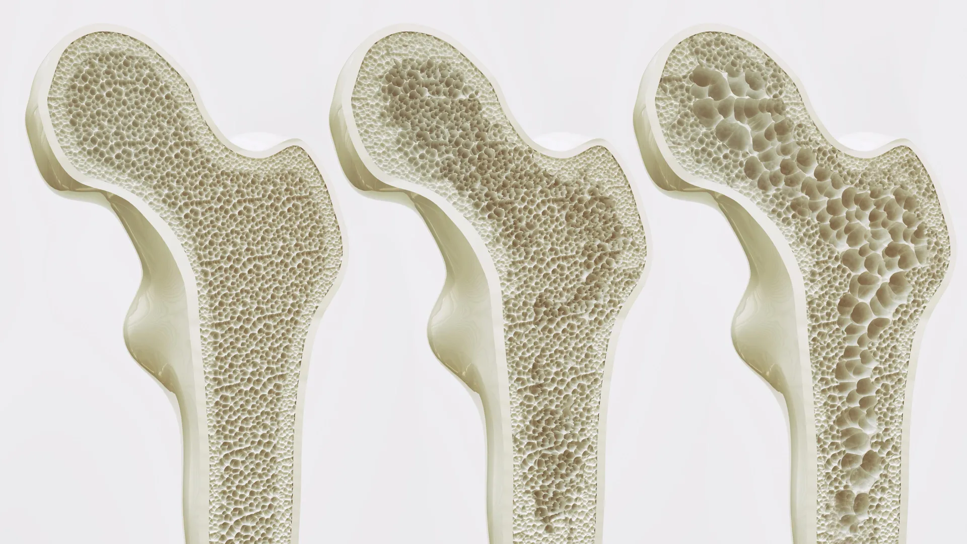
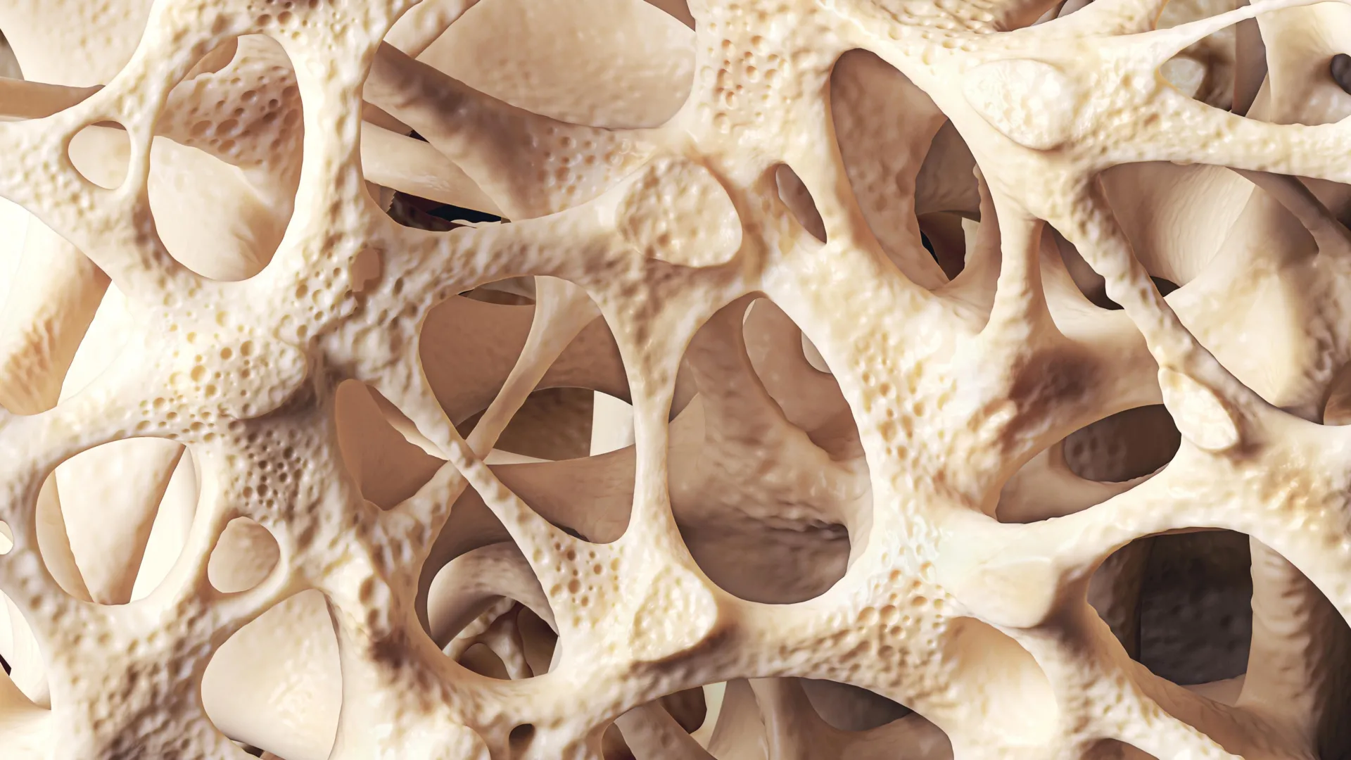
To understand the magnitude of the Basel team’s achievement, one must examine the microarchitectural complexity of the bone marrow itself. The tissue is not a uniform soup of cells; rather, it is divided into distinct, highly specialized microenvironments known as niches. Among the most critical of these is the endosteal niche, located immediately adjacent to the inner surface of the bone tissue.
The endosteal niche plays a fundamental role in regulating hematopoietic stem cells—the master cells responsible for generating all blood lineages. Furthermore, this specific microenvironment is heavily implicated in the pathogenesis of blood cancers, often acting as a protective sanctuary where malignant cells can hide, proliferate, and develop resistance to conventional chemotherapy drugs. Within the human body, the endosteal niche is a densely packed crossroads featuring mineralized bone matrices, blood vessels that supply vital nutrients, resident immune cells, and intricate networks of nerve fibers that modulate cellular activity.
Historically, replicating this multi-layered biological symphony in a laboratory setting proved nearly impossible. For decades, the global scientific community relied on two main methodologies to study bone marrow: animal models—predominantly mice—and simplified two-dimensional cell cultures. While murine models have yielded invaluable insights into mammalian hematopoiesis, significant biological differences exist between mice and humans. Immune system variations, metabolic divergences, and differences in bone architecture mean that therapies showing high efficacy in mice frequently fail during human clinical trials. Meanwhile, traditional two-dimensional cell cultures lack the crucial three-dimensional spatial organization, mechanical cues, and cellular crosstalk that dictate how cells behave in the living body.
Bridging the gap between these inadequate proxies and the realities of human physiology required a radically innovative approach. Led by Professor Ivan Martin and Dr. Andrés García García, the Swiss research team set out to construct a living, breathing human tissue model from the ground up.
Chronology of Innovation: From Stem Cells to Functional 3D Tissues
The development of this groundbreaking bone marrow model represents the culmination of years of meticulous biomaterials engineering and stem cell biology. The project’s foundational steps began with the creation of an artificial bone framework. Rather than using organic bone matrices, the researchers engineered a scaffolding material composed of hydroxyapatite, a naturally occurring calcium phosphate mineral that forms the primary mineral foundation of vertebrate bones and teeth. This scaffold provided the necessary structural rigidity and biochemical affinity required for bone-forming cells to thrive.
With the physical architecture established, the team turned to cellular components. They utilized human somatic cells that had been reprogrammed into induced pluripotent stem cells (iPSCs) using advanced molecular biology techniques. These versatile stem cells hold the remarkable capacity to differentiate into virtually any cell type in the human body when exposed to the appropriate biochemical and physical cues.
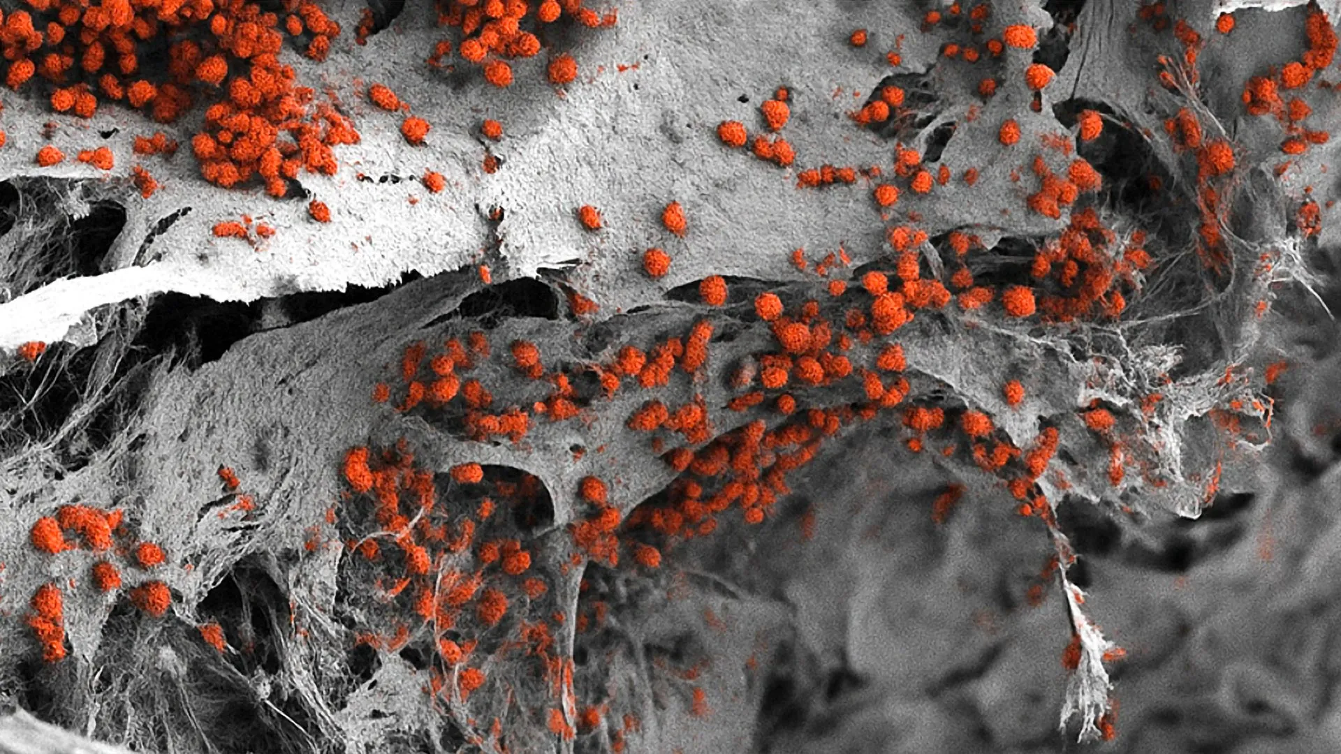
The researchers introduced these reprogrammed human stem cells into the porous hydroxyapatite scaffold. Over a carefully controlled developmental timeline, the cells were guided through precise differentiation pathways. Through the application of specific growth factors and environmental stimuli, the stem cells began to proliferate and differentiate, giving rise to a diverse consortium of specialized cells that mirror those found in native human bone tissue.
Crucially, the resulting structure successfully integrated all the key elements of the endosteal niche. The laboratory-grown tissue featured functional blood vessel networks, active nerve cell extensions, immune cell populations, and mineralized bone matrix components living in symbiosis.
The resulting three-dimensional system is remarkably robust. Measuring approximately eight millimeters in diameter and four millimeters in thickness, it stands as one of the largest and most complex human bone marrow models developed to date. Most impressively, the research team demonstrated that this engineered construct could sustain human blood cell formation—hematopoiesis—continuously in a laboratory environment for several weeks, providing an unprecedented window into the active generation of blood cells outside a living body.
A Paradigm Shift for Animal Testing and Ethical Research
Beyond its immediate physiological significance, the development of this human-cell bone marrow model aligns with a broader ethical and regulatory movement within the global scientific community: the push to advance the principles of the Three Rs—Replacement, Reduction, and Refinement of animal experiments.
For decades, pharmaceutical development and basic hematological research have depended heavily on murine models. While these studies have provided foundational knowledge, the scientific consensus around the limitations of cross-species translation has grown increasingly stark. Regulatory bodies, funding agencies, and academic institutions are under mounting pressure to adopt human-relevant testing systems whenever technologically feasible.
"We have learned a great deal about how bone marrow works from mouse studies," noted Professor Ivan Martin, reflecting on the historical trajectory of the field. "However, our model brings us closer to the biology of the human organism. It could serve as a complement to many animal experiments in the study of blood formation in both healthy and diseased conditions."
By offering a human-specific platform, the University of Basel model allows researchers to bypass the interspecies physiological gaps that plague preclinical trials. Investigators can now observe human hematopoietic processes, immune responses, and pathological disruptions in a context that accurately reflects human genetics and tissue architecture. This capability is expected to significantly reduce the sheer volume of animal subjects required for early-stage hematological research, accelerating the transition toward human-centric biotechnology.
Implications for Drug Development and Oncology
The potential applications of this novel bone marrow model extend across multiple sectors of biomedical science, with particularly profound implications for oncology and pharmaceutical drug discovery.
Blood cancers, such as multiple myeloma, acute myeloid leukemia, and certain lymphomas, originate or reside within the bone marrow niches. Because current in vitro models cannot replicate the protective microenvironment of the endosteal niche, testing how cancer cells interact with bone cells, blood vessels, and extracellular matrices has remained exceptionally difficult. The new Basel platform provides an ideal testing ground for oncologists and pharmacologists to study how malignant cells colonize the bone marrow, evade immune surveillance, and develop resistance to chemotherapy.
Furthermore, the model opens new doors for high-throughput drug screening and toxicity testing. Traditional toxicity assays often fail to predict adverse drug reactions that specifically target human bone marrow precursors—a phenomenon known as bone marrow toxicity or myelosuppression, which is a frequent and dangerous side effect of many cancer therapies. With a reliable human tissue model, pharmaceutical developers can screen new compounds for marrow toxicity much earlier in the pipeline, potentially weeding out hazardous drug candidates before they ever reach human clinical trials.
Despite these promising horizons, the researchers emphasize that technical hurdles remain before the platform can be fully integrated into industrial pharmaceutical pipelines. Dr. Andrés García García pointed out a primary physical constraint of the current design: its scale.
"For this specific purpose, the size of our bone marrow model might be too large," Dr. García García explained. For industrial-scale high-throughput screening—where laboratories need to test thousands of different drug compounds or dosages simultaneously across multi-well plates—the current eight-millimeter architecture is simply too bulky. To meet the demands of modern pharmaceutical drug discovery, future iterations of the platform will need to be miniaturized into high-density formats without sacrificing the complex cellular crosstalk that makes the current model so biologically authentic.
Looking Toward the Future of Personalized Medicine
Perhaps the most transformative long-term implication of this research lies in the realm of personalized medicine. Standardized cancer treatments often follow a one-size-fits-all protocol, despite the fact that individual tumors possess unique genetic mutations and microenvironmental dependencies. What cures one leukemia patient may fail completely in another.
Looking ahead, the research team envisions a future where clinicians could harvest somatic cells from an individual cancer patient, reprogram them using stem cell technology, and construct a patient-specific replica of that individual’s bone marrow in the laboratory.
By utilizing these bespoke tissue models, medical teams could directly test a panel of different chemotherapeutic agents or immunotherapies on the patient’s own living marrow cells. This empirical testing would allow oncologists to identify the most effective therapeutic regimen for an individual patient before administering toxic treatments to their body, dramatically improving treatment efficacy while sparing patients from ineffective therapies and unnecessary side effects.
While realizing the vision of routine personalized bone marrow modeling will require substantial further research, engineering refinements, and rigorous clinical validation, the study published in Cell Stem Cell marks a monumental foundational step. By successfully fusing advanced biomaterials science with human stem cell technology, the University of Basel researchers have unlocked a vital frontier in hematology, offering a clearer, more humane, and deeply human-centric window into the hidden engine of human life.