Stroke remains a leading cause of long-term disability worldwide, claiming the quality of life for millions. According to the World Health Organization, approximately 15 million people suffer a stroke annually, with five million dying and another five million left permanently disabled. In many developed nations, the lifetime risk of experiencing a stroke is estimated at one in four for adults over the age of 25. Despite these staggering figures, modern medicine has historically been limited to acute interventions—such as clot-busting drugs or mechanical thrombectomies—designed to save the patient’s life or prevent further damage. Once the initial window for intervention closes, the resulting neurological deficit, often manifesting as hemiplegia, aphasia, or cognitive impairment, has traditionally been viewed as irreversible.
A collaborative research initiative spearheaded by the University of Zurich (UZH) Institute for Regenerative Medicine is challenging this medical status quo. By utilizing human-derived neural stem cells, researchers have demonstrated that it is possible to facilitate biological repair in damaged brain tissue, effectively reversing motor impairment in animal models.
The Mechanism of Cellular Regeneration
The research, led by Christian Tackenberg, Scientific Head of the Neurodegeneration Group at UZH, and postdoctoral researcher Rebecca Weber, focused on the potential of neural stem cells to act as a catalyst for recovery. Unlike adult neurons, which have limited regenerative capacity, neural stem cells possess the innate plasticity to differentiate into various cell types required by the central nervous system.
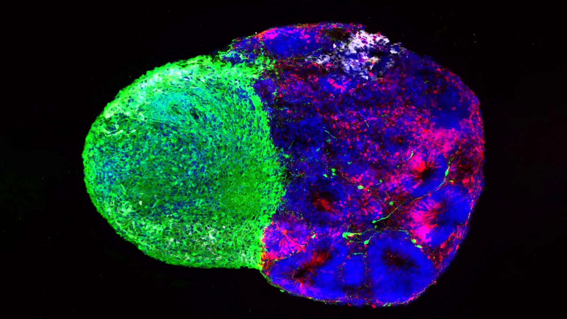
In two comprehensive studies conducted in partnership with Ruslan Rust from the University of Southern California, the team employed induced pluripotent stem cells (iPSCs). These cells are derived by "reprogramming" mature somatic cells—such as skin cells—back into a versatile, embryonic-like state. This process allows scientists to create patient-specific cells, theoretically reducing the risk of immune rejection, although the mouse models used in this study required genetic modification to ensure acceptance of the human-derived grafts.
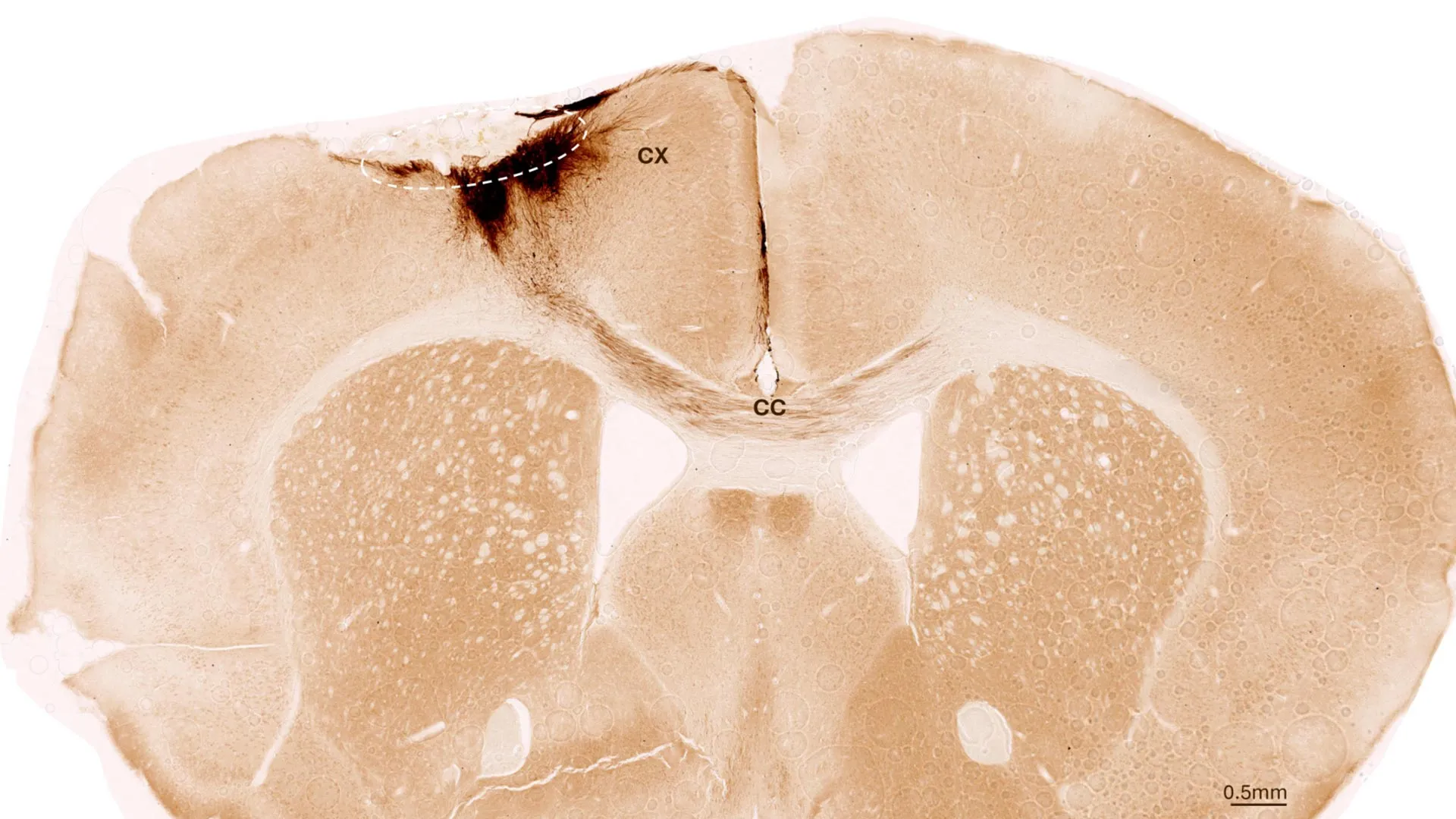
The researchers induced permanent ischemic strokes in mice, meticulously replicating the oxygen deprivation and subsequent cellular necrosis observed in human patients. One week post-stroke, the team transplanted the neural stem cells directly into the peri-infarct area—the tissue surrounding the primary site of injury. Through advanced longitudinal imaging and biochemical analysis, the researchers observed the graft’s survival over a five-week period. The results were significant: the stem cells did not merely survive; they integrated into the existing neural architecture, forming functional synapses with endogenous neurons.
Beyond Replacement: A Holistic Healing Response
Perhaps the most compelling finding of the UZH study is that the therapeutic effect extends far beyond the simple replacement of dead neurons. The transplantation triggered a cascade of systemic neuro-regenerative processes that stabilized the environment of the damaged brain.
Specifically, the research team noted the following physiological improvements:
- Angiogenesis: The development of new blood vessels within the damaged tissue, which improves oxygenation and nutrient delivery to recovering areas.
- Inflammatory Modulation: A reduction in the chronic, neurotoxic inflammatory response that typically follows a stroke, which often exacerbates secondary cell death.
- Blood-Brain Barrier Integrity: The restoration of the blood-brain barrier, a critical vascular boundary that, when breached, allows harmful blood-borne substances to enter the brain, causing further edema and inflammation.
The culmination of these processes was the functional recovery of the mice. Utilizing AI-assisted gait analysis, the researchers documented a clear improvement in motor coordination and movement patterns in the treated subjects compared to the control group. This indicates that the therapy does not just provide a cosmetic fix but facilitates the actual restoration of neural networks required for movement.
The Significance of Temporal Strategy
A critical takeaway from the study involves the timing of the intervention. The research team discovered that waiting seven days post-stroke before performing the transplant yielded better outcomes than immediate intervention.
In the clinical context, this discovery is transformative. Emergency stroke care is a high-pressure environment where time is brain; every second saved during the acute phase reduces the risk of long-term damage. However, requiring a transplant to occur within hours of a stroke would place an immense logistical burden on hospital systems. By identifying a one-week "therapeutic window," the researchers have provided a more realistic timeline for clinical application. This delay allows for the stabilization of the patient, the preparation of specialized cellular products, and the careful planning of the surgical or minimally invasive procedure required to deliver the cells.
Furthermore, the team prioritized safety and regulatory viability by developing a production protocol free from animal-derived reagents. This "xeno-free" approach is a prerequisite for human clinical trials, as it minimizes the risk of pathogen transmission and immune reactions, significantly streamlining the path toward FDA and EMA approval.
Challenges and the Path to Clinical Trials
Despite the success in preclinical models, the transition from laboratory mice to human patients is fraught with technical and safety challenges. The primary concern among neuroscientists remains the potential for uncontrolled cellular proliferation. If stem cells do not stop dividing after integration, they could form tumors or masses that would prove catastrophic in the confined space of the human cranium.
To mitigate this, Tackenberg and his colleagues are currently refining a "safety switch" mechanism—a genetic circuit built into the stem cells that can trigger their destruction or cessation of growth if complications arise. Additionally, the team is moving away from direct, invasive brain grafting. They are investigating endovascular delivery methods, where stem cells would be injected into the vascular system, allowing them to migrate to the site of the stroke via the bloodstream. This approach would be significantly less invasive, potentially turning a complex neurosurgical procedure into a routine interventional radiology operation.
The global medical community is watching these developments closely. With clinical trials for iPSC-based treatments for Parkinson’s disease already underway in Japan, the precedent for human application of stem cell therapy is well-established. Experts in the field of neurology suggest that if the UZH team can successfully demonstrate the safety of their endovascular delivery system, stroke could follow Parkinson’s as the next major neurological condition addressed by regenerative medicine.
Broader Implications for Healthcare
The implications of this research are vast. Stroke currently costs the global economy hundreds of billions of dollars annually in medical expenses and lost productivity. If a regenerative treatment can restore even a fraction of function to patients currently suffering from permanent paralysis or speech impairment, the societal impact would be immeasurable.
However, researchers remain cautious about managing public expectations. "We need to minimize risks and simplify a potential application in humans," Tackenberg emphasized. The journey from a successful mouse model to a standardized, safe, and effective human therapy typically spans years of rigorous clinical phase testing, focusing on safety, dosage, and long-term efficacy.
The collaboration between the University of Zurich and the University of Southern California stands as a testament to the power of international, multi-disciplinary research. By combining expertise in cellular biology, artificial intelligence, and vascular medicine, the team has successfully bridged the gap between theoretical regenerative biology and functional, observable recovery. While the road to widespread clinical use remains long, the identification of a viable, timed, and potentially non-invasive approach provides the most compelling evidence yet that the brain, once thought to be incapable of healing, may eventually be repaired by the very cells that once built it.