Scientists at the Mark and Mary Stevens Neuroimaging and Informatics Institute (Stevens INI) at the Keck School of Medicine of USC have identified a critical, previously under-researched component of cognitive resilience: the condition of superficial white matter. By examining the local communication pathways that sit just beneath the brain’s outer gray matter, researchers have provided new evidence that these nerve fiber networks may act as a structural buffer, potentially mitigating the cognitive decline typically associated with gray matter atrophy. The study, published in Alzheimer’s & Dementia: The Journal of the Alzheimer’s Association, shifts the traditional focus of neurodegenerative research from a singular reliance on gray matter integrity to a more holistic view of brain connectivity.
The Anatomy of Local Communication
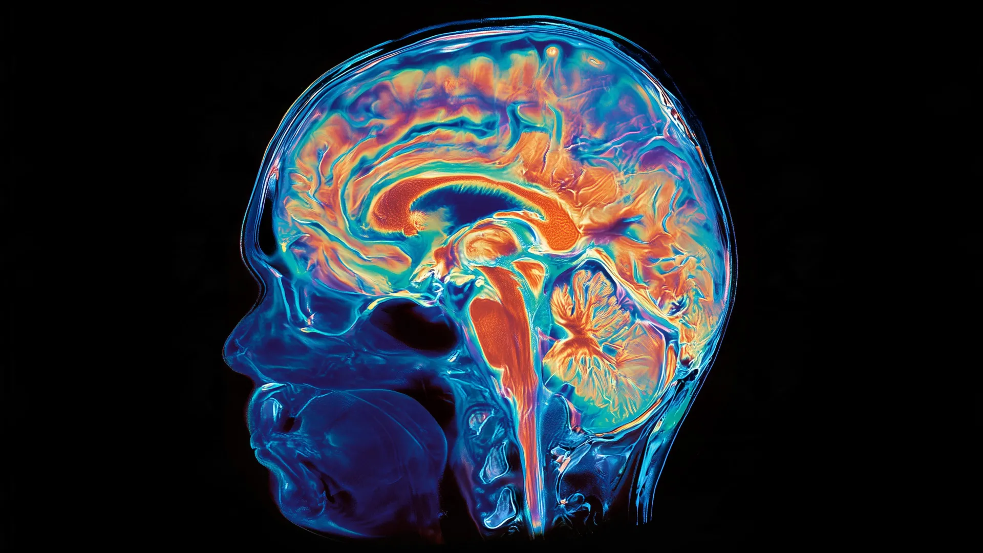
To understand the significance of this discovery, one must look at the brain’s architectural landscape. Gray matter, the wrinkled, outer mantle of the cerebral cortex, is the command center for information processing. Beneath this layer lies superficial white matter (SWM), a thin, complex web of short, U-shaped nerve fibers that bridge neighboring cortical regions. If the brain’s long-distance white matter tracts are the superhighways of the central nervous system, superficial white matter serves as the local road network—the critical infrastructure that allows for nuanced, high-speed communication between adjacent functional areas.
The Stevens INI study posits that these two systems are inextricably linked. When the "local roads" are in poor condition—characterized by a loss of neurites, the tiny projections that send and receive cellular signals, or an increase in interstitial free water—the impact of gray matter degradation becomes significantly more pronounced. Conversely, when these local circuits remain robust, they appear to offer a degree of insulation against the functional consequences of neuronal loss.
Study Methodology and Global Context
The research team leveraged data from the Harmonized Diagnostic Assessment of Dementia for the Longitudinal Aging Study in India (LASI-DAD), a landmark project designed to capture the realities of aging in diverse settings. The study cohort comprised 459 adults aged 60 and older. This demographic is particularly notable; more than 50% of the larger LASI-DAD population possesses low literacy levels, and approximately 60% reside in rural environments. Historically, neuroimaging research has been heavily skewed toward high-income, highly educated populations in Western nations, leading to a potential "WEIRD" (Western, Educated, Industrialized, Rich, and Democratic) bias in medical literature. By shifting the focus to an underrepresented population in a low-to-middle-income country, the USC team has provided a more granular look at how environmental and social variables might influence brain health.
The researchers employed advanced diffusion MRI technology to map these microscopic connections. Unlike conventional MRI, which provides a structural snapshot, diffusion MRI tracks the Brownian motion of water molecules through brain tissue. By analyzing the density of neurites and the presence of free water, scientists can effectively quantify the "integrity" of these delicate fibers, offering a window into the biological state of the brain that standard clinical scans often miss.
Chronology of Cognitive Findings
The cognitive assessments performed on the cohort included a comprehensive battery of tests focusing on language fluency, memory, executive function, and visuospatial orientation. The data revealed a clear, statistically significant correlation between the health of the superficial white matter and language performance. Participants with more intact local fiber networks consistently outperformed their peers in frontotemporal tasks—specifically those related to vocabulary recognition and verbal fluency.
The most striking finding, however, emerged from the intersection of these variables. While gray matter atrophy remains the most reliable biomarker for predicting cognitive decline across the general population, the team observed that the "slope" of this decline was modulated by SWM health. In individuals with compromised superficial white matter, the cognitive deficits resulting from gray matter loss were severe. In those where the superficial white matter remained healthy, the cognitive impact of the same degree of gray matter loss was significantly blunted. This suggests a form of "structural resilience," where the efficiency of local communication helps the brain compensate for the loss of information-processing units.
Expert Perspectives and Theoretical Implications
"Gray matter and superficial white matter are physically close and may play different roles: gray matter processes information, while superficial white matter helps nearby brain regions communicate," said Yingxu Liu, PhD, a postdoctoral scholar at the Stevens INI and the study’s lead author. "Our findings suggest that cognitive health depends not only on how much gray matter is preserved, but also on the condition of the wiring that connects it."
The implications for clinical practice are substantial. If superficial white matter can be identified as a primary site of resilience, it may eventually lead to new therapeutic targets aimed at preserving these pathways in patients at risk for dementia or Alzheimer’s. Leon Aksman, PhD, assistant professor of research neurology at the Stevens INI and the study’s senior author, noted that this finding could explain the wide variability in patient outcomes. "Two people with a similar degree of gray matter loss may not experience the same cognitive effects if the local connections surrounding that gray matter differ in health," Aksman said. "Following participants over time will be essential to test whether preserving these connections can help maintain cognition."
Addressing Global Disparities in Neurobiology
A compelling secondary finding of the study was the increased strength of the association between SWM health and language ability among individuals with no formal education or those living in rural areas. While the researchers were careful to clarify that these social factors do not inherently cause biological tissue changes, they underscore the complex interplay between life experience and brain architecture. The study suggests that brain aging is not a predetermined biological countdown but a process heavily influenced by social, educational, and environmental factors—the "cognitive reserve" that individuals build throughout their lives.
Arthur W. Toga, PhD, director of the Stevens INI, emphasized the necessity of this inclusive research design. "A fuller understanding of brain aging requires research that reflects the world’s social, cultural, and geographic diversity," Toga stated. By bridging the gap between sophisticated neuroimaging and diverse, global community-based data, the study challenges the medical community to look beyond the "standard" brain and consider how different life trajectories impact the underlying neurobiology of aging.
Future Directions and Research Limitations
Despite these significant findings, the research team acknowledges several limitations. As a cross-sectional study, the data represents a single moment in time. This creates a "chicken-or-the-egg" dilemma: researchers cannot yet definitively state whether superficial white matter deterioration is a precursor to gray matter loss, a concurrent symptom, or a secondary effect of early-stage cognitive decline.
Future studies will need to be longitudinal, tracking cohorts over several years to observe the temporal order of these neurological changes. Furthermore, the research team plans to integrate additional biological markers, including vascular health, inflammatory markers, and the accumulation of Alzheimer’s-related proteins such as beta-amyloid and tau. Understanding how these systemic factors interact with local fiber networks will be the next major hurdle in deciphering the mechanics of the aging brain.
The research was made possible through the support of the National Institute on Aging, the National Institute of Mental Health, the National Institute of Neurological Disorders and Stroke, and the Office of the Director of the National Institutes of Health. As the global population continues to age, the work conducted at the Stevens INI provides a vital framework for understanding how we might protect the mind by strengthening its local connections, potentially delaying the onset of dementia and enhancing the quality of life for millions worldwide. By focusing on the structural interplay between the brain’s processing units and its local communication network, scientists have opened a new, promising avenue in the ongoing battle against neurodegenerative disease.