For decades, the foundational tenets of cellular biology have taught that glucose serves one primary, indispensable purpose: acting as the universal metabolic fuel that drives the energetic machinery of nearly every living cell. Generations of biology students have memorized the pathways of glycolysis, the Krebs cycle, and oxidative phosphorylation, all revolving around the catabolism of glucose to harvest ATP. However, groundbreaking new research from Stanford Medicine has upended this long-standing biochemical dogma. According to a landmark study published in the journal Cell Stem Cell, glucose functions not merely as an energetic substrate, but as a master regulatory molecule that directly commands tissue differentiation—the precise biological process through which uncommitted stem cells transform into the specialized, functional cell types that comprise human tissues.
The revelation that glucose actively dictates cell fate by binding directly to proteins—rather than being broken down for calories—stunned the scientific community. The implications of this discovery stretch far across modern medicine, offering fresh theoretical frameworks for understanding and potentially treating complex pathologies marked by metabolic and developmental dysfunction, including diabetes mellitus and various forms of cancer.
A Serendipitous Discovery Rooted in High-Throughput Screening
The path to rewriting biochemistry textbooks began not with a targeted hypothesis about glucose signaling, but through an exploratory, broad-spectrum molecular screen. Dr. Paul Khavari, chair of dermatology at Stanford Medicine, and Dr. Vanessa Lopez-Pajares, a research scientist and lead author of the study, set out to identify the biomolecules responsible for driving cellular differentiation in human skin.
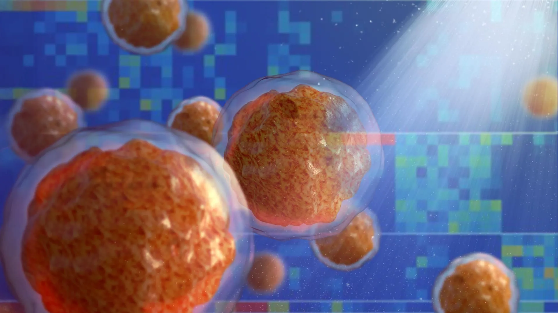
Utilizing an advanced combination of mass spectrometry and high-throughput screening technologies, the research team mapped the dynamic rise and fall of thousands of distinct biomolecules within human skin stem cells. Their objective was to observe how the molecular landscape shifts as these uncommitted progenitor cells mature into keratinocytes, the predominant cell type found in the epidermis, the outermost protective layer of human skin. The underlying hypothesis was straightforward: biomolecules that experience a significant surge in abundance during the transition phase likely play a causal role in steering the cellular differentiation process.
When the analytical data was processed, the researchers identified 193 suspect molecules. Many of these candidates were already known in scientific literature for their involvement in cellular maturation. However, the identity of the second-most elevated molecule in the dataset generated immediate disbelief.
"When we saw glucose at the top of that list, we were stunned," Khavari recalled, reflecting on the initial moments of discovery. "We had expected glucose levels to decrease during differentiation because the cells begin to divide less rapidly, and their energy requirements are less. They are on the path to senescence and death. Yet glucose levels in the cells increase significantly as they move from epidermal stem cells to differentiated keratinocytes."
Proving the Unthinkable: Years of Rigorous Verification
Recognizing the controversial nature of their findings—which directly challenged foundational biochemical principles—the research team exercised extreme caution. Rather than rushing to publication, Khavari and Lopez-Pajares spent several years designing and executing an extensive series of follow-up experiments to confirm the validity of their data.
"At first we just didn’t believe it," Khavari admitted. "Pero los resultados de los experimentos de seguimiento exhaustivos fueron claros." The team systematically ruled out experimental artifacts by measuring cellular uptake using fluorescent and radioactive glucose analogs. Furthermore, they deployed sophisticated biological sensors inside the cells engineered to emit green or red fluorescence upon encountering physiologically relevant concentrations of glucose.
As cellular differentiation proceeded in human skin samples, the intracellular sensors glowed with increasing intensity. To ensure this phenomenon was not an isolated quirk of human epidermal tissue, the researchers expanded their investigation. They analyzed developing fat cells, bone cells, and white blood cells, alongside genetically engineered mouse models expressing fluorescent glucose sensors. Consistently, across every tissue type examined, intracellular glucose levels rose markedly as cells progressed toward differentiation.
Subsequent metabolic analyses revealed that this intracellular accumulation was driven by a dual mechanism: an upregulated import of glucose from the extracellular environment coupled with a downregulation of glucose export. Crucially, metabolic assays confirmed that this intracellular surge was not accompanied by an increase in glycolysis or catabolic breakdown; the glucose remained intact.
Decoupling Energy from Regulation Through Non-Metabolizable Analogs
To definitively separate glucose’s traditional metabolic role from its newly discovered regulatory function, the researchers utilized human skin organoids—sophisticated, engineered skin tissues grown in vitro that closely mimic the structural and cellular organization of native human skin.
When these organoids were subjected to abnormally low glucose environments, their ability to mature and differentiate properly was severely impaired. Transcriptomic analysis revealed that low glucose levels disrupted the expression of more than 3,000 distinct genes, many of which encoded proteins intimately linked to epidermal development and barrier formation.
The critical turning point in the research occurred when the glucose-starved organoids were supplied with a synthetic glucose analog—a molecule structurally identical to glucose in ways that allowed cellular uptake and protein binding, but chemically altered so that cells could not break it down for energy. To the astonishment of the research team, these non-metabolizable analogs rescued the differentiation defect, supporting normal tissue maturation just as effectively as standard, metabolizable glucose.
"That was really the biggest shock," Khavari noted, "because we were stuck in the mindset that glucose is an energy source and nothing else. But these glucose analogs support differentiation just as well as regular glucose."
Mechanistic Insights: How Glucose Controls Gene Expression
With the physiological phenomenon validated, the research team delved deeper into the intracellular mechanisms governing glucose-driven differentiation. They discovered that the intracellular accumulation of glucose is mediated by an upregulation in the production of specialized transmembrane proteins responsible for transporting glucose from the extracellular matrix into the cytoplasm.
Once inside the cell, intact glucose molecules do not float passively. Instead, they physically bind to hundreds of intracellular proteins. Among these targets is a critical regulatory protein known as IRF6 (Interferon Regulatory Factor 6), a transcription factor heavily involved in tissue development. The binding of glucose induces a conformational shift in the IRF6 protein, altering its three-dimensional shape and enhancing its capacity to bind to genomic DNA. This structural change modifies the expression profile of numerous downstream genes responsible for driving cellular differentiation.
Khavari likened this sweeping biochemical interaction to a systemic alert system rather than a finely tuned whisper. "We’re seeing glucose acting like a broadcast signal in the cell, in contrast to the highly specific signaling cascades that drive many cellular functions," he explained. "When glucose levels rise in a cell, they rise everywhere, all at once. It’s like a fire alarm going off in a firehouse. Everyone in the firehouse activates in response."
Broader Implications for Diabetes, Wound Healing, and Oncology
The unveiling of glucose’s "double life" as a master transcriptional regulator provides critical insights into several long-standing medical mysteries and offers novel avenues for therapeutic intervention.
For decades, clinicians have observed that individuals suffering from poorly managed diabetes mellitus—a condition characterized by chronically elevated blood glucose levels—frequently experience impaired wound healing, compromised tissue regeneration, and various dermatological complications. While these symptoms were historically attributed to microvascular damage and chronic inflammation, the new Stanford findings suggest that systemic glucose imbalances may directly interfere with the precise differentiation programs required for effective tissue repair and cellular turnover.
Conversely, embryonic stem cells—which possess the remarkable pluripotency to differentiate into virtually any cell type in the human body—lose this developmental plasticity when exposed to abnormally high glucose concentrations. This cellular hypersensitivity implies that localized metabolic states actively police the boundary between stemness and specialization.
The implications for oncology are perhaps even more profound. Cancer is fundamentally a disease characterized by failed cellular differentiation, where malignant cells become locked in a proliferative, immature state, refusing to mature and undergo normal senescence or apoptosis. Interestingly, various glucose analogs have previously shown therapeutic promise in preclinical and clinical trials as potential anticancer agents. While these compounds were originally designed and administered under the assumption that they would starve tumor cells of energy by blocking glycolysis, the Stanford study suggests an alternative mechanism of action. Rather than merely starving cancer cells, these analogs may be forcing immature, highly proliferative cancer cells to undergo forced differentiation, halting uncontrolled tumor growth.
Future Directions and the Emerging Field of Small-Molecule Regulation
As the scientific community digests these findings, the Stanford Medicine team is already looking toward the future, hoping to map the exact pathways through which glucose signaling operates across both healthy and diseased cellular landscapes.
"This finding is a springboard for research on dysregulation of glucose levels, which affects hundreds of millions of people," Khavari stated. "Pero también es probable que sea importante en el desarrollo del cáncer porque el cáncer es una enfermedad de diferenciación fallida. Este es un campo completamente nuevo y en crecimiento. People have thought that small biomolecules like glucose were quite passive in the cell. This is another piece of evidence to pay close attention to other roles these molecules might play."
The study, which was formally published online on March 21 in Cell Stem Cell, received primary financial backing from the National Institutes of Health under research grants R01AR043799, AR045192, K01AR070895, and P30CA124435, alongside vital support from the U.S. Department of Veterans Affairs Office of Research and Development.
By proving that one of biology’s most ubiquitous metabolites doubles as an instructional master switch for cellular identity, this research marks a paradigm shift in molecular biology. It prompts researchers across disciplines to reconsider the vast, unmapped functional repertoires of simple biological molecules long assumed to be thoroughly understood.