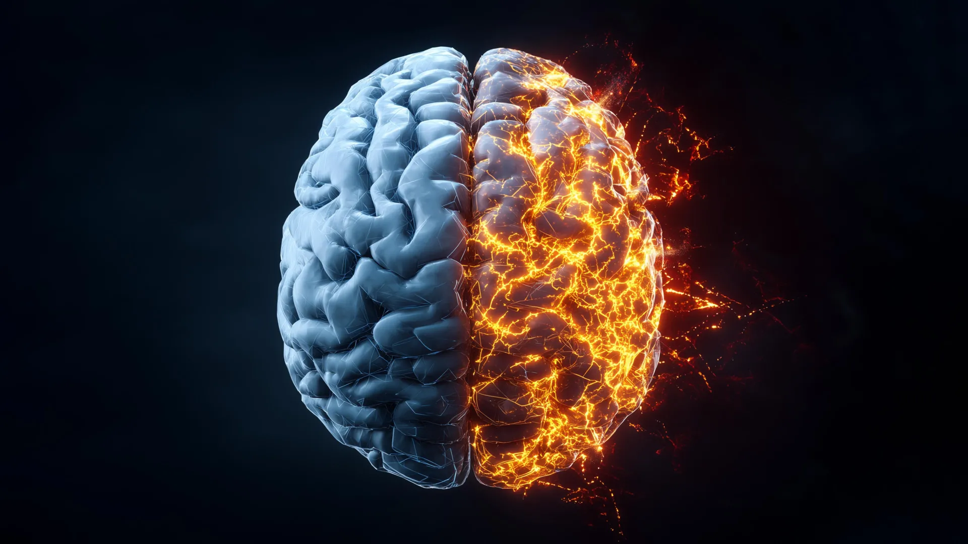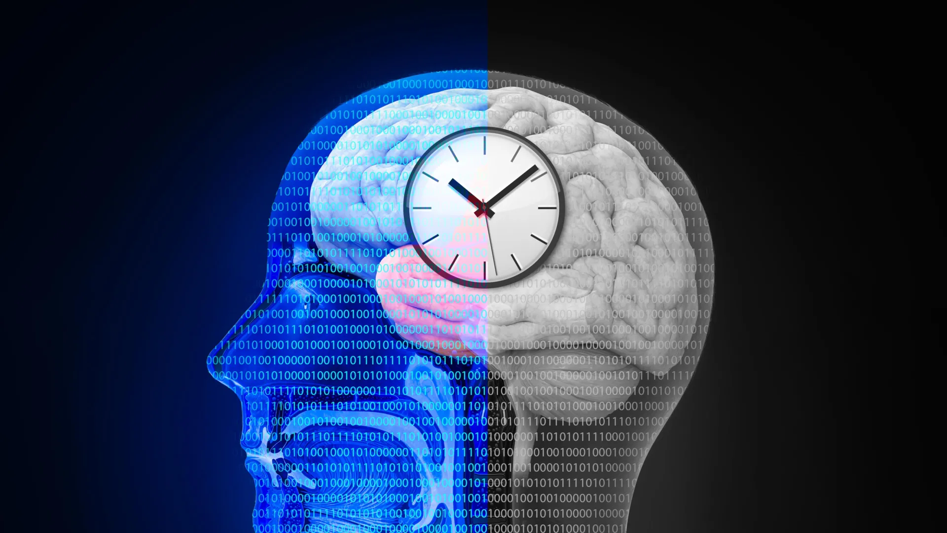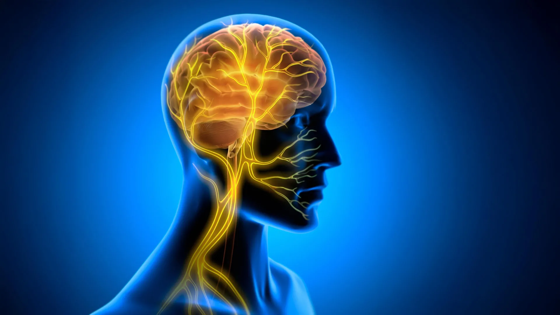
A groundbreaking study published in the open-access journal PLOS Medicine has unveiled significant evidence that various neurodegenerative, psychiatric, and addiction-related conditions are tethered to distinct patterns of accelerated brain aging. Led by Shile Qi of the Nanjing University of Aeronautics and Astronautics in China, the research utilizes predictive age difference (PAD) metrics to demonstrate that the human brain does not age uniformly across all diagnostic categories. By comparing chronological age against "brain age" derived from structural magnetic resonance imaging (MRI), the research team has mapped how different disorders leave unique signatures on the physical structure of the brain.
The Methodology of Predictive Age Difference
The core of this investigation relies on the concept of predictive age difference (PAD). In neuroimaging research, scientists have developed algorithms trained on vast datasets of healthy individuals to predict an individual’s age based solely on the structural integrity, volume, and atrophy patterns of their brain tissue. When a subject’s brain scan suggests an age significantly higher than their actual chronological age, the resulting positive PAD score indicates accelerated brain aging.
For this study, the research team analyzed high-resolution structural MRI data from a massive cohort of 45,900 control subjects, establishing a robust baseline for normative aging. This data was then compared against 2,698 individuals diagnosed with a spectrum of conditions, including Alzheimer’s disease (AD), mild cognitive impairment (MCI), schizophrenia, bipolar disorder, major depressive disorder, alcohol and tobacco addiction, attention-deficit/hyperactivity disorder (ADHD), and autism spectrum disorder (ASD). This comparative analysis allowed the researchers to isolate whether specific pathologies correlate with a measurable shift in the brain’s "biological clock."
Chronology and Evolution of Brain Aging Research
The study of brain aging through neuroimaging has evolved rapidly over the last two decades. In the early 2000s, brain imaging was primarily used to detect macro-scale lesions or tumors. However, the advent of machine learning in the 2010s allowed researchers to move beyond simple morphology toward predictive modeling.
The current study represents a culmination of years of data integration. By aggregating 45,900 control scans, Qi and his colleagues have bypassed the limitations of smaller, localized studies that often lacked statistical power. The project, supported by the Key Research and Development Plan of Jiangsu Province and the National Natural Science Foundation of China, highlights a shift in focus: from asking "is the brain damaged?" to asking "how much has the aging process been accelerated by these specific conditions?"
Findings: Hierarchy of Neurobiological Impact
The research results reveal a clear hierarchy of impact. Neurodegenerative conditions—specifically Alzheimer’s disease and mild cognitive impairment—exhibited the strongest associations with elevated PAD values. This finding is consistent with the clinical progression of these diseases, where progressive neuronal loss and atrophy are hallmark features.
However, the study extends beyond neurodegeneration. Psychiatric conditions, including schizophrenia, bipolar disorder, and major depressive disorder, also showed measurable increases in PAD. This suggests that the biological burden of these conditions is not merely functional or chemical but involves structural modifications that mirror the aging process. Interestingly, the study noted that while addiction—specifically to alcohol—showed significant association with increased PAD, no statistically significant difference in PAD was found between the control group and individuals with ADHD or ASD. This suggests that the neurological signatures of neurodevelopmental conditions may not follow the same trajectory of "accelerated aging" as degenerative or psychiatric illnesses.
Spatial Distribution: Mapping the Damage
One of the most innovative aspects of the study is the mapping of PAD across specific brain regions. The researchers found that different disorders target distinct neurological architecture:
- Prefrontal Cortex: This region demonstrated elevated PAD across the majority of the conditions studied, confirming its role as a hub for cognitive decline and psychiatric dysfunction.
- Frontal and Temporal Lobes: These areas were particularly affected in individuals with psychiatric disorders, potentially explaining the emotional and cognitive deficits associated with these conditions.
- Dementia-Specific Patterns: In patients with Alzheimer’s and MCI, the researchers observed significantly higher PAD in the frontal and occipital cortex, providing a visual confirmation of the widespread degradation characteristic of these diseases.
- Addiction Signatures: The brain regions associated with addiction—the default mode network, the salience network, the putamen, and the thalamus—showed unique aging patterns. These networks are critical for reward processing, decision-making, and self-referential thought, all of which are frequently compromised in chronic addiction.
Biological Mechanisms and Gene Transcription
To understand why these structural changes occur, the research team correlated their imaging findings with gene expression data. They discovered that the patterns of brain aging identified in the scans were linked to distinct biological pathways. This implies that the accelerated aging observed is not a generic response to stress or disease, but rather a manifestation of specific underlying biological processes—such as inflammation, oxidative stress, or disrupted cellular repair—that vary depending on the condition.
Clinical Implications and Future Biomarkers
While the study is strictly correlational, its implications for clinical practice are substantial. Currently, diagnosing conditions like Alzheimer’s or schizophrenia often relies on symptomatic assessment, which can be subjective and slow. The ability to use PAD as a quantitative, objective biomarker could revolutionize early detection.
"Different neurological disorders appear to leave different signatures on the brain aging clock," the authors noted. If these signatures can be refined, clinicians might one day use a standard MRI scan to determine not only the presence of a disorder but the specific "biological age" of the brain, allowing for more personalized treatment plans.
However, experts caution that these results must be interpreted with nuance. Many patients suffer from comorbidities; for instance, individuals with schizophrenia often face higher rates of substance abuse, making it difficult to determine whether accelerated aging is a direct result of the primary diagnosis or a secondary effect of lifestyle factors and long-term medication use.
The Path Forward: Limitations and Challenges
The researchers acknowledge that the study does not establish causality. The data provides a snapshot of the brain at a specific point in time, and longitudinal studies will be necessary to observe how these PAD values evolve as a disease progresses. Furthermore, while the sample size of 45,900 is impressive, future research must ensure that data is representative of diverse populations, as aging processes can be influenced by environmental, nutritional, and socioeconomic factors that may not be fully captured in existing brain imaging databases.
Despite these challenges, the research provides a vital roadmap for future investigation. By shifting the focus toward the "brain clock," the scientific community now has a more granular understanding of how diverse pathologies converge on the physical structure of the human brain. As the global population ages and the incidence of neurological conditions continues to rise, the ability to identify these signatures could be the key to developing therapeutic interventions that slow, or perhaps one day halt, the process of accelerated brain aging.
The study stands as a testament to the power of big data in medicine. By synthesizing thousands of scans into a coherent model of brain health, Qi and his team have provided a framework that could influence neurobiology for years to come. Whether through the development of new diagnostic tools or the exploration of the genetic pathways identified in this study, the findings underscore a critical reality: the brain’s age is not merely a number, but a complex, readable record of a life lived and the pathologies that have challenged it.