Scientists at the Indiana University School of Medicine have successfully developed a pioneering imaging technique designed to overcome decades-long technological barriers in studying mouse bone marrow. By utilizing the advanced multiplex imaging capabilities of the Phenocycler 2.0 platform, researchers have charted a new path for investigating the deeply complex, gelatinous, and hard-encased tissue that serves as the primary factory for blood and immune cells. This breakthrough, recently detailed in the peer-reviewed journal Leukemia, bridges a critical gap in biomedical research, offering unprecedented resolution and panoramic visibility into the cellular microenvironment of bone marrow. The development arrives at a pivotal moment in translational medicine, providing pharmaceutical developers and academic researchers with a sophisticated analytical lens to accelerate therapies targeting hematological malignancies, severe autoimmune conditions, and degenerative musculoskeletal disorders.
Main Facts of the Research and Technological Innovation
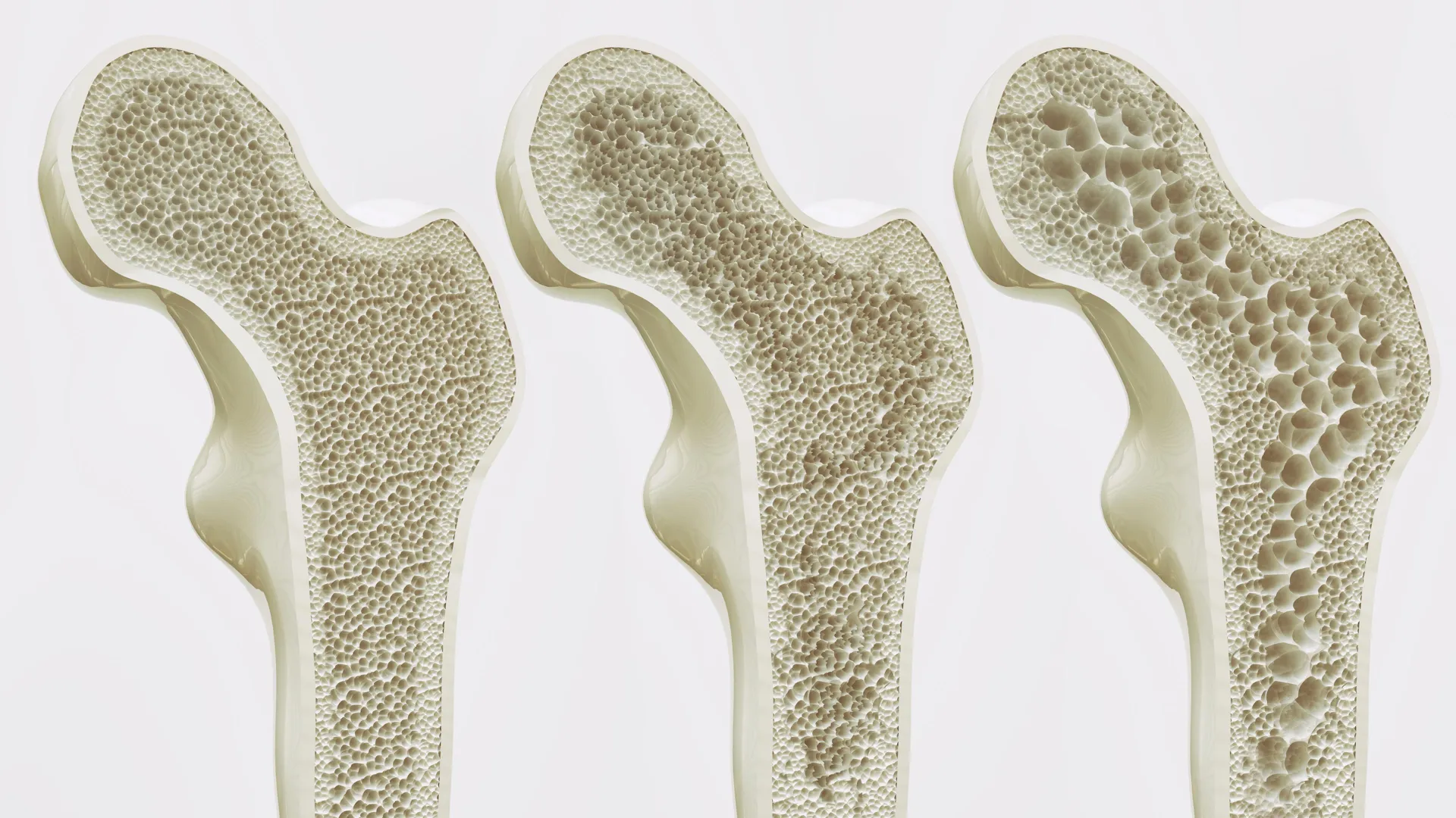
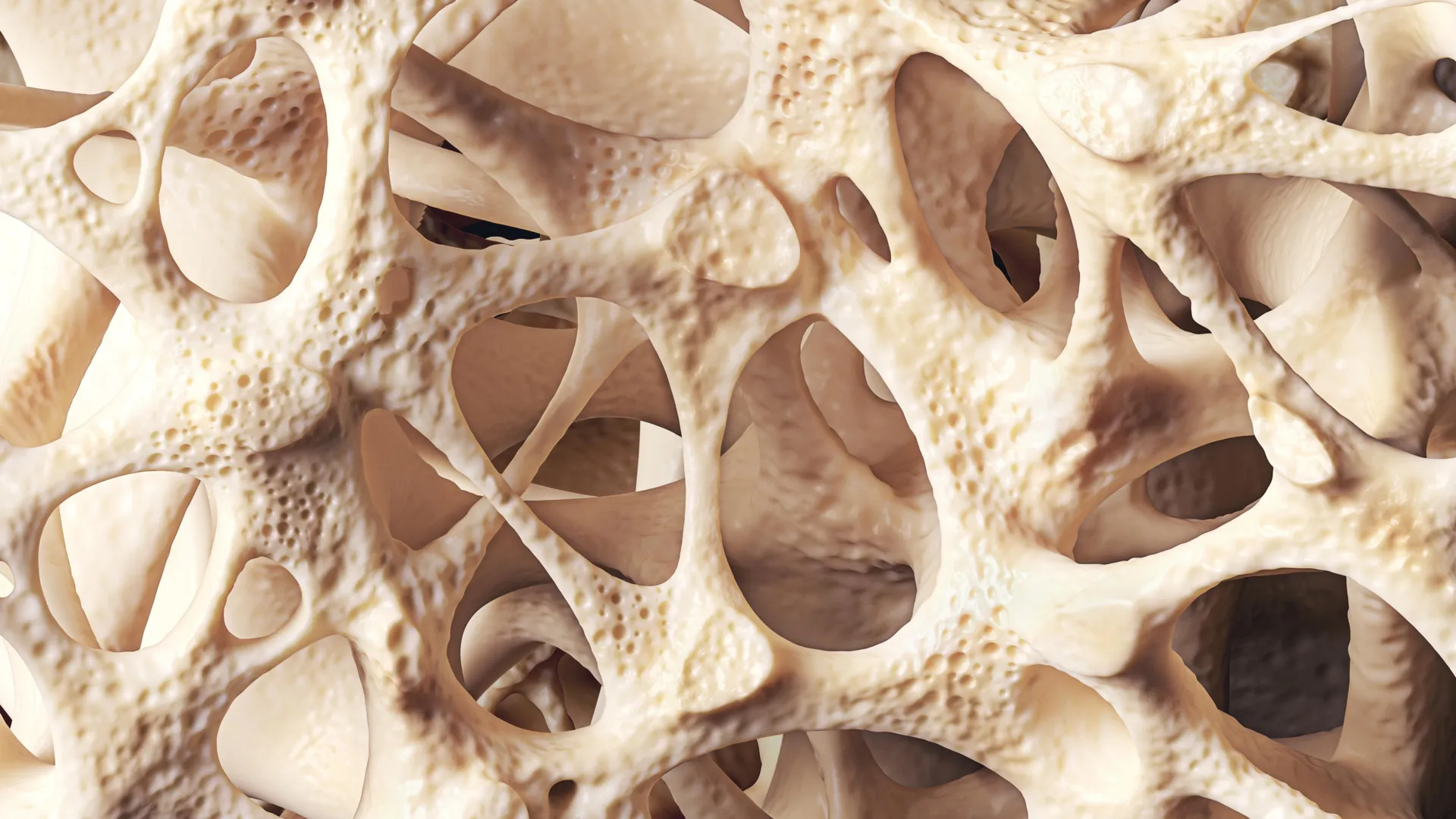
At the core of this scientific advancement is the successful adaptation of the Phenocycler 2.0 multiplex imaging tool to intact murine bone marrow tissue. Historically, bone marrow has presented a notoriously stubborn challenge to microscopists and cellular biologists. Its unique physical composition—a soft, gelatinous core protected by a rigid, mineralized outer shell of cortical and trabecular bone—makes standard tissue sectioning, preservation, and visualization exceptionally difficult without introducing structural artifacts or destroying cellular architecture.
Traditionally, researchers relied heavily on two primary methodologies to analyze bone marrow: flow cytometry and standard fluorescence microscopy. While valuable, both approaches carry inherent and severe limitations. Flow cytometry demands the mechanical and enzymatic dissociation of tissues into single-cell suspensions. Consequently, this destruction of the native tissue architecture strips away vital spatial context, rendering it impossible to determine precisely where specific cells resided relative to one another, blood vessels, or bone surfaces.
Standard fluorescence imaging preserves spatial context to a degree, but it is typically restricted by spectral overlap to detecting only three to four cellular markers simultaneously. This narrow window prevents researchers from observing the vast, intricate networks of interacting cell types that govern hematopoiesis—the production of all cellular blood components—and immune responses.
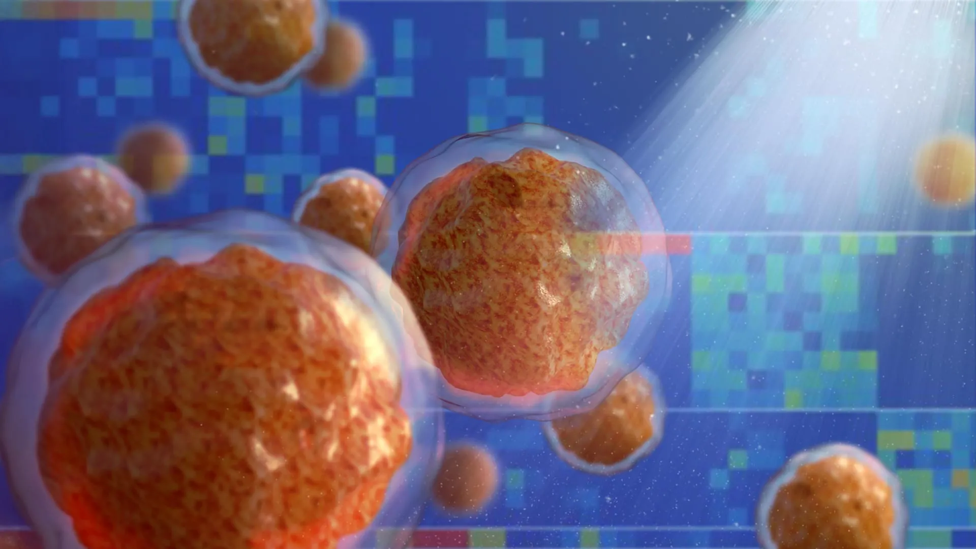
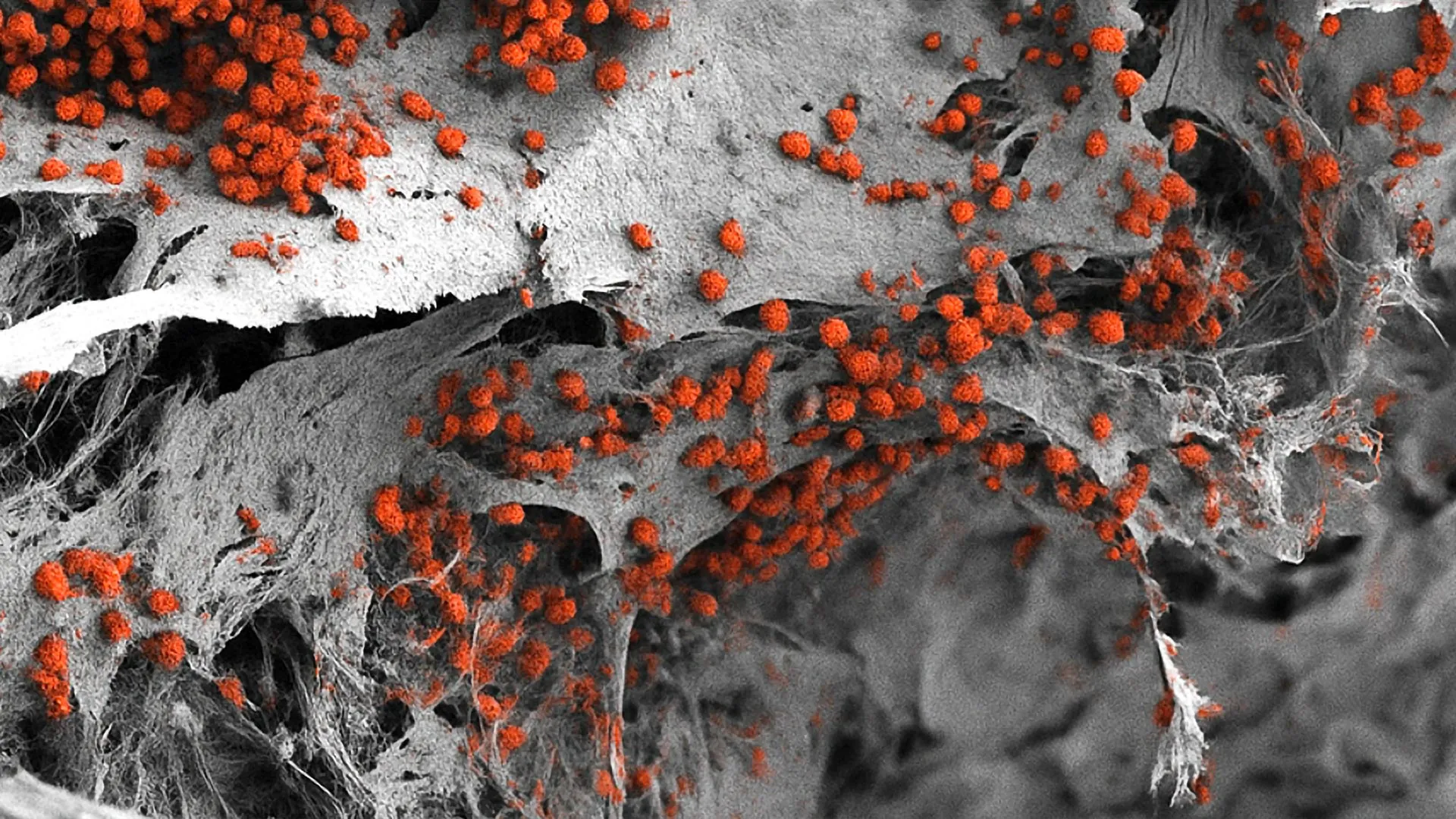
The newly refined methodology at the Indiana University School of Medicine shatters these technical boundaries. By applying the Phenocycler 2.0 system, the research team successfully visualized an unprecedented 25 distinct cellular markers concurrently within completely intact, undisturbed bone marrow tissue. This quantum leap in multiplexing capacity allows scientists to map multiple lineages of hematopoietic stem cells, mature immune cells, stromal elements, and vascular networks in a single, high-resolution view. By maintaining the spatial topography of the tissue while multiplying the visible markers by more than sixfold compared to traditional modalities, the IU team has created a powerful window into the microscopic choreography of bone marrow pathology.
Background Context and Chronology of the Project
The journey toward this methodological breakthrough represents a convergence of advanced spatial biology instruments and decades of institutional expertise in hematology at Indiana University. The project was spearheaded by the IU Cooperative Center of Excellence in Hematology (IUCCEH), a hub dedicated to fostering cutting-edge research in blood disorders and bone marrow biology.
While the Phenocycler 2.0 platform has seen prior deployment across the biomedical research community, its application had largely been restricted to structurally straightforward solid organs, such as the spleen, kidneys, lymph nodes, and certain tumor biopsies. Adapting the technology to withstand the rigors of decalcified, mineralized bone environments containing gelatinous marrow required meticulous protocol optimization. The IUCCEH research team spent months refining tissue preparation, antibody conjugation, and cyclic staining protocols to prevent tissue degradation while ensuring clear signal-to-noise ratios across all 25 concurrent markers.
The collaborative nature of the study brought together multidisciplinary experts spanning orthopedic surgery, pediatric research, hematology, and commercialization. Sonali Karnik, PhD, assistant research professor of orthopedic surgery at the IU School of Medicine, served as co-lead author alongside a dedicated team of investigators. The study also drew critical leadership and oversight from Reuben Kapur, PhD, director of the Herman B Wells Center for Pediatric Research and co-director of the IUCCEH, who served as co-senior author.
Following the successful completion of the validation studies and the publication of their findings in Leukemia, the institutional momentum behind the discovery accelerated rapidly. Recognizing the profound commercial and clinical potential of the imaging workflow, the Indiana University Innovation and Commercialization Office formally filed a provisional patent for the methodology. This legal step paves the way for potential industry partnerships, licensing agreements, and eventual commercial kits that could standardize bone marrow multiplexing for laboratories worldwide.
Supporting Data and Collaborative Contributions
The breadth of the study is reflected in its extensive roster of contributors and the rigorous validation steps undertaken to prove the reliability of the 25-marker panel. The research team systematically tested and verified antibodies against a vast array of hematopoietic populations, ensuring that marker signals did not cross-react or degrade during the cyclic staining and bleaching phases characteristic of the Phenocycler technology.
In addition to Drs. Karnik and Kapur, the comprehensive study involved a robust team of IU School of Medicine researchers, including Connor Gulbronson, Paige C. Jordan, Rahul Kanumuri, Baskar Ramdas, Ramesh Kumar, Melissa L. Hartman, Izza Khurram, Drew M. Brown, Karen E. Pollok, Pratibha Singh, and Melissa A. Kacena. Each contributor brought specialized expertise ranging from bone biology and flow cytometry to advanced computational image analysis, which was necessary to stitch together and interpret the massive datasets generated by 25-channel imaging.
Financial backing for the project was provided by the National Institutes of Health (NIH), underscoring the federal recognition of bone marrow research as a high-priority area for translational medicine. The sustained support from NIH grants highlights the urgency with which funding agencies view the need for better tools to dissect complex tissue microenvironments, particularly those tied to treatment-resistant blood cancers and degenerative bone diseases.
Official Responses and Perspectives from Leadership
The implications of this technical leap have resonated deeply within the academic and clinical leadership at Indiana University, who view the innovation as a transformative asset for both basic science and applied drug discovery.
"Bone marrow is difficult to study because it is gelatinous and encased in hard bone," explained Dr. Sonali Karnik, emphasizing the physical hurdles that historically frustrated researchers. "Since bone marrow plays an important role in blood and immune cell formation and houses valuable stem cells, our unique imaging approach offers a useful tool for a variety of research applications."
Dr. Reuben Kapur expanded on the broader utility of the breakthrough, particularly regarding its reliance on murine models to mirror human pathology. "Because mouse models are widely used to study human diseases, this technique offers a promising new method for investigating a range of conditions like autoimmune diseases, leukemia and other disorders involving bone marrow," Dr. Kapur noted. By establishing a reliable framework for multiplex imaging in mice, researchers can now track subtle cellular shifts in real time across disease progression and therapeutic intervention.
Institutional colleagues have likewise praised the collaborative synergy that allowed the IUCCEH team to conquer a challenge that had stymied other laboratories. By pairing orthopedic expertise with hematological and pediatric research infrastructure, the team built a versatile pipeline capable of addressing questions previously thought too complex for spatial biology platforms.
Broader Impact and Future Implications for Medicine
The successful deployment of this imaging technique carries far-reaching implications across multiple fields of biomedicine, drug development, and clinical diagnostics.
Advancing Oncology and Leukemia Research
Hematological malignancies such as acute myeloid leukemia (AML), multiple myeloma, and various lymphomas originate and evolve within the bone marrow microenvironment. Tumor cells often hijack normal hematopoietic niches, outcompeting healthy stem cells and evading chemotherapeutic agents through intimate cell-to-cell contacts with stromal cells. By enabling researchers to visualize 25 or more markers simultaneously in intact tissue, the IU methodology allows oncologists and pharmacologists to map the exact spatial topography of tumor cells relative to blood vessels, immune cells, and protective bone niches. This high-definition mapping can reveal mechanisms of drug resistance, expose vulnerable microenvironmental dependencies, and guide the rational design of combination therapies that disrupt survival signals within the marrow.
Unlocking Autoimmune and Musculoskeletal Disorders
Beyond oncology, diseases such as rheumatoid arthritis, lupus, and various bone-wasting disorders involve chronic inflammation and pathological remodeling within the bone marrow and adjacent skeletal structures. Understanding how autoreactive immune cells are generated, maintained, or recruited within marrow niches requires precise spatial data. The new imaging tool equips immunologists to track immune cell maturation and signaling networks with unprecedented clarity, potentially illuminating novel therapeutic targets for conditions that currently lack curative treatments.
Next-Phase Technological Expansion
The research team is far from resting on its initial success. Having proven the viability of the 25-marker panel, the IU innovators are actively working to expand the scope and scale of the platform. Current developmental efforts focus on incorporating additional structural and functional features into the panel, including bone matrices, neural networks, muscle tissues, and an even broader array of immune and signaling cell types. By expanding the panel into a truly multi-systemic toolkit, the researchers aim to capture the dynamic interplay between skeletal, neural, and hematopoietic systems within a single imaging run.
As the provisional patent process moves forward and collaborative inquiries increase from academic institutions and pharmaceutical partners alike, the Indiana University School of Medicine stands at the forefront of a spatial biology revolution. By lifting the veil on the hidden architecture of bone marrow, this breakthrough imaging technique promises to accelerate the translation of laboratory discoveries into life-saving therapies for patients worldwide.