

Scientists at the Johns Hopkins Kimmel Cancer Center and the Johns Hopkins Bloomberg School of Public Health have unlocked a critical biological mechanism that fuels the progression of a rare and aggressive form of kidney cancer. Published on April 22 in the journal Cell Reports, the study reveals that abnormal proteins produced by rearranged fusion genes assemble into microscopic liquid droplets within cellular nuclei. These condensates act as hyperactive control centers, modulating gene expression to accelerate tumor proliferation and metastasis. Backed by funding from the National Institutes of Health (NIH), the discovery not only illuminates the pathology of translocation renal cell carcinoma (tRCC) but also establishes a promising blueprint for targeted pharmacological interventions where standard treatments currently do not exist.
The Chronology of the Discovery: From Genetic Swaps to Condensate Physics
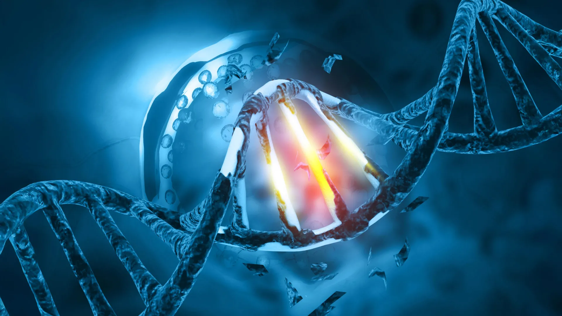
The roots of this breakthrough stretch back years, building upon foundational knowledge in molecular oncology regarding chromosomal translocations. Translocation renal cell carcinoma arises when genetic material is incorrectly repaired following a chromosomal break. During this process, a segment of the TFE3 gene from the end of a chromosome fuses with the beginning portion of one of roughly 20 distinct partner genes, most frequently NONO, SFPQ, or PRCC. While oncologists and geneticists have long recognized that these genetic fusions trigger tRCC, the precise biochemical trajectory from a mutated gene to unchecked cellular growth remained elusive.
To demystify this process, senior author Danfeng "Dani" Cai, Ph.D., an assistant professor of biochemistry and molecular biology at the Johns Hopkins Bloomberg School of Public Health, and her investigative team embarked on a high-resolution imaging initiative. Utilizing cells harvested from patients diagnosed with the rare malignancy, researchers engineered glowing molecular tags onto the aberrant TFE3 fusion proteins.
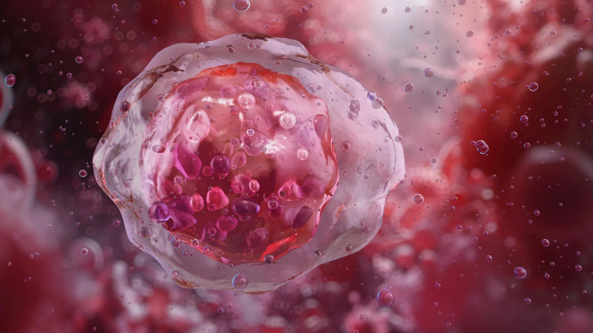
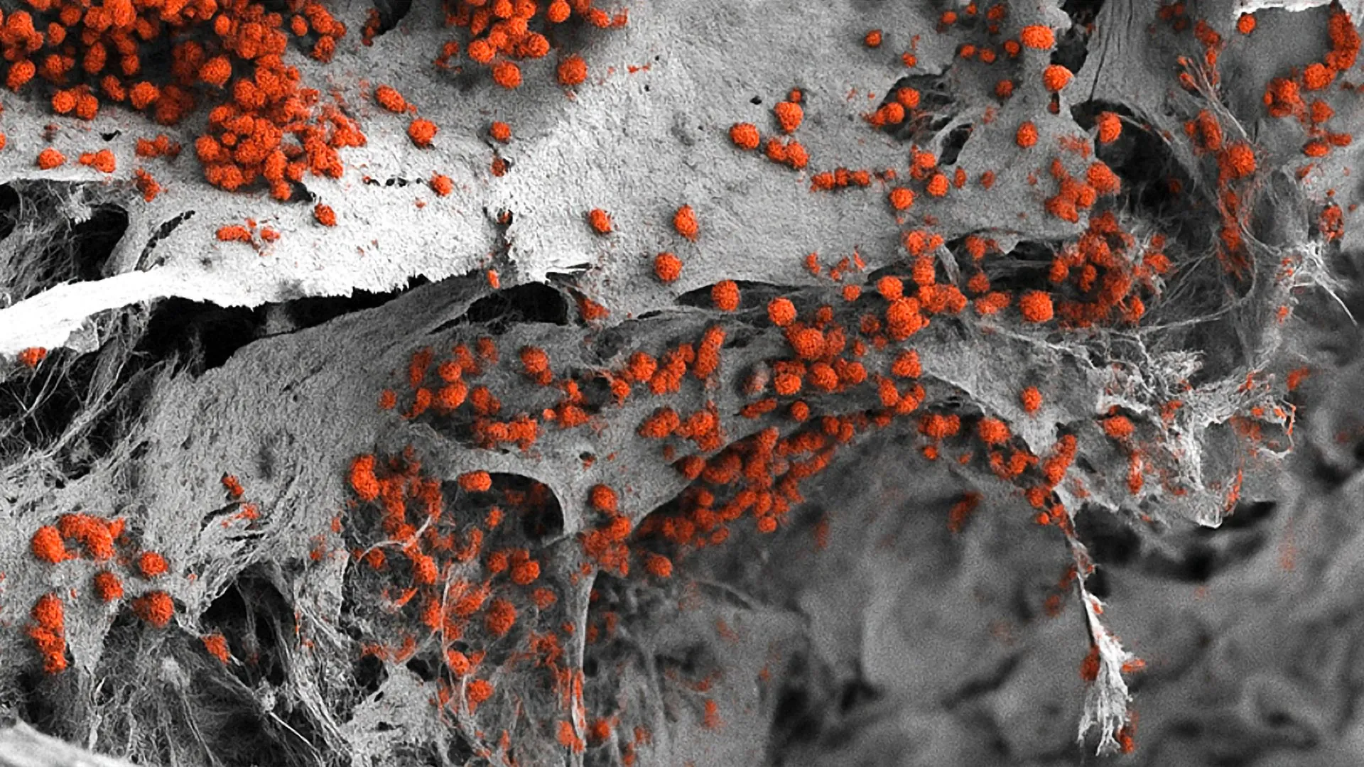
Using advanced microscopy, the team observed that these tagged proteins did not disperse randomly throughout the cell. Instead, they concentrated within the cell nucleus—the vault where genomic DNA is stored—forming distinct punctate structures. Further analysis confirmed that these dots were liquid condensates, specialized biochemical compartments formed via liquid-liquid phase separation. Within these droplets, the fusion proteins colocalized with active transcription markers and transcriptional machinery, strongly implying a direct role in driving gene transcription.
Decoding the Chromatin Landscape
To understand how these condensates physically manipulate cellular DNA, Dr. Cai partnered with co-author Eneda Toska, Ph.D., an assistant professor of oncology at the Johns Hopkins Kimmel Cancer Center. Genomic DNA within human cells is densely packed and organized around histone proteins into a structure known as chromatin, which structurally resembles beads on a string. Tightly wound regions of chromatin generally repress gene expression by denying transcriptional machinery access to the DNA, whereas open regions facilitate active transcription.
Drs. Cai and Toska mapped the genomic interactions of the TFE3 fusion proteins to observe how they modify this architectural landscape. Their investigations revealed that the fusion proteins induce targeted chemical modifications that dynamically open and close specific chromatin sites. By reshaping the chromosomal landscape, these proteins unlock access to genes that govern cell proliferation and cellular motility—two biological functions that malignant tumors hijack to grow and metastasize.
"We found that these fusion proteins open and close different sites on the chromatin by making chemical modifications," Dr. Toska explained. "They bind, regulate and redesign the chromosome landscape, interacting with target genes that promote cell proliferation and movement."
Deconstructing the Molecular Machinery
To confirm whether the liquid condensate structure was fundamentally responsible for this tumorigenic gene regulation, the researchers performed a series of targeted structural mutations. By systematically editing out specific segments of the TFE3 fusion proteins, the team sought to identify the precise physical domains required to maintain droplet formation.
The breakthrough came when they removed a small segment responsible for creating a coiled-coil—a structural motif where alpha-helices wrap around each other—located in the bridge connecting the TFE3 tail to its fusion partner. Without this structural motif, the modified fusion proteins lost their ability to phase-separate into liquid droplets. Crucially, once the droplets failed to form, the proteins simultaneously lost their capacity to activate cancer-promoting genes.
This structural vulnerability points directly toward a novel therapeutic strategy. If scientists can design small-molecule inhibitors or drugs capable of chemically disrupting these liquid condensates, they may be able to neutralize the oncogenic drivers of tRCC without necessarily having to target every downstream signaling pathway individually.
Supporting Data and Prevalence in Oncology
Translocation renal cell carcinoma represents a distinct clinical challenge within modern oncology. While clear cell renal cell carcinoma accounts for the vast majority of adult kidney cancers, tRCC disproportionately affects younger populations, including children, adolescents, and young adults. Among the myriad genetic partners capable of fusing with TFE3, the research team concentrated their efforts on the two most statistically prominent drivers: NONO and SFPQ. Combined, these two fusion variants account for approximately 40 percent of all documented TFE3 fusions in clinical tRCC cases.
Furthermore, the implications of this study extend well beyond the boundaries of kidney oncology. Phase separation and liquid-liquid condensate formation have emerged over the last decade as a fundamental organizing principle in cell biology, but their direct role in human pathology is only recently coming to light. Other malignancies driven by chromosomal translocations—such as Ewing sarcoma, a bone-and-soft-tissue cancer, and various forms of acute leukemia—depend on aberrant fusion proteins to subvert normal cellular control.
"Other cancers, such as Ewing sarcoma and leukemia, are caused by fusion genes as well," noted Dr. Cai. "It’s possible that these fusion genes form similar droplets, or condensates, that regulate genes in these cancers and could react to similar treatment strategies."
Official Responses and Collaborative Framework
The breadth of the study required a multidisciplinary collaboration spanning biophysics, molecular biology, clinical oncology, and computational genomics. The research team integrated expertise from multiple departments within Johns Hopkins University, alongside key contributions from the National Cancer Institute (NCI).
Co-authors contributing to the published findings included Choon Leng So, Ye Jin Lee, Wanlu Chen, Binglin Huang, Emily De Sousa, Yangzhengyu Gao, Marie Elena Portuallo, Sumaiya Begum, Kasturee Jagirdar, Vito Rebecca, and Hongkai Ji from the Johns Hopkins Bloomberg School of Public Health; Bujamin Vokshi from the Johns Hopkins University School of Medicine; and W. Marston Linehan from the National Cancer Institute.
Financial support for the investigation was sustained through a diverse portfolio of federal grants and institutional awards. Primary funding was provided by the National Institutes of Health, specifically the National Institute of General Medical Sciences (grant R35GM142837) and the National Cancer Institute (grants K22CA245487, R01CA276187, and K01CA245124). Additional backing originated from the National Human Genome Research Institute (grants R01HG013409 and R01HG010889), a Department of Defense Kidney Cancer Idea Development Award (grant W81XWH2210900), a Jayne Koskinas Ted Giovanis grant, and a Johns Hopkins Provost Catalyst Award.
In accordance with institutional disclosure policies, study co-author Dr. Toska reported receiving research grants from AstraZeneca, alongside consulting fees from AstraZeneca and Menarini.
Broader Impact and Clinical Implications
The absence of standardized, effective treatment regimens for patients diagnosed with advanced or metastatic translocation renal cell carcinoma has long frustrated clinicians. Traditional chemotherapy and standard immunotherapy approaches often yield suboptimal results in these specific genetically driven tumors, leaving patients and physicians with limited clinical options.
By reframing the pathology of tRCC through the lens of biophysical chemistry—specifically the formation of pathologic phase-separated condensates—this study transitions the therapeutic search from abstract protein inhibition to targeted structural disruption. The research team’s immediate future objectives involve cataloging all remaining protein components localized within these nuclear droplets. Pinpointing every cooperating molecule inside the condensate will enable high-throughput drug screening initiatives to identify pharmacological agents capable of dissolving or preventing the formation of these structures.
If validated in upcoming preclinical and clinical trials, the strategy of targeting liquid-liquid phase separation could revolutionize the therapeutic landscape not only for rare pediatric and adult kidney cancers, but potentially for an array of recalcitrant, fusion-driven malignancies across human oncology.