
In a landmark study that bridges the gap between molecular genetics and clinical psychiatry, researchers at McGill University and the Douglas Research Centre have identified specific cellular malfunctions within the human brain that characterize major depressive disorder. The study, recently published in the prestigious journal Nature Genetics, represents a significant leap forward in understanding the biological architecture of mental illness. By utilizing high-resolution genomic mapping, the research team discovered that two distinct types of brain cells—excitatory neurons and microglia—exhibit abnormal activity patterns in individuals who suffered from depression. This discovery not only provides a clearer picture of the physiological disruptions associated with the condition but also challenges long-standing societal stigmas by reinforcing the fact that depression is a measurable biological disorder.
Major depressive disorder (MDD) is a global health crisis, affecting more than 264 million people according to the World Health Organization. It remains a leading cause of disability worldwide, contributing significantly to the global burden of disease. Despite its prevalence, the underlying biological mechanisms have remained partially obscured for decades, often leading to a "one-size-fits-all" approach to treatment that fails a significant portion of the patient population. The findings from McGill University offer a roadmap for moving away from generalized treatments toward precision medicine, where therapies could be designed to target the specific cellular pathways identified in this research.
The Technological Vanguard: Single-Cell Genomics
The breakthrough was made possible through the application of advanced single-nucleus chromatin accessibility profiling and RNA sequencing. Unlike traditional methods that analyze bulk brain tissue—which can mask the unique signatures of individual cells—these single-cell techniques allow scientists to zoom in on the genetic "switches" within thousands of individual cells. By examining both the RNA (which indicates which genes are currently active) and the DNA (the underlying code and its regulatory mechanisms), the team was able to pinpoint exactly where the molecular machinery was failing.
Dr. Gustavo Turecki, the study’s senior author and a professor at McGill University’s Department of Psychiatry, emphasized the precision of this approach. Dr. Turecki, who also serves as the Canada Research Chair in Major Depressive Disorder and Suicide, noted that this is the first time researchers have been able to map gene activity alongside the mechanisms that regulate the DNA code specifically within the context of depression. This dual-layered analysis provided a high-definition view of the disruptions occurring in the brain’s regulatory landscape.
The Critical Role of the Douglas-Bell Canada Brain Bank
A pivotal component of this discovery was the access to rare and high-quality brain tissue. The research team relied on post-mortem samples provided by the Douglas-Bell Canada Brain Bank (DBCBB). Established in 1980, the DBCBB is one of the world’s most significant repositories of human brain tissue, containing over 3,000 samples from individuals who lived with various neurodegenerative and psychiatric conditions.
The study analyzed samples from 100 individuals: 59 who had been diagnosed with major depressive disorder and 41 who served as a control group. The ability to compare these two cohorts at a cellular level was essential for identifying the specific anomalies associated with depression. The use of post-mortem tissue is often the only way to study the human brain with this level of molecular detail, as live imaging technologies like MRI or PET scans, while useful, do not yet have the resolution to observe individual gene expressions within specific cell types.
Identifying the Culprits: Excitatory Neurons and Microglia
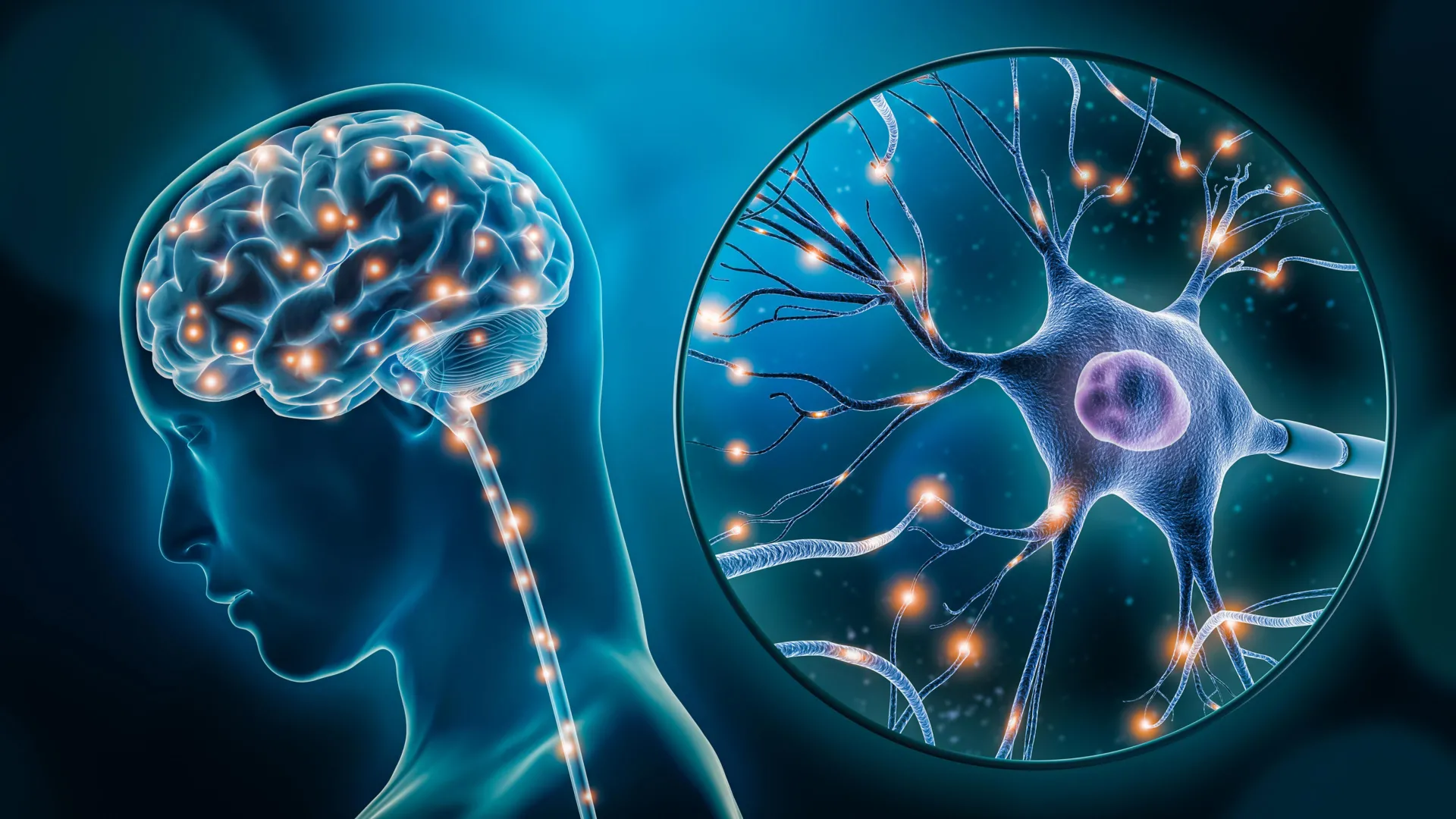
The research highlighted two primary cell types that behave differently in the depressed brain. The first are excitatory neurons, which are responsible for transmitting "go" signals throughout the brain. These neurons play a fundamental role in regulating mood, cognitive function, and the body’s response to stress. In the samples from individuals with depression, the researchers found that the genes responsible for maintaining synaptic plasticity—the brain’s ability to adapt and form new connections—were significantly altered. This suggests that in a depressed state, the brain’s communication networks may become rigid or inefficient.
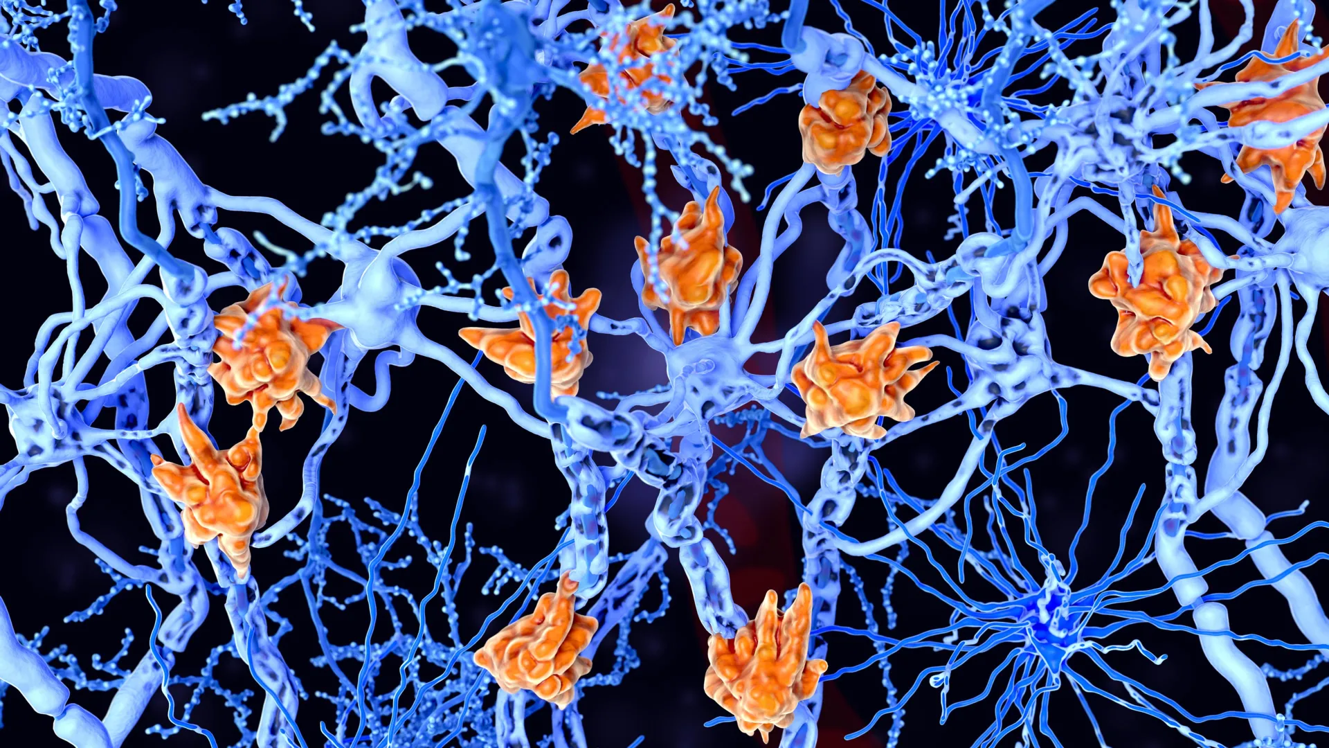
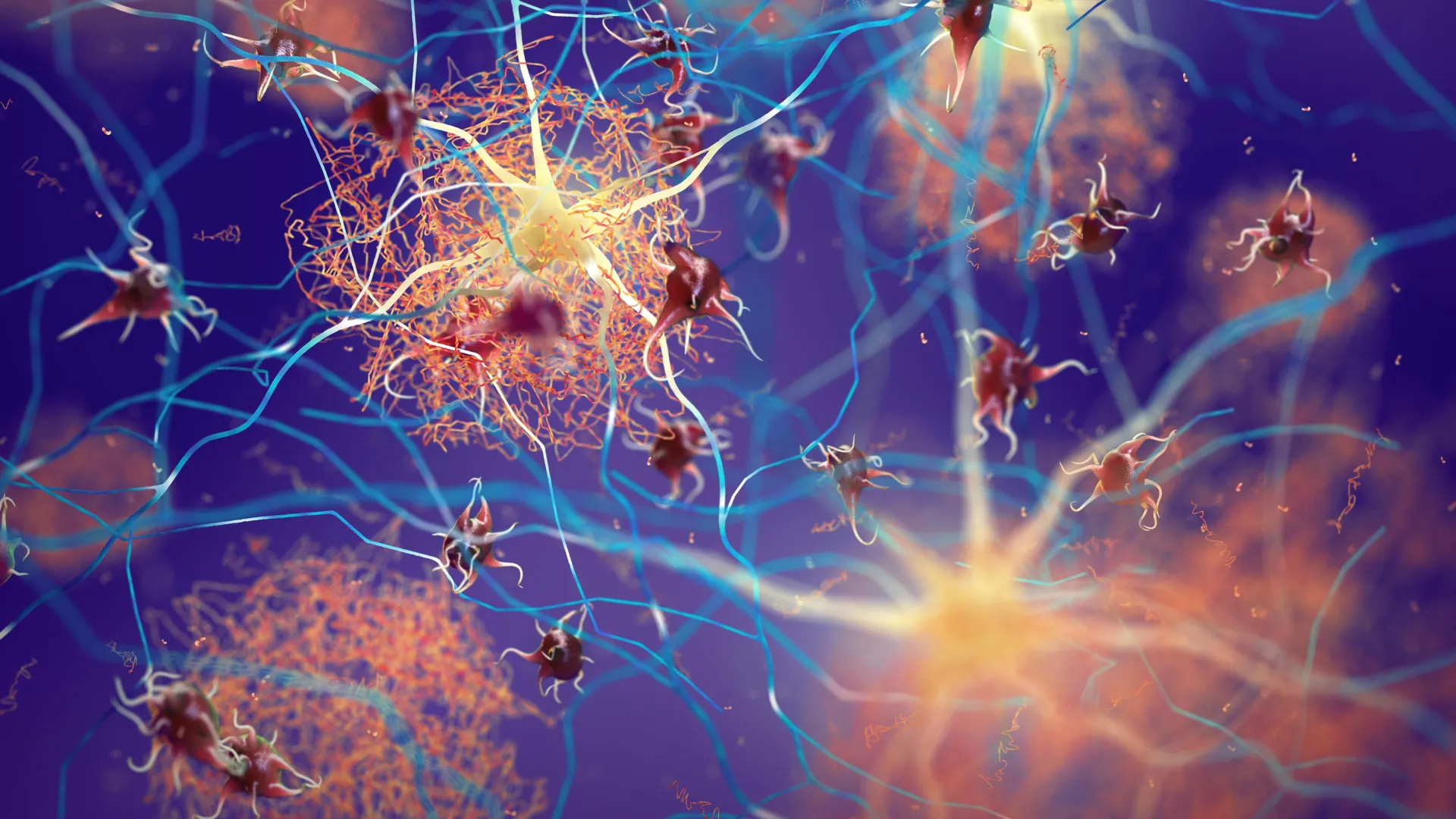
The second key finding involved microglia, which are the primary immune cells of the central nervous system. Historically viewed merely as the brain’s "housekeepers," microglia are now understood to play a sophisticated role in managing neuroinflammation and pruning synapses. The study revealed that a specific subtype of microglia showed altered genetic activity in depressed individuals. This discovery aligns with a growing body of evidence suggesting that neuroinflammation and immune system dysregulation are central components of psychiatric disorders. When microglia do not function correctly, they may trigger inflammatory responses that damage neuronal health and disrupt mood regulation.
A Chronology of Discovery and Research Evolution
The path to this discovery has been built over decades of evolving neurobiological theory. In the mid-20th century, the "monoamine hypothesis" dominated the field, suggesting that depression was simply a "chemical imbalance" of neurotransmitters like serotonin and dopamine. While this led to the development of SSRIs (Selective Serotonin Reuptake Inhibitors), it did not explain why many patients failed to respond to these medications.
By the early 2000s, research began to shift toward the role of neuroplasticity and the "stress-diathesis model," which looks at how genetic predispositions interact with environmental stressors. The Douglas Institute has been at the forefront of this shift. Over the last decade, the integration of "big data" and genomics allowed researchers to move beyond single genes to look at entire genomic networks.
The timeline for this specific study involved several years of meticulous tissue processing and data analysis. Between 2018 and 2023, the team at McGill focused on refining single-nucleus sequencing protocols to ensure that the delicate RNA within post-mortem samples remained intact. The publication in Nature Genetics in 2024 marks the culmination of this rigorous process, providing a definitive link between specific cell-type dysfunctions and the clinical manifestation of depression.
Supporting Data and Global Context
The necessity for this research is underscored by the current limitations in psychiatric care. Clinical data indicates that approximately 30% to 50% of patients with depression do not achieve full remission with first-line antidepressant treatments. Furthermore, "treatment-resistant depression" affects millions, leading to higher rates of hospitalization and suicide.
Data from the World Federation for Mental Health suggests that by 2030, depression will be the leading cause of the global disease burden. The economic impact is equally staggering; in the United States alone, the economic burden of major depressive disorder was estimated at $326 billion annually as of 2020. By identifying that the cellular disruption lies within excitatory neurons and microglia, the McGill study provides a biological rationale for why traditional monoamine-based therapies may be insufficient for many, as those treatments do not directly address the immune-inflammatory pathways or the specific synaptic failures identified in the study.
Reactions from the Scientific Community
While the study was conducted by a specific team at McGill, its implications have resonated throughout the global neuroscience community. Experts in the field of transcriptomics have hailed the study as a "proof of concept" for the power of single-cell analysis in psychiatry.
Inferred reactions from the broader medical community suggest a sense of cautious optimism. Dr. Gustavo Turecki’s findings reinforce the narrative that mental health is physical health. For years, patient advocacy groups have campaigned for depression to be treated with the same medical rigor as diabetes or cardiovascular disease. This study provides the empirical evidence needed to support that shift in perspective. Clinicians have noted that while new treatments may be years away, having a "cellular map" allows for more targeted drug screening, potentially shortening the timeline for drug development.
Broader Impact and the Future of Treatment
The long-term implications of this research are profound. By identifying microglia as a key player, the study opens the door for the exploration of anti-inflammatory drugs as adjunct therapies for depression. Similarly, understanding the genetic switches in excitatory neurons could lead to the development of "neuro-regenerative" therapies that aim to restore synaptic health rather than just modulating neurotransmitter levels.
Furthermore, this research contributes to the de-stigmatization of mental illness. When depression is viewed as a measurable cellular dysfunction, it moves the conversation away from "willpower" or "emotional resilience" and places it firmly in the realm of biological medicine. This can lead to better insurance coverage for treatments, increased public funding for research, and a more empathetic societal approach to those suffering from the condition.
The research team at McGill and the Douglas Institute now plans to expand their investigation. The next phase of the research will involve functional studies, where scientists will attempt to "correct" these cellular disruptions in laboratory models to see if it reverses depressive-like behaviors. They also aim to determine if these cellular signatures can be detected through more accessible means, such as blood tests or advanced neuroimaging, which could eventually lead to a biological diagnostic tool for depression.
Acknowledgments and Institutional Support
The study, titled "Single-nucleus chromatin accessibility profiling identifies cell types and functional variants contributing to major depression," was a collaborative effort involving several prominent researchers, including lead author Anjali Chawla. The success of the project was bolstered by significant financial support from the Canadian Institutes of Health Research (CIHR), the Brain Canada Foundation, and the Fonds de recherche du Québec – Santé (FRQS). Additionally, the Healthy Brains, Healthy Lives (HBHL) initiative at McGill University provided the interdisciplinary framework necessary to handle the massive datasets generated by the genomic sequencing.
As the scientific community continues to digest these findings, the work of Turecki and his colleagues stands as a testament to the power of modern genomics. It provides a bridge between the microscopic world of the cell and the macroscopic reality of human suffering, offering hope that for the millions living with depression, a more precise and effective era of treatment is on the horizon.


Leave a Reply