
The fundamental understanding of how high-energy radiation interacts with biological matter has taken a significant leap forward as an international team of researchers successfully filmed the "roaming" behavior of atoms immediately preceding a molecular explosion. This discovery, centered on a complex process known as electron-transfer-mediated decay (ETMD), reveals that the damage caused by radiation is not merely a static electronic event but is instead a dynamic process steered by the physical movement of atomic nuclei. By observing these "roaming" atoms in real-time, scientists have identified a previously hidden driver of radiation damage that could redefine our approach to radiobiology, cancer treatment, and the development of protective materials.
The Atomic Mechanics of Radiation Damage
Radiation damage is a ubiquitous concern in both medical and industrial contexts. When high-energy particles, such as X-rays or gamma rays, penetrate a living cell, they do not simply pass through harmlessly. Instead, they deposit energy into the atoms and molecules that constitute the cell’s machinery. This energy creates "excited" states, where electrons are pushed into higher energy levels or stripped away entirely, leaving behind volatile ions.
The traditional view of radiation damage often focuses on the direct hit—an X-ray photon striking a DNA strand and snapping its chemical bonds. However, a significant portion of radiation-induced biological damage is indirect, caused by the secondary particles and decay processes that follow the initial ionization. Among these, electron-transfer-mediated decay (ETMD) has emerged as a critical, yet difficult-to-study, mechanism. In ETMD, an atom that has been ionized by radiation stabilizes itself by "stealing" an electron from a neighboring atom. The energy released during this transfer is then used to eject a low-energy electron from a third neighbor. This chain reaction results in multiple ionized particles and a cascade of low-energy electrons, which are particularly effective at breaking chemical bonds in their immediate vicinity.
A Breakthrough in Real-Time Observation
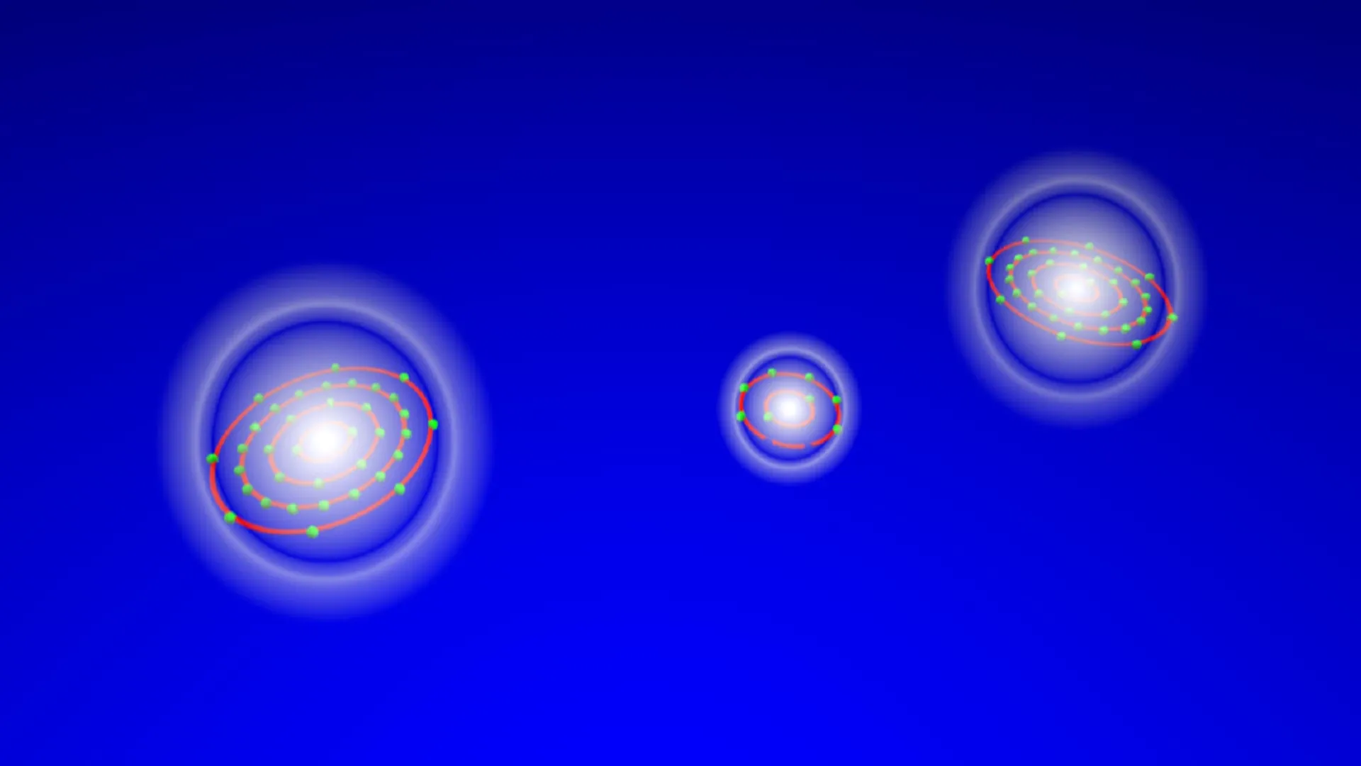
To capture this elusive process, researchers from the Molecular Physics Department, working alongside international collaborators, designed an experiment using a model system consisting of a neon atom weakly bound to two krypton atoms, known as a NeKr2 trimer. This specific configuration was chosen because it represents the simplest possible system capable of undergoing the three-body interaction required for ETMD.
The experiment was conducted at two of the world’s most advanced synchrotron facilities: BESSY II in Berlin and PETRA III in Hamburg. Using soft X-rays, the team targeted the neon atom, knocking out one of its inner-shell electrons and initiating the decay process. To observe what happened next, they utilized a COLTRIMS (Cold Target Recoil Ion Momentum Spectroscopy) reaction microscope. This sophisticated instrument allows scientists to detect the fragments of a molecular explosion and reconstruct the exact positions and momenta of the atoms at the moment of decay.
The results were transformative. Rather than the atoms remaining in a fixed geometry until the moment of explosion, the researchers observed the atoms "roaming"—shifting and reorganizing their spatial arrangement over a period of up to one picosecond. In the realm of atomic physics, a picosecond (one-trillionth of a second) is an immense duration, providing ample time for the nuclei to explore various configurations before the final electronic decay occurs.
Chronology of the Atomic Roaming Process
The study’s timeline of events provides a detailed look at the life cycle of a radiation-induced decay. The process begins with the "trigger" event: the absorption of an X-ray photon by the neon atom, creating a vacancy in its electronic structure.
In the initial femtoseconds (quadrillionths of a second) following excitation, the system remains relatively close to its original, ground-state configuration. However, as time progresses into the picosecond range, the internal forces of the trimer begin to shift. The researchers noted that the atoms do not follow a single, linear path toward decay. Instead, they engage in a "roaming" motion, exploring a wide range of configuration spaces.
At approximately 0.5 picoseconds, a dominant pathway emerges where one krypton atom moves significantly closer to the excited neon atom. This proximity facilitates the transfer of an electron, which is a prerequisite for ETMD. Simultaneously, the second krypton atom may shift farther away or move into a position where it can more easily absorb the released energy and emit a secondary electron. By the time the decay finally occurs, the "shape" of the molecule is often radically different from its starting point. This structural evolution directly dictates the efficiency and the speed of the decay process.
Supporting Data and Simulation Insights
The experimental findings were corroborated by extensive ab initio simulations. These theoretical models tracked thousands of potential atomic pathways, calculating the probability of decay at every possible geometric arrangement. The simulations confirmed that the "roaming" motion is not a random byproduct but a fundamental steering mechanism.
The data revealed that the decay rate is highly sensitive to the distance between the neon and krypton atoms. As the atoms roam and the distance fluctuates, the probability of ETMD increases or decreases by orders of magnitude. This means that the nuclear motion—the physical movement of the "heavy" parts of the atom—effectively controls the timing of the electronic explosion.
Furthermore, the researchers identified that the energy of the emitted electrons is directly tied to the geometric configuration at the moment of decay. In a biological context, this is a crucial finding, as the energy of secondary electrons determines how far they can travel through cellular fluid and how much damage they can inflict on nearby proteins or DNA.
Official Responses and Scientific Significance
The lead authors of the study have emphasized that this work changes the way scientists must think about radiation chemistry. Florian Trinter, one of the lead authors, noted that the ability to "literally watch" the atoms move before decay provides an intuitive understanding that was previously missing. "The decay is not just an electronic process," Trinter stated, "it is steered by nuclear motion in a very direct and intuitive way."
Senior author Till Jahnke echoed this sentiment, highlighting the importance of the configuration space. "The atoms explore large regions of configuration space before the decay finally takes place," Jahnke explained. "This shows that nuclear motion is not a minor correction—it fundamentally controls the efficiency of non-local electronic decay."
The scientific community has responded with high interest, as the study provides the most detailed real-space and real-time view of ETMD to date. By establishing a precise benchmark for a three-atom system, the research provides a foundation for more complex studies involving liquids and biological tissues.
Broader Impact on Medicine and Biology
The implications of this research extend far beyond the laboratory. Understanding the mechanics of ETMD is essential for accurately modeling how radiation affects the human body. In clinical settings, such as proton therapy or traditional X-ray radiation for cancer, the goal is to maximize damage to tumor cells while minimizing damage to healthy tissue.
Because ETMD produces low-energy electrons, it is a major contributor to "local" radiation damage. These electrons have a short range, meaning they deposit all their destructive energy in a very small volume. If scientists can predict and eventually influence the conditions under which ETMD occurs, it may be possible to enhance the effectiveness of radiation therapies or develop new radioprotective drugs that stabilize molecular structures against "roaming" during exposure.
Moreover, the study sheds light on the behavior of radiation in water—the primary component of living cells. In aqueous environments, atoms and ions are constantly moving and interacting with surrounding water molecules. The discovery that "roaming" motion governs decay suggests that the fluid nature of the cellular environment plays a much more active role in radiation damage than previously accounted for in static models.
Future Research and Theoretical Extensions
The successful imaging of atomic roaming in a NeKr2 trimer is just the beginning. The research team aims to extend these insights to more complex systems, including solvated ions and large biomolecules. The ultimate goal is to create a comprehensive "movie" of radiation damage in a living cell, tracking the energy flow from the initial X-ray strike to the final chemical break.
This work also supports the development of new theoretical models. Current software used to calculate radiation doses in hospitals often relies on simplified assumptions about how molecules break down. By integrating the data from this study, theorists can develop more accurate simulations that account for nuclear motion and roaming. This could lead to more precise dosing in radiotherapy, reducing side effects for patients.
In conclusion, the observation of atoms "roaming" before decay marks a milestone in molecular physics. It reveals that the destructive power of radiation is a complex dance between electrons and nuclei, where the physical path taken by an atom determines the ultimate fate of the molecule. As we continue to peel back the layers of these ultrafast processes, our ability to harness radiation for medicine—and protect against its harmful effects—will only grow more sophisticated. The "hidden driver" of radiation damage has been brought into the light, providing a new lens through which we can view the invisible interactions that shape the building blocks of life.


Leave a Reply