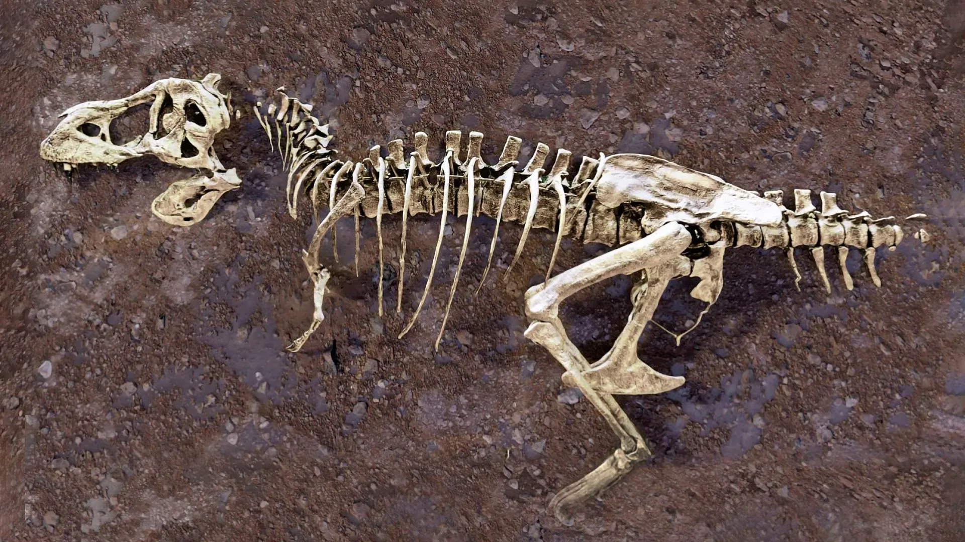
The discovery of a fractured rib within the skeletal remains of Scotty, the world’s largest and oldest known Tyrannosaurus rex, has provided paleontologists with an unprecedented medical record from the Late Cretaceous period. While skeletal remains are the primary window into the anatomy of dinosaurs, the preservation of soft tissue is an occurrence so rare that it is often dismissed as statistically improbable. However, recent advancements in neutron imaging at the Department of Energy’s Oak Ridge National Laboratory (ORNL) have unveiled a hidden network of mineralized blood vessels inside the rib, capturing a biological "snapshot" of a healing injury that occurred 66 million years ago. This discovery not only provides a unique view of dinosaur physiology but also demonstrates how modern particle physics is revolutionizing the field of paleontology.
A Chronology of Discovery: From the Frenchman River to the Particle Accelerator
The journey of Scotty—a specimen that has become a cornerstone of North American paleontology—began in 1991, when researchers from the Royal Saskatchewan Museum unearthed the remains in the Frenchman River Valley. This region of Saskatchewan, Canada, is recognized as a premier site for fossil discovery, offering a geological record that sits just beneath the threshold of the Cretaceous-Paleogene (K-Pg) extinction event.
For decades, the bones were studied using traditional morphological analysis. It was not until 2020 that the investigation took a technical turn. Jerit Mitchell, then an undergraduate physics student at the University of Regina (U of R) working under the guidance of professor Mauricio Barbi, initiated a study to look beyond the surface of the bone. Using micro-CT scanning at the Canadian Light Source, Mitchell detected anomalies within the rib that suggested the presence of internal structures.
By 2026, the project had scaled significantly. Researchers transitioned from standard X-ray techniques to the high-intensity environments of the High Flux Isotope Reactor (HFIR) and the Spallation Neutron Source (SNS) at ORNL. These facilities provided the necessary resolution to map the interior of the fossil without the destructive requirements of invasive sampling. The progression from simple observation to high-energy neutron tomography marks a new epoch in how researchers approach the "hidden" biology of prehistoric specimens.
The Science of Seeing the Unseen: Neutrons vs. X-Rays
To understand the magnitude of this breakthrough, one must distinguish between the imaging technologies employed by the research team. Traditional X-ray imaging, including the computed tomography (CT) scans familiar in medical settings, relies on the absorption of photons by dense materials. While effective for visualizing calcium-rich bone, X-rays often struggle to differentiate between the subtle densities of mineralized soft tissues and the surrounding rock matrix.
Neutron imaging offers a distinct advantage: neutrons interact primarily with the nuclei of atoms rather than their electron clouds. They are exceptionally sensitive to light elements, particularly hydrogen, which is a key component of organic molecules and water—the primary building blocks of biological soft tissue.
At ORNL, the team utilized two primary instruments to achieve their results:
- MARS (Multimodal Advanced Radiography Station): Located at the HFIR, this instrument produces a beam of cold neutrons. These neutrons are ideal for highlighting hydrogen-rich signatures, allowing researchers to isolate soft tissue remnants from the surrounding mineralized bone matrix with high precision.
- VENUS (Virtual Environment for Neutron Sciences): Located at the SNS, this instrument utilizes high-energy neutrons capable of deeper penetration. This allowed the team to image the entire, massive rib structure of Scotty, creating a 3D volumetric model that revealed the vascular network in its entirety.
By integrating these data sets with X-ray synchrotron radiation—which excels at visualizing the heavy-metal staining often found in fossils—the team was able to construct a multi-layered, cellular-level medical record of the dinosaur.
Analyzing the Injury: A Moment Frozen in Time
The biological narrative revealed by the rib is one of struggle and biological resilience. The fossilized blood vessels discovered within the fracture zone indicate that after the rib was broken, the dinosaur’s body initiated a robust inflammatory response. Iron-rich blood flooded the site of the injury, and new vascular pathways were constructed to support the metabolic demands of bone repair.
"It is like winning the lottery," remarked Mauricio Barbi. The preservation of these vessels is largely attributed to the specific taphonomic conditions of the Frenchman River Valley. After the dinosaur’s death, the carcass was deposited in a salty marsh. The anaerobic, mineral-rich environment of the sediment likely slowed the process of microbial decay, allowing the delicate vascular network to undergo permineralization—a process where minerals replace organic matter at the cellular level before the structure can collapse.
The fact that the injury remained in a state of healing indicates that Scotty succumbed to other factors before the bone could achieve full structural integrity. This provides researchers with a rare "biological timestamp," showing the physiological state of a apex predator in its final days.
Implications for Paleontology and Evolutionary Biology
The study of Scotty’s rib transcends mere curiosity; it provides a framework for analyzing pathologies across the dinosauria clade. Marcella Berg, an assistant professor at U of R, emphasizes that these methods allow scientists to build a comparative database of dinosaur health. "By piecing the clues together, we understand the past and how things could evolve in the future," she noted.
The broader implications of this research are threefold:
- Standardization of Non-Invasive Diagnostics: The successful application of neutron imaging proves that high-energy physics facilities can and should be integrated into standard paleontological research pipelines. This eliminates the need for "destructive sampling," where researchers must cut or crush samples to study their interiors.
- Revisiting Museum Collections: As Jerit Mitchell noted, thousands of fossils reside in global collections that have not been scrutinized with modern imaging technology. The "secrets" hidden in these bones—ranging from gut contents to skin impressions and healed fractures—are now accessible for the first time.
- Understanding Extinction Dynamics: By analyzing the prevalence of injuries and disease in specimens like Scotty, researchers can gain insight into the health and environmental stressors faced by dinosaur populations during the final stages of the Cretaceous period.
Expert Perspectives and Future Directions
Hassina Bilheux, the lead instrument scientist for the VENUS beamline, highlights the interdisciplinary nature of the work. "People often think of neutrons as tools for studying batteries or advanced materials, but they’re just as innovative for answering questions about ancient life," she said. This sentiment reflects a growing trend in the scientific community: the application of "hard science" tools to solve "soft science" mysteries.
The U of R team is currently preparing to expand their research to include other specimens in their collection, including fossilized amber and preserved dinosaur scales. The goal is to develop a comprehensive, non-invasive diagnostic protocol that can be shared with institutions worldwide. By comparing the healing patterns found in Scotty with those of other theropods, the team hopes to determine if such injuries were common in large-bodied predators, potentially revealing details about their territorial behavior, social interactions, and the dangers inherent in hunting massive prey.
As the data from the VENUS and MARS instruments continues to be synthesized, the scientific community anticipates a new wave of studies that utilize these high-resolution 3D models. For the researchers involved, the work is more than just an examination of a long-dead creature; it is a testament to the fact that, even after 66 million years, the history of life on Earth remains partially written, waiting for the right tools to bring it to light. The story of Scotty’s rib is only the beginning of a larger effort to decode the biological histories hidden in the stones of our planet’s past.