The landscape of regenerative medicine and hematological oncology may be on the verge of a profound transformation. In a landmark preclinical study published on February 25 in Nature Immunology, an international team of investigators led by Weill Cornell Medicine has identified a singular molecular switch that is strictly essential for blood stem cells to transition from a dormant state into an active, highly regenerative phase. This pivotal discovery revolves around FLI-1, a DNA transcription-regulating protein that controls the activity of thousands of genes. By demonstrating how transient activation of FLI-1 can "wake up" hibernating stem cells without triggering oncogenic mutations, the research team has unlocked a potential methodology to dramatically enhance the efficacy, speed, and safety of bone marrow transplants and gene-targeted interventions.
The Biological Paradox of Stem Cell Dormancy
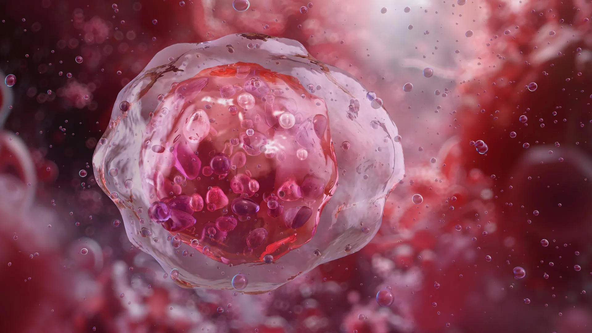
To understand the magnitude of this breakthrough, one must examine the delicate biological balancing act governing somatic stem cells. Stem cells are the fundamental, immature building blocks responsible for the maintenance, repair, and regeneration of virtually all tissues in the mammalian body. Under normal physiological conditions, adult blood stem cells—primarily resident within the spongy microenvironment of the bone marrow—reside in a quiescent, slowly dividing state. This evolutionary hibernation protects the precious stem cell pool from DNA replication errors, exhaustion, and premature aging.
However, when the body experiences acute trauma, hemorrhage, or systemic stress, these quiescent cells must rapidly switch gears. They are stimulated, or mobilized, to exit the bone marrow sanctuary, enter the systemic circulation, and migrate to sites of injury or localized depletion. Once deployed, they multiply exponentially and differentiate into mature, functional components of the circulatory and immune systems, including red blood cells, platelets, and white blood cells.
For decades, clinicians and cellular biologists have wrestled with a frustrating paradox: while this quiescent state is necessary for long-term stem cell preservation, it presents a formidable barrier in clinical settings. When harvested for transplants or extracted for ex vivo genetic modification, these resting cells are often sluggish, resistant to expansion, and slow to engraft in a recipient host. This inertia is particularly pronounced when cells are harvested from patients who have already endured intensive rounds of systemic chemotherapy or radiation therapy, treatments notoriously damaging to bone marrow niches.
Unraveling the Genetic Machinery: The Role of FLI-1
To decipher the complex molecular machinery driving this cellular awakening, the Weill Cornell Medicine research team deployed advanced single-cell profiling techniques and high-throughput computational analyses. Their goal was to map the precise differential gene activity distinguishing quiescent blood stem cells from their actively proliferating counterparts.
Through rigorous genomic interrogation, the investigators zeroed in on FLI-1, a transcription factor protein known to govern the transcriptional output of thousands of distinct genes. The absence or suppression of FLI-1 was shown to lock blood stem cells in their dormant state, effectively severing their biochemical dialogues with neighboring cells in the bone marrow. Most critically, a lack of FLI-1 shuts down communication with specialized endothelial cells that line the local blood vessels—a specialized microenvironmental hub known as the vascular niche.
Conversely, when FLI-1 activity is introduced and restored, it immediately re-establishes these vital physiological connections. FLI-1 acts as a conductor, orchestrating the co-adaptability between the stem cell and its vascular niche. By pushing the stem cell into an activated, highly regenerative metabolic and transcriptional state, FLI-1 dramatically amplifies the cell’s capacity to proliferate, expand its numerical volume, and successfully engraft within a new host organism.
A Safe Chronological Approach: Overcoming Oncogenic Risks
Despite its powerful regenerative properties, manipulating FLI-1 historically carried significant scientific hesitation. In the fields of hematology and oncology, it is well-documented that genetic mutations leading to the chronic overactivity or dysregulation of FLI-1 are established drivers of certain types of leukemias and blood cancers. Indiscriminate or prolonged upregulation of this potent transcription factor could theoretically plunge normal cells down a malignant pathway.
To circumvent this formidable safety hurdle, the research team engineered an innovative, transient delivery method inspired by the technological framework of modern modified mRNA-based vaccines. Rather than permanently altering the genome or sustaining chronic protein expression, the investigators utilized modified messenger RNA to stimulate the blood stem cells with transient bursts of FLI-1 for a strictly controlled window of just a few days.
This temporal precision proved to be the key to the entire methodology. By administering the signal temporarily, the stem cells are effectively coaxed out of their biological hibernation, undergo rapid expansion, and acquire robust functional and durable engraftment capabilities upon transfer to a recipient—all while exhibiting zero evidence of malignant transformation or tumor formation.
Solving a Long-Standing Hematological Mystery
Beyond its immediate therapeutic implications, the study successfully resolved a long-standing biological puzzle that had perplexed the hematology community for years: why human umbilical cord blood-derived stem cells possess significantly greater regenerative potential than adult stem cells isolated from peripheral blood.
The investigators demonstrated that this disparity in potency is directly associated with baseline differences in FLI-1 activity. Umbilical cord blood stem cells maintain a molecular profile that naturally optimizes their interactions with the regenerative vascular niche, a property that adult stem cells lose over time. By artificially modulating FLI-1, researchers can essentially imbue adult stem cells with the enhanced regenerative vigor typically seen in neonatal tissues.
Furthermore, the study shed new light on the bi-directional signaling dynamics that govern the bone marrow microenvironment. Bioinformatics analyses led by the research team proved that stem cell activity is neither entirely cell-autonomous nor strictly dictated from the top-down by endothelial signals. Instead, successful regeneration is contingent upon a dynamic, reciprocal biochemical dialogue and co-adaptability between the stem cell and its vascular niche.
Expert Insights and Official Perspectives
The clinical implications of this discovery are vast, touching upon numerous branches of modern medicine where hematopoietic cell replenishment is the primary therapeutic modality.
"The approach we outlined in this study could substantially improve the efficiency of marrow transplants and marrow-cell-targeted gene therapies, especially in cases where the donor has a very limited supply of viable blood stem cells," stated study senior author Dr. Shahin Rafii. Dr. Rafii holds multiple prestigious titles at Weill Cornell Medicine, serving as the director of the Hartman Institute for Therapeutic Organ Regeneration and the Ansary Stem Cell Institute, chief of the division of regenerative medicine, and the Arthur B. Belfer Professor in Genetic Medicine. He is also affiliated with the Englander Institute for Precision Medicine and the Sandra and Edward Meyer Cancer Center.
Marrow transplants are a cornerstone of modern oncology, essential for rescuing patients with hematological malignancies—such as leukemias, lymphomas, and multiple myelomas—whose native bone marrow has been obliterated by high-dose conditioning regimens. However, finding a matching donor can be exceptionally difficult, and harvested grafts often contain insufficient quantities of functional stem cells to guarantee rapid and safe immune reconstitution.
Similarly, advanced gene therapies for severe monogenic blood disorders, such as beta-thalassemia and sickle cell disease, rely heavily on harvesting a patient’s own autologous blood stem cells, inserting a healthy therapeutic gene via viral vectors, and expanding these vulnerable, manipulated cells in an ex vivo laboratory environment before re-infusing them into the patient. The fragile nature of these cells during ex vivo culture has historically created major bottlenecks in manufacturing and clinical delivery. A safe, reliable pharmacological or mRNA-based switch to drive rapid, secure expansion would streamline these complex therapeutic protocols.
Co-first author Dr. Tomer Itkin, formerly an instructor of biology in medicine in the Rafii laboratory and currently the director of Tel Aviv University’s Neufeld Cardiovascular Research Institute alongside his role as an assistant professor at the Sagol Center for Regenerative Medicine, emphasized the safety profile of the intervention. "The stem cells we prime with FLI-1 modified mRNA in this way wake up from hibernation, expand and functionally and durably engraft in the recipient host, without any evidence of cancer," Dr. Itkin noted.
Adding to the computational depth of the project, co-first author Sean Houghton, a bioinformatics analyst in the Rafii laboratory during the research and currently a senior bioinformatics analyst for the Englander Institute for Precision Medicine at Weill Cornell Medicine, highlighted the collaborative nature of the microenvironmental signaling. "We showed that stem cell activity is not autonomous but also is not fully determined by endothelial cell vascular niche signals—it depends instead on signaling and adaptability between the two."
Broader Impact and Future Directions
The publication of these findings marks the transition from foundational discovery to translational development. Buoyed by the success of their preclinical models, the Weill Cornell Medicine team is actively planning further developmental phases, which will involve scaling up their modified mRNA-based methodology to safely and transiently introduce FLI-1 into human blood stem cells.
The ultimate objective of this translational pipeline is the initiation of human clinical trials. If successfully translated to the clinic, this approach could fundamentally alter the standard of care for bone marrow transplantation, mitigate the risks associated with graft failure, enhance the viability of limited donor supplies, and accelerate the commercial and clinical viability of complex gene therapies. By unlocking the master regenerative switch of the blood system, researchers are moving closer to a future where diverse hematological disorders can be treated with long-term, stable, and completely safe blood production.
The research detailed in this study was made possible through substantial financial backing and grant support from the National Institutes of Health (NIH), specifically the National Heart, Lung, and Blood Institute (NHLBI), the National Institute of Diabetes and Digestive and Kidney Diseases (NIDDK), and the National Institute of Allergy and Infectious Diseases (NIAID), under grant numbers R35HL150809, R01DK136327, and U01AI138329. Additional philanthropic and institutional support was provided by the Hartman Institute for Therapeutic Organ Regeneration, the Ansary Stem Cell Institute, and the Selma and Lawrence Ruben Daedalus Fund for Innovation at Weill Cornell Medicine. In accordance with institutional disclosure policies, Dr. Shahin Rafii is noted as an unpaid co-founder of Angiocrine Bioscience.