Researchers in Austria have unveiled a pioneering technological framework that bridges the gap between subatomic particle physics and advanced computation, introducing a design for a quantum computer-integrated electron microscope. Developed through a collaborative consortium spanning TU Wien, the University of Vienna, JKU Linz, and the University of Innsbruck, this novel diagnostic tool aims to overcome fundamental physical limitations that have constrained transmission electron microscopy for decades. By harnessing the principles of quantum entanglement and algorithmic data processing, the system is engineered to extract hidden data payloads from standard imaging electrons, effectively transforming weak scatter signals into high-contrast structural maps while dramatically reducing the cumulative radiation dose delivered to fragile specimens.
The Main Facts of the Quantum-Electron Hybrid System
At its operational core, conventional transmission electron microscopy relies on bombarding a target sample with a concentrated beam of high-energy electrons. As these particles traverse the material, they interact with its atomic structure, altering their trajectories or velocities. Standard detectors simply count the spatial distribution of these transmitted or scattered electrons to reconstruct a grayscale projection of the interior layout. However, this classical counting method discards a vast array of quantum-mechanical properties inherently borne by every individual electron passing through the column.
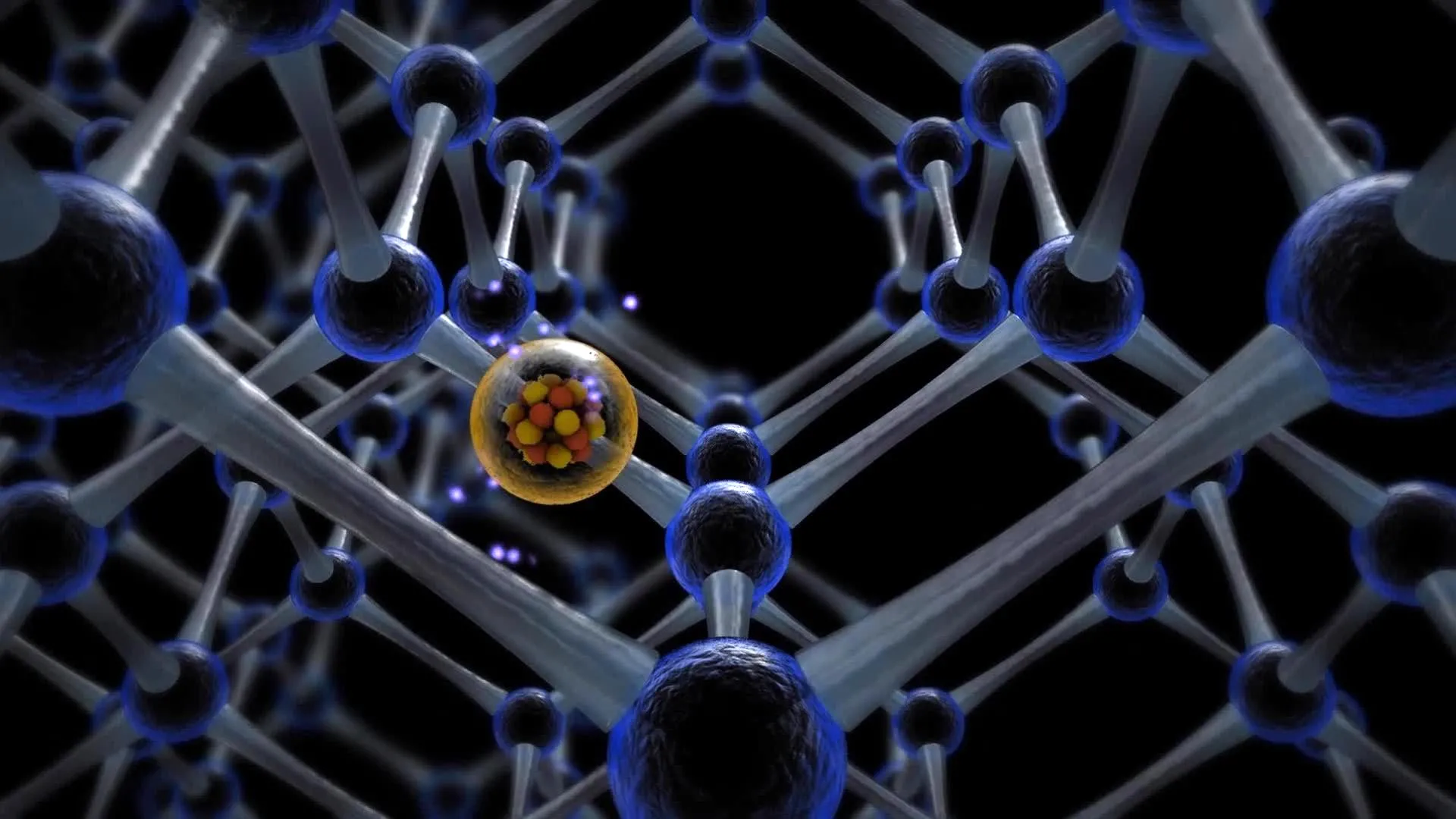
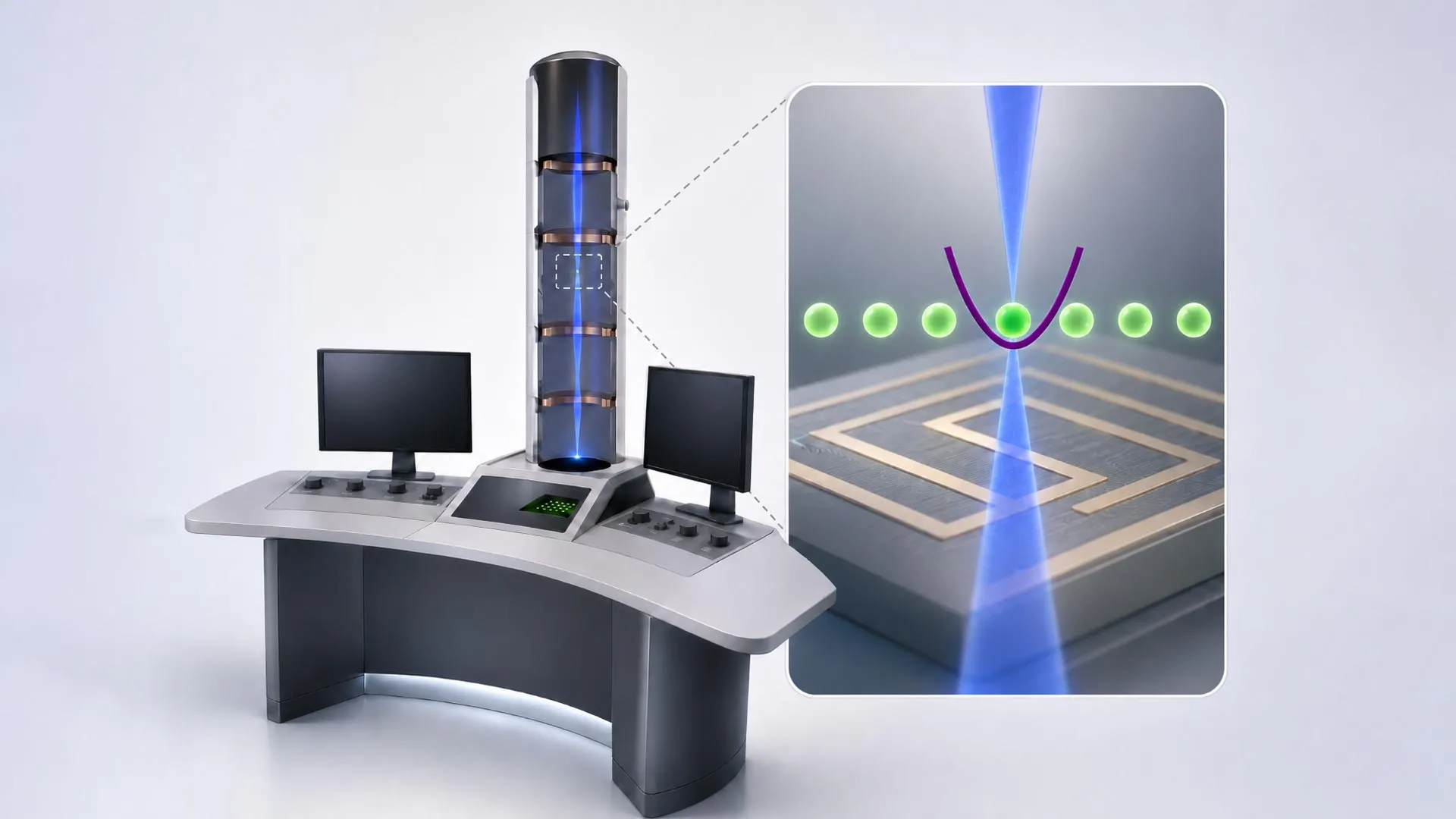
The Austrian research collaborative has designed an apparatus that intercepts this lost quantum information. By positioning a specialized trapped-ion quantum computer directly along the path of the electron beam, the system facilitates direct physical interactions between the imaging electrons and the stationary ions. This interaction establishes quantum entanglement—a phenomenon wherein two separate physical systems become deeply interconnected, sharing a unified quantum state regardless of spatial separation.
When an electron interacts with a trapped ion, information regarding the electron’s phase, momentum, and subtle scattering interactions is transferred and securely stored within the quantum register of the ion. Subsequent electrons passing through the column undergo similar entangling operations. Through a sequence of precisely tailored quantum logic gates, the integrated quantum processor aggregates these incremental data fragments. This collective processing converts what would normally register as random background noise in a conventional detector into a coherent, high-strength signal. Consequently, scientists can acquire pristine structural data using a fraction of the electron dose traditionally required, addressing a critical bottleneck in structural biology and materials science.
Historical Context and the Evolution of Electron Microscopy
The development of electron microscopy began in the early 1930s, spearheaded by physicists Ernst Ruska and Max Knoll, who realized that accelerated electrons possess a de Broglie wavelength significantly shorter than that of visible light. This fundamental physical property allowed microscopists to bypass the optical diffraction limit, opening the door to imaging at magnifications millions of times greater than conventional light microscopes. Over the subsequent decades, incremental engineering improvements—such as aberration correctors, field-emission guns, and high-stability power supplies—pushed resolution down to the sub-angstrom scale, allowing researchers to visualize individual atomic columns and lattice defects.
Despite these monumental hardware advancements, a stubborn obstacle remained: the destructive nature of the electron beam. High-energy electrons carry substantial momentum and kinetic energy. When directed onto delicate, beam-sensitive materials—most notably biological macromolecules, native proteins, and soft-matter polymers—the collision cascade induces severe radiolytic damage, bond breakage, and molecular rearrangement. In cryo-electron microscopy, a technique that earned the Nobel Prize in Chemistry in 2017, samples are flash-frozen in vitreous ice to mitigate structural degradation, yet they remain highly susceptible to radiation damage.
For decades, the trade-off between signal-to-noise ratio and radiation damage appeared immutable. Improving image contrast necessitated a higher electron dose, which in turn destroyed the very structures researchers sought to analyze. The new quantum-enhanced approach directly targets this physical impasse. By mathematically decoupling the signal-to-noise ratio from the raw electron dose, the technology challenges the standard statistical constraints that have governed microscopy since its inception.
Chronology of the Collaborative Research Effort
The genesis of this interdisciplinary breakthrough traces back to the formation of the quantA Cluster of Excellence, a major research initiative funded by the Austrian Science Fund (FWF) designed to foster synergy between quantum information science and practical technological applications.
The foundational theoretical work began when researchers at TU Wien, integrating expertise from theoretical physics and atomic physics, started modeling how free electrons could be coupled coherently with bound quantum systems without disrupting the primary imaging pipeline. By formulating the necessary mathematical proofs, the team demonstrated that quantum memory registers could successfully store phase and momentum data scavenged from a passing electron beam.
As the theoretical framework solidified, the consortium expanded to incorporate specialized competencies from partner institutions. The algorithmic architectures required to coordinate multi-electron entanglement and subsequent quantum-state processing were co-developed with Johannes Kofler’s research group at JKU Linz. Simultaneously, physical hardware realization plans took shape, drawing upon the trapped-ion quantum computing hardware expertise led by Philipp Schindler’s team at the University of Innsbruck.
With mathematical validation complete, the project entered its hardware integration phase. Researchers are currently constructing the physical prototype at TU Wien’s University Service Center for Transmission Electron Microscopy (USTEM). This facility brings together the vacuum systems, electromagnetic lenses, and electron optics of a standard microscope alongside the delicate laser-cooling and ion-trapping apparatus required to maintain the quantum computer.
Supporting Data, Technical Parameters, and Theoretical Limits
In conventional particle counting, the statistical precision of an image is governed by Poisson distribution statistics, meaning that the signal-to-noise ratio scales proportionally with the square root of the number of detected electrons ($N$). To double the clarity of a structural image via standard methods, researchers must quadruple the electron dose delivered to the sample. For radiation-sensitive biological specimens, this quadratic penalty represents an insurmountable wall.
The quantum-enhanced approach proposed by the Austrian consortium aims to circumvent classical shot-noise limits by exploiting multi-particle quantum correlations. By storing phase information in a quantum memory array rather than relying strictly on direct spatial intensity measurements, the information yield per particle increases non-linearly. According to preliminary theoretical projections formulated by the team, the system can extract multiple bits of quantum metadata per electron that are ordinarily washed out by thermal fluctuations, chromatic aberrations, and shot noise.
Furthermore, the integration of trapped-ion systems—which are renowned for their exceptionally long coherence times and high gate fidelities—provides a stable platform for storing and manipulating the delicate quantum states transferred from the beam. The system operates by systematically cycling electrons through the interaction zone, performing real-time quantum error correction and state distillation before reading out the final integrated signal via laser fluorescence of the trapped ions.
Official Responses and Statements from the Research Consortium
The interdisciplinary nature of the project has drawn widespread acclaim from the European scientific community, highlighting the power of collaborative grant structures.
"Today, we can image tiny details on the atomic scale," noted Philipp Haslinger from the Institute of Atomic and Subatomic Physics at TU Wien. "However, this requires a large number of electrons. And not every sample can be exposed to so many electrons without being damaged. This is often a problem, particularly when imaging biological samples such as individual proteins."
Detailing the mechanics of the system, Elias Pescoller, doctoral student and first author of the study at TU Wien, explained the integration process: "Our idea is to combine the electrons with a quantum computer. We let them interact with ions that are held in place along the path of the electron beam. This can, for example, create quantum entanglement between the electron and the quantum computer. The electron and the ion then share a joint quantum state."
Pescoller also emphasized the broader paradigm shift represented by the work: "Quantum physics allows us to overcome the statistical limits that constrain conventional electron microscopes."
Addressing the operational advantages during data acquisition, Dennis Rätzel of TU Wien noted, "If we perform very specific quantum-computing operations each time, we can optimally combine the information from several electrons so that we obtain a signal of maximum strength even though we use only a relatively small number of electrons."
Iva Březinová from the Institute for Theoretical Physics at TU Wien underscored the implications for data recovery: "The electrons themselves are used to image small objects, just as in any other electron microscope. But by processing the quantum information carried by these electrons in a quantum computer, we can extract significantly more information from the process. What would previously have been indistinguishable from random noise can thus become a clear signal."
Highlighting the institutional synergy, Thomas Juffmann from the University of Vienna remarked, "It is really exciting that, within the quantA Cluster of Excellence, we can combine the expertise in quantum information, quantum computing and electron microscopy available at the different universities in Austria. This allows us to launch a unique project."
Broader Impacts and Future Implications for Science and Industry
The successful realization of a quantum computer-integrated electron microscope could catalyze a transformative shift across multiple scientific disciplines. In structural biology, the ability to obtain high-resolution tomograms of dynamic macromolecules, membrane proteins, and viral capsids at ultra-low electron doses would reduce structural artifacts caused by beam-induced denaturation. This could grant structural biologists unprecedented insights into native-state protein folding, drug-target interactions, and cellular machinery at the single-molecule level.
In materials science and nanotechnology, the technology holds immense promise for characterizing fragile two-dimensional materials, beam-sensitive perovskites, metal-organic frameworks, and battery interfaces. Many advanced materials degrade rapidly under the high-current densities typically required to resolve light elements or subtle atomic distortions. By extracting maximum intelligence from minimal particle counts, engineers could map atomic vacancies, dopant distributions, and interfacial boundaries with minimal structural perturbation.
Beyond microscopy, the successful coupling of free-electron beams with stationary quantum processors opens up exciting avenues in fundamental physics. The experimental platform established at USTEM could serve as a versatile testbed for studying fundamental quantum electrodynamics, decoherence phenomena in particle beams, and novel forms of quantum-enhanced metrology.
As the construction phase progresses from mathematical validation to physical execution, the global scientific community will closely monitor the Austrian consortium. If the experimental integration of trapped-ion systems within an operational microscope column meets expectations, it will not merely upgrade an existing analytical instrument; it will redefine the fundamental boundaries of how humanity observes the subatomic architecture of the universe.