

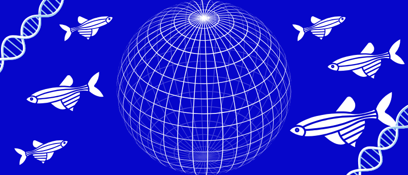
A groundbreaking advancement in developmental biology has emerged from the University of Basel in Switzerland, where a research team has unveiled a novel imaging method capable of visualizing the intricate dance of thousands of genes simultaneously across entire zebrafish embryos. This pioneering technology, dubbed weMERFISH, has enabled the creation of a comprehensive 4D atlas that meticulously maps the activity of genes and the movements of cells as a simple cluster of cells transforms into a fully formed embryo with distinct head, trunk, and tail structures. Published in the prestigious journal Science, this work represents a significant leap forward in understanding the fundamental processes of life, offering an unprecedented window into the complex interplay that orchestrates early development.
For decades, scientists have grappled with one of biology’s most profound questions: how does a singular fertilized egg orchestrate the intricate cascade of events that culminates in a complex, multi-cellular organism? The journey from a rudimentary cell cluster to a fully differentiated embryo is a symphony of genetic instruction and cellular action, involving the precise activation and deactivation of thousands of genes and the coordinated migration, differentiation, and organization of billions of cells. Unraveling this highly complex interplay has been a central challenge in developmental biology, with implications spanning from understanding congenital anomalies to advancing regenerative medicine. This new atlas promises to demystify many of these previously opaque processes.
Prior to this innovation, research into gene activity during embryogenesis was largely confined to two-dimensional slices of tissue. While these methods offered valuable insights into localized gene expression, they inherently lacked the holistic, three-dimensional context necessary to comprehend whole-embryo development. The fragmented views often failed to capture the dynamic, spatially detailed patterns of gene activity, particularly at a subcellular resolution, making it challenging to track how genetic programs drove cellular behaviors across the entire developing organism. Such limitations hindered a complete understanding of how cellular maturation and movement were intrinsically linked to the underlying genetic instructions, leaving many questions about morphogenetic movements and tissue patterning unanswered.
The team, led by Professor Alex Schier at the Biozentrum, University of Basel, sought to overcome these significant hurdles. Their innovative weMERFISH technology now allows for the direct measurement of the activity of nearly 500 genes across entire tissues with subcellular precision. Crucially, by integrating these direct measurements with existing single-cell data, the researchers were able to extrapolate and calculate the spatial patterns of thousands more genes, alongside the activity of approximately 300,000 potential regulatory regions. This unparalleled level of detail has culminated in the creation of the first comprehensive 4D atlas of early embryonic development, providing a dynamic blueprint of how genes and cells collaboratively sculpt the growing embryo.
The Enigma of Embryonic Development: A Biological Imperative
The formation of an embryo is a process of breathtaking complexity and precision. From the moment of fertilization, a single cell embarks on a tightly regulated program of cell division, differentiation, and morphogenesis, giving rise to all the diverse cell types and tissues that constitute a living organism. This journey is governed by the genome, a vast instruction manual encoded in DNA, where genes act as blueprints for proteins and regulatory molecules. The challenge for developmental biologists lies not just in identifying these genes, but in understanding when, where, and how intensely they are expressed, and critically, how their expression patterns coordinate the physical changes and movements of cells that shape the embryo.
Classical embryology, dating back centuries, meticulously documented the visible stages of development through observation and microscopy. With the advent of molecular biology, researchers began to probe the genetic underpinnings, identifying key developmental genes and signaling pathways. Techniques such as in situ hybridization allowed for the visualization of specific gene expression patterns in tissues, while genetic screens in model organisms like the fruit fly, nematode, and zebrafish identified genes critical for various developmental processes. However, these methods often provided static snapshots or focused on a limited number of genes, making it difficult to grasp the overarching, dynamic regulatory networks that control whole-organism development. The sheer scale of genomic information and cellular interactions demanded a more comprehensive and integrated approach, one that could capture both spatial and temporal dimensions across the entire embryo simultaneously.
Bridging the Gap: From 2D Slices to 4D Realities
The transition from single-cell organisms to complex multicellular forms necessitates an exquisite level of spatial and temporal control over gene expression. Previous techniques, while foundational, often presented a fragmented view. Standard immunohistochemistry or traditional RNA in situ hybridization methods typically allowed for the visualization of one or a few genes at a time on thin tissue sections. While powerful for targeted studies, reconstructing the entire spatial landscape of gene activity across a whole embryo from hundreds of such slices was a monumental, often impractical, task, inherently prone to losing critical 3D context and subtle spatiotemporal dynamics.
Furthermore, the rise of single-cell RNA sequencing (scRNA-seq) revolutionized our ability to profile gene expression in individual cells, providing unprecedented insights into cellular heterogeneity and developmental trajectories. However, scRNA-seq inherently dissociates cells from their native tissue context, sacrificing crucial spatial information. The challenge thus became how to combine the gene-expression resolution of scRNA-seq with the spatial and temporal resolution required for whole-embryo studies. The scientific community recognized the urgent need for a technology that could spatially map the transcriptional states of thousands of genes within intact tissues, thereby bridging the gap between gene expression dynamics and actual cellular behavior and morphology. This necessity directly fueled the development of innovative spatial transcriptomics approaches, with weMERFISH standing out as a particularly powerful iteration.
weMERFISH: A Technological Marvel for Spatial Genomics
At the heart of this breakthrough is weMERFISH (weighted Multiplexed Error-Robust Fluorescence in situ Hybridization), a sophisticated imaging technology designed to achieve highly accurate and multiplexed detection of RNA molecules within intact biological samples. Unlike traditional in situ hybridization methods that use a single fluorescent probe per target RNA, MERFISH employs a combinatorial barcoding strategy. Each RNA molecule is labeled with a unique sequence of fluorescent probes, allowing for the simultaneous detection and identification of hundreds or even thousands of different RNA species within the same cell or tissue. The "error-robust" component refers to built-in redundancy and error correction mechanisms that ensure high fidelity in RNA identification, even in densely packed cellular environments. The "weighted" aspect likely refers to optimized probe design and imaging protocols to enhance signal-to-noise ratio and quantitative accuracy.
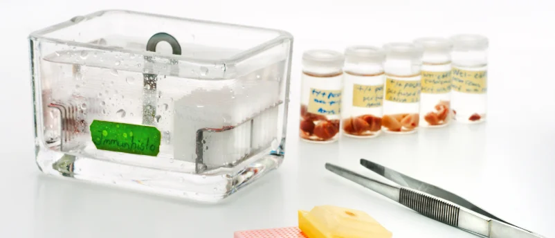
By applying weMERFISH to early zebrafish embryos, the Basel team could directly measure the activity of nearly 500 genes with subcellular resolution. This means they could not only tell which genes were active in which cells, but often, where within the cell (e.g., nucleus vs. cytoplasm) the RNA molecules were localized, providing additional layers of regulatory insight. The zebrafish embryo is an ideal model system for such studies due to its external development, transparency, and rapid developmental timeline, allowing for live imaging and easy manipulation. The ability to integrate these direct, high-resolution spatial measurements with existing single-cell transcriptomic datasets allowed the researchers to infer the spatial patterns of thousands of additional genes that were not directly probed, thereby creating an even richer and more comprehensive map of gene expression across the entire developing organism.
The 4D Atlas: A Global Resource for Developmental Biologists
The culmination of this technological prowess is the creation of a dynamic 4D atlas of early embryonic development. This atlas is not merely a collection of static images; it is an interactive, multidimensional resource that maps gene activity, gene regulation, and cellular movements in both space (three dimensions) and time (developmental progression). As Yinan Wan, the first author of the study, articulated, "A central question has been: How do thousands of genes work together in an embryo, and how is their activity linked to the movement of cells?" The weMERFISH technology provided the means to answer this question with unprecedented detail.
The atlas integrates the direct weMERFISH measurements with previous single-cell data, enabling the calculation of spatial patterns for thousands of genes and the activity of approximately 300,000 potential regulatory regions across the embryo. This vast dataset has been made freely accessible to the global scientific community through a dedicated web platform, MERFISHEYES (http://schier.merfisheyes.com). This open-access approach underscores the team’s commitment to accelerating research worldwide. As Wan emphasized, "The atlas is intended as a resource for developmental biologists around the world," providing an invaluable tool for hypothesis generation, data validation, and the discovery of novel regulatory mechanisms. This commitment to open science magnifies the potential impact of the research, allowing countless other laboratories to leverage this foundational data for their own investigations into embryogenesis, regeneration, and disease.
Key Discoveries: Unveiling Developmental Secrets
The application of the weMERFISH technology and the subsequent creation of the 4D atlas have already yielded several profound insights into the mechanisms governing embryonic development. These discoveries challenge previous assumptions and provide novel perspectives on how complex structures arise.
One of the most striking observations relates to the spatial representation of temporal progression, a phenomenon Wan described by saying, "In a sense, you can see time in space." During the formation of the zebrafish tail, the researchers observed a clear gradient: immature stem cells were localized at the very tip of the tail, while progressively more mature cells, such as specialized muscle cells, were found further anterior along the body axis. This spatial arrangement effectively mirrors the temporal sequence of development, where cells differentiate and mature as they move away from the stem cell niche. This finding provides a powerful framework for understanding how developmental programs unfold sequentially and how cellular identities are established along an axis of growth.
Another significant revelation from the atlas pertains to the intimate relationship between gene expression dynamics and morphogenetic movements – the physical shaping of the embryo. The researchers were surprised to find a strong alignment between changes in gene activity and the ways in which cells move through the embryo. This direct link between molecular programming and macroscopic cellular behavior is crucial. It suggests that the precise timing and location of gene activation directly dictate the forces and movements that drive cell migration, tissue folding, and organ formation. This insight moves beyond simply observing correlation to strongly inferring causation, offering a mechanistic understanding of how genetic instructions translate into physical form.
Perhaps one of the most paradigm-shifting discoveries addresses the formation of sharp boundaries between different tissues, such as the demarcation between muscle and backbone tissue. Historically, it was often hypothesized that such clear boundaries might arise through a process of "cell sorting," where cells intermingle and then actively segregate into distinct populations based on differential adhesion or other physical properties. However, the Basel team’s atlas revealed a different primary mechanism. They identified specific zones of cells where the activity of many genes changes dramatically and distinctly from one side of the boundary to the other. A comparison of early and later developmental stages showed that these genes were initially active on both sides of the nascent boundary but later became restricted to only one side. Crucially, the researchers observed minimal crossing of these boundaries by cells. As Professor Alex Schier eloquently stated, "These boundaries do not arise because cells are intermingled and then sort, but mainly because cells change their genetic program." This finding highlights the paramount role of intrinsic genetic programming in establishing tissue architecture, rather than solely relying on dynamic cell movements and re-arrangements.
Chronology of Innovation and Future Trajectories
The development of weMERFISH and the subsequent creation of the 4D atlas represent the culmination of years of dedicated research and technological innovation within Professor Schier’s laboratory at the Biozentrum. While the precise timeline for weMERFISH development is not detailed, such sophisticated imaging techniques typically involve extensive iterative cycles of biochemical optimization, hardware engineering, and computational algorithm development, often spanning several years. The data acquisition for the zebrafish atlas itself would have involved meticulous sample preparation, high-resolution imaging across multiple developmental stages, and massive computational resources for data processing and analysis, likely over a period of many months, if not years, before the rigorous peer-review process for Science publication.
The publication of this work has been met with significant excitement within the developmental biology community. Leading experts anticipate that the MERFISHEYES atlas will become a fundamental resource, analogous to the Human Genome Project or comprehensive brain atlases, accelerating discovery across numerous subfields. Researchers worldwide can now access and interrogate this rich dataset, testing hypotheses about specific gene functions, regulatory networks, and cellular behaviors with unprecedented spatial and temporal context.
Broader Impact and Future Implications
The combined power of weMERFISH, the MERFISHEYES atlas, and its potential integration with live imaging techniques establishes a formidable new toolkit for researchers. It allows for the unprecedented joint analysis of gene activity, gene regulation, and cell movement across the entire embryo, opening doors to a deeper understanding of vertebrate development.
Looking ahead, Professor Schier’s team plans to expand the atlas to encompass additional developmental stages, further completing the picture of early vertebrate development. This ongoing work will progressively reveal how the intricate genetic programs evolve as the embryo matures, building increasingly complex structures and organs. The long-term vision articulated by Schier is ambitious yet profoundly impactful: "In the long term, we want to understand which combinations of gene activity and cellular behavior are required to form a specific organ or tissue." This fundamental understanding is critical not only for basic biological knowledge but also for translational applications.
The implications of this research are far-reaching. For regenerative medicine, understanding the precise genetic and cellular instructions required to form a heart or a spinal cord could revolutionize approaches to tissue engineering and organ regeneration, offering hope for patients suffering from organ failure or traumatic injuries. By delineating the "recipe" for organ formation, scientists could potentially guide stem cells to differentiate into specific cell types and assemble into functional tissues in vitro or in vivo.
Furthermore, this atlas provides an invaluable resource for understanding the origins of birth defects and developmental disorders. Many congenital anomalies arise from errors in gene expression or cellular movements during embryogenesis. By having a comprehensive baseline of normal development, researchers can more effectively pinpoint deviations in gene activity or cellular behavior that lead to pathological outcomes. This could lead to earlier diagnosis, better prognostic tools, and novel therapeutic interventions.
From an evolutionary perspective, Schier’s musing – "One day we may find out how many ways there are to build a heart or a spinal cord" – underscores the atlas’s potential to inform evolutionary developmental biology. By comparing the developmental programs of different species, scientists can identify conserved mechanisms and evolutionary innovations, shedding light on the diversification of life forms. The ability to precisely map gene activity during the formation of complex structures across various organisms could reveal the genetic toolkits and regulatory logic that have been repurposed or modified throughout evolution.
In conclusion, the development of the weMERFISH technology and the creation of the 4D developmental atlas represent a monumental achievement in biological imaging and developmental biology. By providing an unprecedented, holistic view of gene-cell coordination during embryogenesis, this work not only answers long-standing questions but also lays a robust foundation for future discoveries, promising to transform our understanding of life’s most fundamental processes and pave the way for revolutionary advancements in medicine and biotechnology.


Leave a Reply