An international collaborative research effort, spearheaded by scientists at the Bernhard Nocht Institute for Tropical Medicine (BNITM) in Hamburg, Germany, and the Icahn School of Medicine at Mount Sinai in New York, USA, has leveraged a sophisticated human cerebral organoid model to illuminate the previously elusive mechanisms by which the Ebola virus (EBOV) can persist within the immune-privileged confines of the human brain. This groundbreaking investigation, published recently, provides unprecedented detail into a phenomenon that poses significant risks of inflammatory disease, long-term relapse, and critically, the potential for re-initiation of devastating EBOV disease outbreaks. Understanding this long-term survival strategy of the virus within its host is paramount for developing effective therapeutic interventions and enhancing global health security.
The Persistent Threat: Understanding Ebola Virus Disease and its Aftermath
Ebola virus disease (EVD), caused by various species of the Ebolavirus genus, is a severe, often fatal illness with a notorious history of devastating outbreaks across Africa. As a member of the Filoviridae family, which also includes Marburg virus (MARV) and Sudan virus (SUDV), EBOV is characterized by its pleomorphic structure and a single-stranded, negative-sense RNA genome. The disease typically manifests with sudden onset of fever, intense weakness, muscle pain, headache, and sore throat, followed by vomiting, diarrhea, rash, impaired kidney and liver function, and in some cases, both internal and external bleeding. The average case fatality rate of EVD is around 50%, though this can vary significantly depending on the specific virus species and the quality of healthcare. For those who survive the acute phase of EVD, a complex and often debilitating array of symptoms, collectively known as Post-Ebola Syndrome (PES), can persist for months or even years. PES can affect nearly every organ system, leading to chronic fatigue, muscle and joint pain, vision problems, hearing loss, and neurological complications, including severe meningoencephalitis, which is inflammation of the brain and its surrounding membranes.
The persistence of EBOV in immune-privileged organs such – such as the testes, eyes, and particularly the brain – has been a major clinical and epidemiological concern. These organs are characterized by barriers that naturally limit immune surveillance to protect their delicate tissues from damaging inflammatory responses. While crucial for normal function, this reduced immune response inadvertently creates sanctuaries where viruses can evade detection and elimination, leading to persistent viral particles. The mechanisms underpinning this viral sanctuary and the subsequent long-term persistence have remained largely a mystery, hampering efforts to fully understand PES and prevent future outbreaks originating from survivors.
A New Frontier in Research: The Power of Human Cerebral Organoids
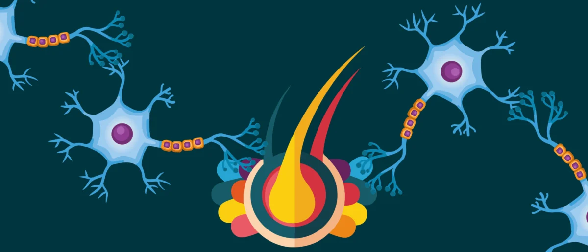
Traditional research models, particularly animal models, have provided valuable insights into EVD, but often fall short in fully replicating the intricate human host-pathogen interactions, especially within the complex architecture of the central nervous system. This is where the innovative application of human cerebral organoids has revolutionized the field. Cerebral organoids are three-dimensional, self-organizing cell cultures derived from human induced pluripotent stem cells (iPSCs). These "mini-brains" recapitulate key aspects of human brain development, structure, and cellular diversity, including neurons, astrocytes, oligodendrocytes, and microglia – the primary immune cells of the brain. This makes them an exceptionally powerful tool for studying neurological diseases, neurodevelopmental disorders, and, critically, human-specific neurotropic viral infections without the ethical and logistical challenges associated with animal experimentation.
For this study, researchers meticulously generated these cerebral organoids from human iPSCs, providing an unprecedented platform to directly observe and analyze filovirus infection and persistence within a human central nervous system context. This model allowed them to investigate in detail how Ebola virus and other filoviruses interact with human brain cells over extended periods, an endeavor previously fraught with technical and biological limitations.
Lina Widerspick, the first author of the publication and a former researcher at the BNITM, underscored the significance of this model: "These cerebral organoids enable us to investigate in detail the mechanisms that Ebola virus and other filoviruses use to persist in the human central nervous system. Through experiments in this model system, we can gain insights that help us improve our understanding of the long-term effects of persistence, like the severe and sometimes fatal inflammation seen in Ebola virus disease survivors with meningoencephalitis." Her statement highlights the direct clinical relevance of the organoid model, bridging the gap between laboratory findings and patient outcomes.
Unveiling the Mechanisms of Viral Persistence
Using their advanced cerebral organoid model, the research team made several critical discoveries regarding the persistence of filoviruses. They observed productive persistent infection with EBOV, SUDV, and MARV within the organoids for an extended period, up to 120 days. This long duration of active infection is crucial, as it mirrors the timeline observed in some EVD survivors experiencing long-term complications.
A significant finding was the diverse range of cerebral cell types susceptible to EBOV infection. The virus was found to infect neurons, the fundamental signaling units of the brain, as well as astrocytes, which support neuronal function; oligodendrocytes, responsible for myelin production; and microglia, the brain’s resident immune cells. This widespread cellular tropism suggests that EBOV can establish a broad foothold within the brain, making complete viral clearance challenging.
Further investigation into the mechanisms facilitating this persistence revealed two key modes of viral spread. Beyond the traditional method of budding from the host cell, where new virions are released into the extracellular space to infect new cells, the researchers also observed significant cell-to-cell transmission. This direct transfer of viral particles between adjacent cells offers a stealthier means of propagation, potentially allowing the virus to spread while largely evading detection by the immune system, which primarily targets extracellular viral particles. This cell-to-cell spread could be a critical factor in establishing and maintaining long-term viral reservoirs within the brain.

Despite the immune-privileged status of the brain, the organoids did produce pro-inflammatory cytokines in the later stages of infection. These chemical messengers are typically deployed by the immune system to combat pathogens. However, a critical observation was that this elevated immune response, while present, proved insufficient to curb the persistent infection. This paradoxical situation – inflammation without viral clearance – is consistent with clinical observations in EVD survivors. Co-senior author César Muñoz-Fontela from BNITM explained, "We therefore conclude that a persistent Ebola virus infection in immune-privileged tissues can lead to local inflammation. This observation is consistent with the fact that some Ebola virus disease survivors develop inflammation of the eye, meninges, or brain months after infection with Ebola virus." This direct correlation between experimental findings and clinical manifestations strengthens the validity and impact of the study.
Adding another layer of complexity to the persistence puzzle, the researchers linked the formation of defective viral genomes (DVGs) and particles to EBOV persistence. They found mutations in EBOV genomes within late-stage, persistently infected cerebral organoids. DVGs are truncated or altered viral genomes that often arise during viral replication. While they are typically non-infectious on their own, they can interfere with the replication of full-length infectious viruses and modulate host immune responses. The presence of these mutations and DVGs suggests a sophisticated viral strategy: by generating defective genomes, the virus might be able to limit intracellular detection, thereby reducing the host cell’s alarm signals and allowing a low-level, persistent infection to continue under the radar.
Historical Context: The Shadow of Ebola Outbreaks
The threat of EVD is not new. The first recognized outbreaks occurred in 1976 in simultaneous events in Sudan and the Democratic Republic of Congo (then Zaire). Since then, numerous outbreaks have occurred, primarily in sub-Saharan Africa. The most significant to date was the 2014-2016 West African Ebola epidemic, which affected Guinea, Liberia, and Sierra Leone. This epidemic was unprecedented in scale, duration, and geographic spread, resulting in over 28,000 cases and more than 11,000 deaths. It exposed critical weaknesses in global health infrastructure and preparedness, and importantly, brought the issue of long-term survival and post-Ebola syndrome to the forefront of medical research.
During and after the West African epidemic, clinicians noted a growing number of survivors experiencing persistent symptoms, including severe neurological issues. These observations underscored the urgent need to understand how the virus could linger in the body, well beyond the acute phase of illness. Subsequent outbreaks, such as those in the Democratic Republic of Congo, continued to highlight the challenges of managing survivors and preventing potential re-emergence from persistent reservoirs. For instance, in 2021, a new Ebola case was identified in Guinea, linked to a survivor from the 2014-2016 outbreak, tragically demonstrating the real-world implications of viral persistence and the potential for rekindling outbreaks.
The Global Health Imperative: Addressing Post-Ebola Syndrome
The findings from this study are critically important for addressing the global health imperative of Post-Ebola Syndrome. For EVD survivors, the burden of PES can be immense, impacting their quality of life, mental health, and economic stability. Understanding the precise mechanisms of viral persistence in the brain, particularly how it leads to inflammation and neurological damage, is the first step towards developing targeted therapies that can alleviate or prevent these debilitating long-term effects. Currently, treatment for PES symptoms is largely supportive, but a deeper understanding of the underlying viral processes could pave the way for specific antiviral or anti-inflammatory interventions.
Moreover, the risk of viral re-initiation of outbreaks from survivors is a serious public health concern. While rare, documented instances of EBOV transmission from survivors have occurred. Identifying how and where the virus hides, and the conditions under which it might become active again, is crucial for developing strategies to monitor survivors, provide appropriate care, and mitigate the risk of new outbreaks. This research directly contributes to that understanding, offering a scientific basis for improved surveillance and public health interventions.
Future Directions and the Promise of Organoid Research
This work not only significantly advances our understanding of EBOV persistence in the brain but also powerfully demonstrates the expanding and promising role that cerebral organoids can play in investigating intricate host-virus interactions. The implications extend far beyond Ebola, suggesting that organoid models could become indispensable tools for studying a wide array of neurotropic viruses and other infectious diseases.
One of the most immediate and significant implications is the potential for optimizing antiviral development. By providing a human-relevant model system, researchers can more effectively screen potential antiviral compounds, identify those that can penetrate immune-privileged sites like the brain, and evaluate their efficacy in clearing persistent infections. This could dramatically accelerate the pipeline for developing much-needed therapies for EVD and other viral diseases. Furthermore, the use of organoids has the potential to substantially reduce the reliance on animal models in infectious disease research, addressing ethical concerns and improving the translational relevance of preclinical studies.
However, the researchers emphasize that more work is needed. To fully comprehend the complex relationship between the virus and its host, further long-term studies are essential. Understanding the dynamics of persistence over even longer periods, and how environmental or host factors might influence viral latency and reactivation, remains a critical area of investigation. Additionally, a more diverse and comprehensive investigation of other filoviruses, beyond EBOV, SUDV, and MARV, is necessary to provide a complete characterization of filoviral persistence mechanisms across the family. Such broad studies could reveal common strategies employed by these highly pathogenic viruses, leading to more universal therapeutic approaches.
The international collaboration between BNITM and the Icahn School of Medicine at Mount Sinai exemplifies the power of global scientific partnerships in tackling complex health challenges. Their collective expertise, combined with cutting-edge organoid technology, has unveiled fundamental aspects of Ebola virus biology that were previously inaccessible. This foundational research holds the promise of transforming our approach to EVD, moving from merely treating acute illness to comprehensively managing long-term health consequences for survivors and preventing future epidemics. The journey to fully eradicate the threat of Ebola is long, but this study marks a significant stride forward in that critical endeavor.