Leaf through a textbook, watch a wellness influencer or listen in at the gym, and it can feel as though the human body has already been mapped to exhaustion. Every muscle named, every nerve traced. Everything understood and readily available. Most people recognize at least a few anatomical terms – "traps," "glutes," "biceps." After centuries of dissection, microscopy, and medical imaging, it seems reasonable to assume the work is done. Surely anatomy, as a discipline, must be complete?
It isn’t. Not even close.
The enduring perception of anatomical completeness stems from foundational texts that, while revolutionary for their time, created an illusion of definitive knowledge. The publication of Andreas Vesalius’s De Humani Corporis Fabrica in 1543 marked a seismic shift in anatomical understanding. For centuries, medical knowledge had been heavily reliant on the writings of ancient physicians, particularly Galen of Pergamon. Galen, a Greek physician in the Roman Empire, had conducted extensive dissections, but primarily on animals, inferring human anatomy from these observations. Vesalius, however, championed direct observation through human dissection. His meticulous illustrations and detailed descriptions challenged long-held Galenic doctrines, correcting numerous errors and establishing anatomy as an empirical science grounded in visible evidence. This work, a monumental undertaking for its era, solidified the idea that the human body was finally being systematically documented.
Three hundred years later, the publication of Gray’s Anatomy by Henry Gray in 1858 further reinforced this impression. Gray’s comprehensive and accessible textbook, accompanied by detailed anatomical plates, became an indispensable resource for medical students and practitioners. It presented a seemingly complete catalog of the body’s structures, meticulously indexed and organized, solidifying the notion that anatomy had reached a state of near-perfection. The widespread adoption and continuous revision of Gray’s Anatomy over subsequent editions cemented its status as the authoritative text, leading many to believe that the fundamental mapping of the human body was a closed chapter.
However, the seemingly neat and orderly presentation of anatomical knowledge in textbooks belies a far more complex and dynamic reality. These educational tools, designed for clarity and pedagogical effectiveness, often present a generalized, universalized model of the human body. They offer a "typical" or "standard" anatomy, which, while essential for foundational learning, is a simplification. Real human anatomy is inherently variable, a spectrum rather than a single, fixed blueprint.
The Historical Foundations: A Legacy of Limitation
The development of anatomical knowledge, particularly in its early topographical stages – the mapping of structures in relation to one another – was significantly shaped by the challenging and often illicit methods of acquiring human cadavers. For centuries, obtaining bodies for dissection was fraught with legal and ethical hurdles. The demand from burgeoning medical schools and anatomists outstripped legitimate supply, leading to the rise of "resurrectionists," or body snatchers. These individuals, often driven by financial incentives, exhumed recently buried bodies.
The social stratification of the time meant that the poor, the institutionalized, and those without family to guard their graves were disproportionately targeted. These vulnerable populations, often marginalized in life, became the primary subjects for anatomical study in death. The bodies obtained were frequently in various states of decomposition, having been subjected to the rigors of burial and the passage of time. Furthermore, the conditions under which early anatomists worked were far from ideal. Poor lighting, limited preservation techniques, and the inherent challenges of working with decaying tissue significantly impacted the quality of observations.
The demographic profile of the cadavers available for dissection was also inherently skewed. Many bodies were likely malnourished or suffered from chronic diseases, leading to atypical presentations of tissues and organs. The bodies of women, in particular, were often dissected but infrequently documented or described in detail, contributing to a male-centric view of anatomical norms. Sample sizes were typically small and opportunistic, meaning that anatomists were limited to the bodies they could acquire, rather than conducting systematic studies across diverse populations. Consequently, the anatomical "norm" that emerged from these studies was constructed from a narrow, socially stratified, and often compromised sample.
This is not to diminish the extraordinary technical skill and keen observational abilities of early anatomists. Their dedication to direct observation, particularly in challenging circumstances, was groundbreaking. However, the inherent limitations of their methods and the societal contexts in which they operated inevitably shaped what they could see and, crucially, what they might have missed. The idealized diagrams and descriptions that eventually found their way into textbooks were, therefore, built upon a foundation that was not as universally representative as it might have seemed.

A Slowdown and a Renaissance
Following the intense periods of anatomical discovery and cataloging in the 18th and 19th centuries, the pace of anatomical investigation began to slow considerably in the 20th century. By the 1960s, the publication of new cadaveric studies had dwindled worldwide. The prevailing assumption was that the human body had been thoroughly mapped, its structures comprehensively understood and documented. Medical education, while continuing to thrive, largely shifted its focus from generating new anatomical observations to teaching and reinforcing the established body of knowledge. This perceived stability, however, masked a deeper issue: much of the accepted anatomical understanding had been inherited and accepted rather than continuously tested and re-evaluated.
However, this period of relative stasis was not to last. A confluence of factors in recent decades has triggered a significant renaissance in anatomical study. Advancements in medical imaging technologies, such as high-resolution MRI, CT scans, and ultrasound, have provided unprecedented non-invasive views of living human anatomy, allowing for the study of structures in their dynamic, functional context. Simultaneously, there has been a renewed interest in rigorous cadaveric research, employing modern techniques and a greater emphasis on detailed documentation.
Furthermore, a growing awareness of the vastness of anatomical variation has challenged the notion of a single, universal human form. Researchers are now actively exploring the spectrum of human anatomy, recognizing that deviations from the textbook norm are not anomalies but rather integral aspects of human biological diversity. This renewed scientific curiosity, coupled with improved technological capabilities, has revealed that the map of the human body is far from complete, with many structures and variations still awaiting detailed description and understanding.
Beyond the "Standard" Human Body: Embracing Variation
One of the most profound paradigm shifts in modern anatomy has been the recognition that anatomical variation is not the exception, but the rule. Textbooks, for pragmatic reasons, present a generalized "typical" human body to facilitate learning. However, in reality, human anatomy exists along a complex spectrum, influenced by a multitude of factors.
These variations manifest across several dimensions simultaneously. There are inherent differences between males and females, influenced by genetics and hormonal profiles. The body undergoes significant transformations throughout the lifespan, from development in utero and childhood growth to the aging process, each stage presenting distinct anatomical characteristics. Furthermore, genetic predispositions and environmental influences, including diet, lifestyle, and geographic location, contribute to population-level anatomical differences.
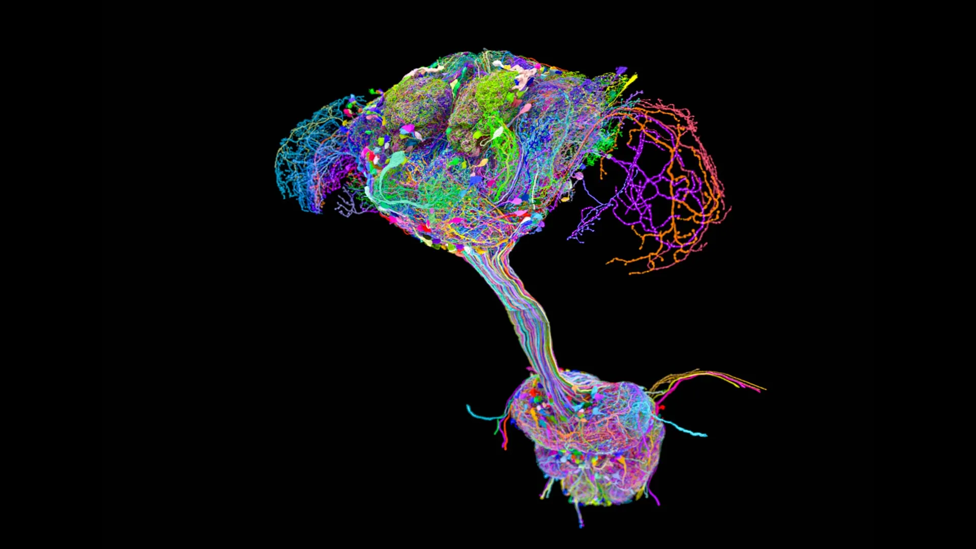
Beyond these broad patterns lies an enormous degree of individual variation. The branching patterns of blood vessels can differ significantly from person to person, with arteries and veins taking alternative routes. Muscles may be absent in some individuals, while others might possess duplicated or accessory muscles. Even the intricate folding patterns of the cerebral cortex, crucial for cognitive function, exhibit considerable inter-individual variability. The "standard" anatomy depicted in textbooks should, therefore, be understood not as a universal blueprint but as a simplified reference point within a wide biological range.
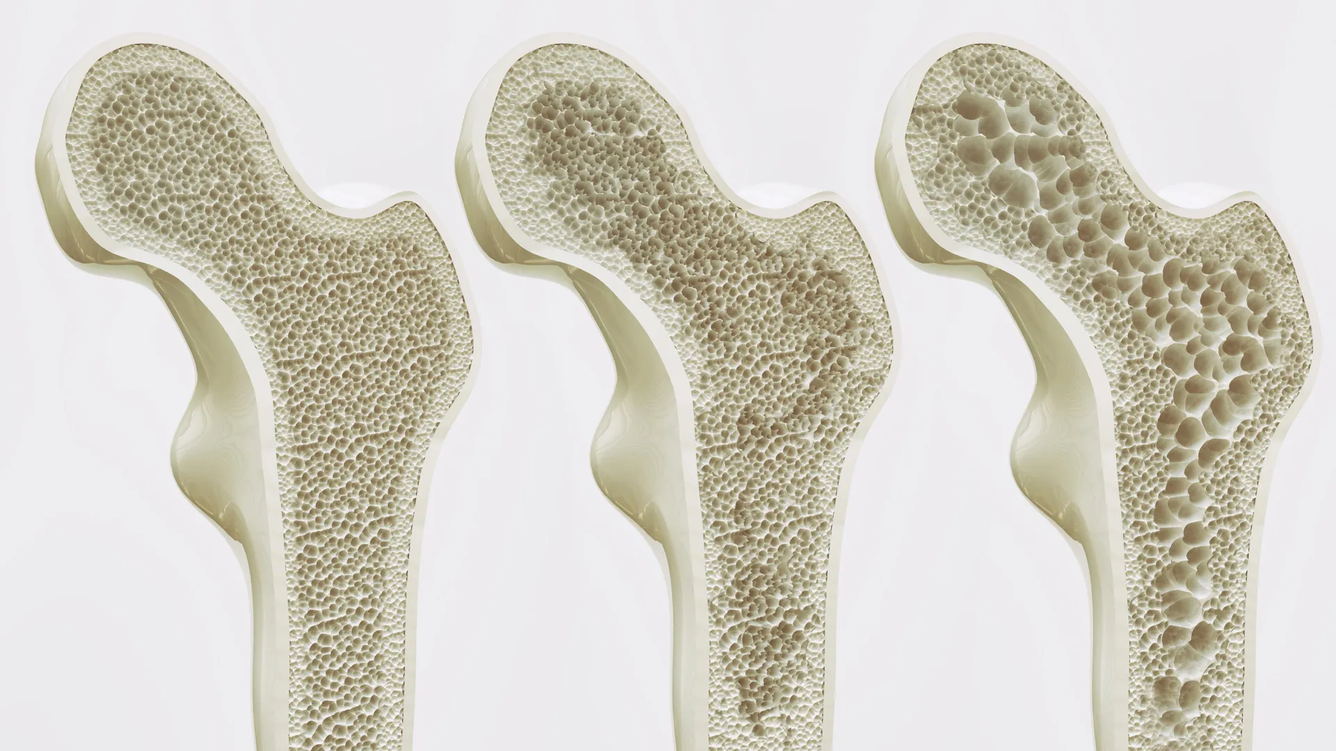
The implications of this anatomical diversity extend far beyond the confines of an anatomy lecture hall or surgical theater. Subtle differences in the alignment of joints, for instance, can influence an individual’s susceptibility to conditions like osteoarthritis. Variations in vascular anatomy can play a critical role in determining an individual’s risk profile for stroke or aneurysm. Understanding this intricate tapestry of anatomical diversity is therefore central not only to the precision required in surgical interventions but also to the accuracy of medical imaging interpretation, the development of biomechanical models, and the study of disease pathogenesis.
Unveiling the Unseen: New Discoveries and Future Frontiers
Even after centuries of dedicated study, the human body continues to yield new anatomical insights. Structures that were once overlooked, poorly described, or even unknown are being brought to light through advanced research techniques. Recent years have seen the identification of previously unrecognized lymphatic vessels in the dura mater, the protective outer membrane of the brain, which play a crucial role in immune surveillance and fluid drainage. Similarly, overlooked ligaments in the knee joint have been re-examined, revealing their importance in joint stability.
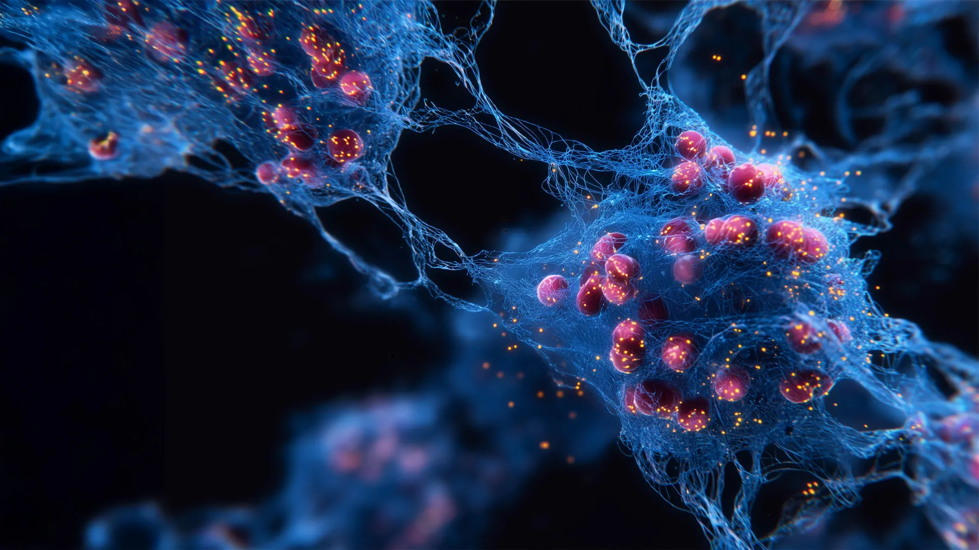
Familiar tissues are also being understood in entirely new ways. For example, research into the fascia – the connective tissue that surrounds muscles, bones, and organs – is revealing its complex role in proprioception (the sense of the relative position of one’s own parts of the body and strength of effort being employed in movement) and its potential involvement in chronic pain conditions. The "map" of the human body is not static; it is a dynamic document that is constantly being revised and expanded.
This ongoing exploration underscores a fundamental truth: the more closely we study the human body, the more we realize how much there is still to learn. While it is beneficial for individuals to possess a greater understanding of their own bodies to advocate for their health and engage more confidently with healthcare providers, it is crucial to remember that the canonical anatomy presented in educational materials is a model, a representation designed for teaching. It is not a perfect or exhaustive depiction of biological reality. The pursuit of anatomical knowledge is an ongoing journey, revealing the profound complexity and enduring mystery of the human form. The work of mapping the human body, it seems, is far from over.