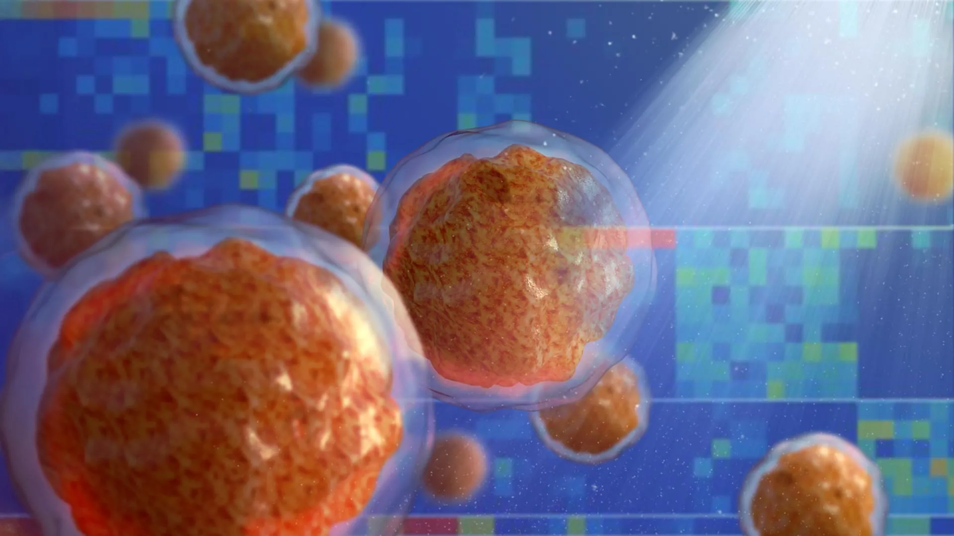
The human body’s hematopoietic system, often referred to as the "blood factory," is one of the most complex and dynamic environments in the biological world. Located within the core of our bones, this tissue is responsible for the continuous production of billions of new blood cells every day, including oxygen-carrying red cells, clotting platelets, and the diverse array of white cells that constitute the immune system. For the first time in medical history, researchers have successfully recreated this intricate network in a laboratory setting using exclusively human cells. This achievement, led by a team at the University of Basel and University Hospital Basel, represents a paradigm shift in how scientists study blood-related diseases and test new pharmacological interventions.
The bone marrow is not merely a passive reservoir for cells; it is a highly specialized organ comprising bone cells, intricate nerve networks, a dense web of blood vessels, and various signaling molecules. Because of its deep-seated location and biological complexity, it has historically been one of the most difficult tissues to replicate or study in its natural state. When this system functions correctly, it remains largely invisible to the individual. However, when the delicate balance of blood production is disrupted—as is the case in leukemia, lymphoma, or various types of anemia—the consequences are life-threatening. Understanding the underlying mechanisms of these disruptions requires a model that can accurately mirror the human environment, a need that this new research aims to fulfill.
The Vital Role of Bone Marrow in Human Health
To appreciate the magnitude of this breakthrough, one must understand the biological significance of the bone marrow "niche." Within the marrow, there are specific microenvironments, or niches, that regulate the behavior of hematopoietic stem cells (HSCs). These stem cells are the precursors to all blood cells. The "endosteal niche," located near the internal surface of the bone, is of particular interest to oncologists and hematologists. This specific area is known to be a sanctuary for both healthy stem cells and malignant cancer cells.
In many forms of blood cancer, the endosteal niche provides a protective environment that allows cancer cells to remain dormant and resist conventional treatments like chemotherapy. Until now, scientists lacked a human-based platform that integrated all the necessary components of this niche—bone cells, blood vessels, immune cells, and nerves—into a single, functional 3D system. Previous research models were either too simplified, consisting of only one or two cell types in a petri dish, or relied on animal models, which often fail to replicate the specific molecular signaling pathways unique to human biology.
Overcoming the Limitations of Animal Models
For decades, the gold standard for studying bone marrow and blood diseases has been the use of mice and other animal subjects. While animal research has provided foundational knowledge regarding hematopoiesis, the translation of these findings to human clinical applications is often fraught with challenges. Mouse bone marrow, while structurally similar, possesses different genetic expressions and cytokine sensitivities than human marrow. This biological gap frequently leads to "translational failure," where a drug that appears effective and safe in mice fails to produce the same results in human clinical trials.
The researchers at the University of Basel, led by Professor Ivan Martin and Dr. Andrés García García, sought to bridge this gap. Their findings, recently published in the prestigious journal Cell Stem Cell, detail the creation of a model that brings researchers closer to the actual biology of the human organism. By removing the cross-species variables inherent in animal testing, the team has created a more reliable platform for observing how human diseases progress and how human cells respond to new chemical compounds.
Engineering the Endosteal Niche: A Technical Triumph
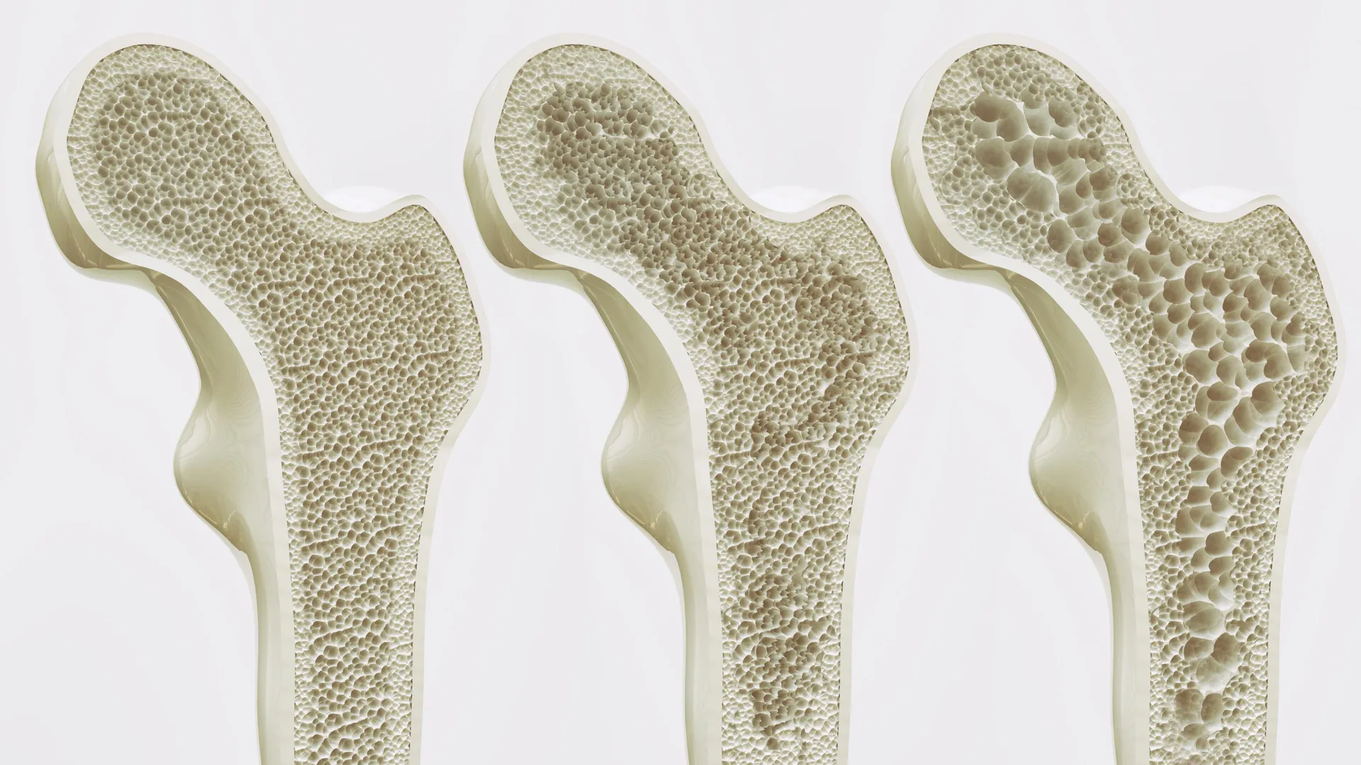
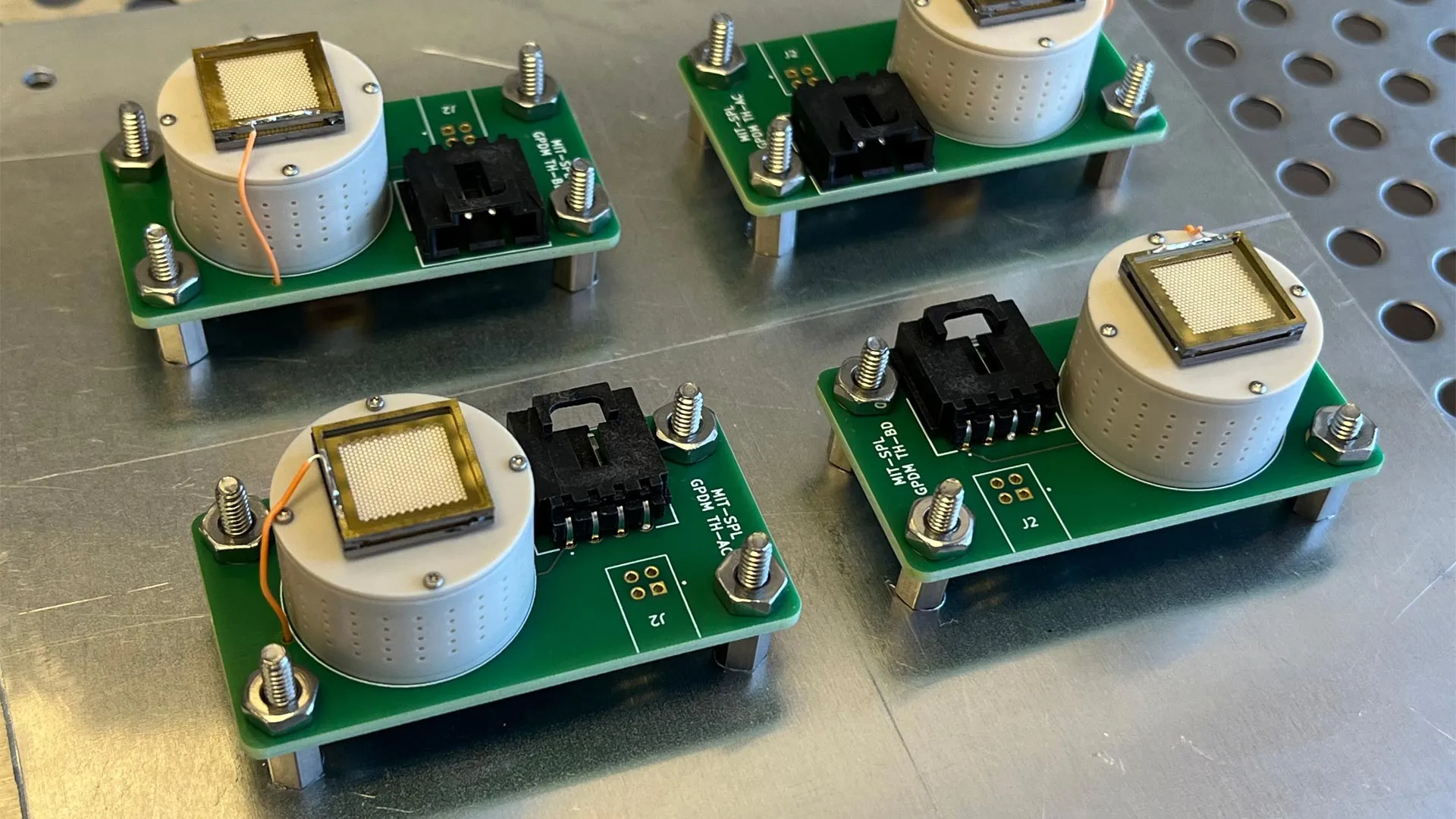
The development of the 3D bone marrow model was a multi-stage process that combined advanced bioengineering with molecular biology. The foundation of the system is an artificial bone framework made of hydroxyapatite. Hydroxyapatite is a naturally occurring mineral form of calcium apatite that makes up the primary inorganic constituent of human bones and teeth. By using this material as a scaffold, the researchers provided the cells with a familiar, biocompatible environment that mimics the physical stiffness and chemical composition of real bone.
The most innovative aspect of the study, however, lies in the use of induced pluripotent stem cells (iPSCs). These are adult cells that have been genetically reprogrammed to an embryonic-like state, giving them the ability to differentiate into almost any cell type in the human body. By applying specific molecular signals and growth factors to these stem cells within the hydroxyapatite scaffold, the team was able to guide their development into a diverse array of specialized tissues.
Over a period of several weeks, the stem cells transformed into bone-forming cells (osteoblasts), blood vessel cells (endothelial cells), and even nerve cells. This self-organizing process resulted in a three-dimensional structure that matches the human endosteal niche with unprecedented accuracy. The model’s dimensions are also a point of scientific interest; measuring approximately eight millimeters in diameter and four millimeters in thickness, it is significantly larger and more complex than previous "organ-on-a-chip" attempts.
Supporting Data and Functional Longevity
One of the primary challenges in tissue engineering is maintaining the viability and functionality of the lab-grown tissue over time. In this study, the researchers demonstrated that their 3D bone marrow model could maintain active human blood cell formation for several weeks. This longevity is crucial for long-term studies, such as observing the slow progression of a disease or the delayed side effects of a medication.
Analysis of the model showed that the cells within the system communicated through the same signaling pathways used in the human body. For example, the interaction between the mesenchymal stromal cells and the hematopoietic stem cells within the lab-grown niche mirrored the "cross-talk" observed in vivo. This data suggests that the model is not just a physical replica, but a functional biological system capable of mimicking the life cycle of blood cells.
Official Responses and the Scientific Perspective
The implications of this research have been met with enthusiasm within the academic community. Professor Ivan Martin emphasized the dual benefit of the model as both a scientific tool and an ethical advancement. "We have learned a great deal about how bone marrow works from mouse studies," Martin stated. "However, our model brings us closer to the biology of the human organism. It could serve as a complement to many animal experiments in the study of blood formation in both healthy and diseased conditions."
This sentiment aligns with the global "3R" principle—Replacement, Reduction, and Refinement—which encourages scientists to find alternatives to animal testing. By providing a high-fidelity human alternative, the University of Basel team is contributing to a broader movement within the pharmaceutical and medical research industries to move toward more ethical and human-relevant testing methodologies.
However, the team also acknowledged the hurdles that remain. Dr. Andrés García García pointed out that while the current model is ideal for studying complex biological interactions, its size might be a drawback for certain industrial applications. "For the specific purpose of drug development, the size of our bone marrow model might be too large," García García explained. For high-throughput drug screening, where thousands of compounds are tested simultaneously, the system would need to be miniaturized into a "micro-niche" format to allow for rapid, automated testing.
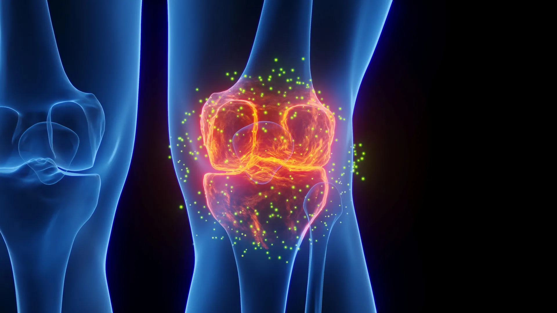
Implications for Oncology and Personalized Medicine
The most immediate impact of this 3D model is likely to be felt in the field of hemato-oncology. Blood cancers such as multiple myeloma and acute myeloid leukemia are notoriously difficult to treat because of their ability to "hide" within the bone marrow. By seeding this lab-grown marrow with cancer cells from patients, researchers can study how these malignancies interact with the surrounding tissue.
This opens the door to the era of personalized medicine. In the future, a doctor could take a small sample of a patient’s own cells, grow a personalized 3D bone marrow model in the lab, and then test various combinations of chemotherapy or immunotherapy on that specific patient’s tissue. This would allow clinicians to identify the most effective treatment strategy without the "trial and error" approach that often characterizes current cancer care, potentially saving lives and reducing the debilitating side effects of ineffective treatments.
Economic and Ethical Impact of Reducing Animal Testing
Beyond the clinical benefits, the transition toward human-cell-based models has significant economic implications. The cost of maintaining animal facilities and the high failure rate of animal-tested drugs contribute billions of dollars to the cost of drug development. A human-based model that can more accurately predict drug efficacy and toxicity could significantly streamline the pipeline from the laboratory to the pharmacy.
Furthermore, public and regulatory pressure to reduce animal testing is at an all-time high. In late 2022, the United States passed the FDA Modernization Act 2.0, which removed the requirement for animal testing for new drug protocols if alternative methods are available. The development of the Basel bone marrow model arrives at a pivotal moment, providing the very type of alternative that regulatory bodies are now actively seeking.
Future Directions and the Challenge of Scalability
As the researchers look toward the future, the next steps involve refining the model for broader use. This includes integrating a more robust immune system component and exploring how the model responds to systemic factors like hormones or aging. The challenge of scalability remains a primary focus; creating a version of the system that can be easily reproduced in laboratories worldwide is essential for its widespread adoption.
While the study represents an early step, it is a foundational one. The ability to grow a "blood factory" in a lab using only human cells was once the realm of science fiction. Today, it is a reality that promises to deepen our understanding of human biology, accelerate the discovery of new medicines, and offer a new glimmer of hope for patients battling blood diseases. The work of Martin, García García, and their colleagues at the University of Basel stands as a testament to the power of interdisciplinary science in solving some of the most enduring mysteries of the human body.